The Optic Nerve in Glaucoma
Douglas R. Anderson
THE NORMAL RETINA AND OPTIC NERVE
The approximately 1 to 1.5 million axons that form the optic nerve arise from the retinal ganglion cells and course toward the optic disc in a well-known pattern (Fig. 1). From the nasal retina the fibers take a straight course toward the disc. Axons originating temporal to the fovea arc around the macula to enter the upper and lower poles of the optic nerve head. The macula fibers pass directly to the temporal quadrant of the disc in the papillomacular bundle.
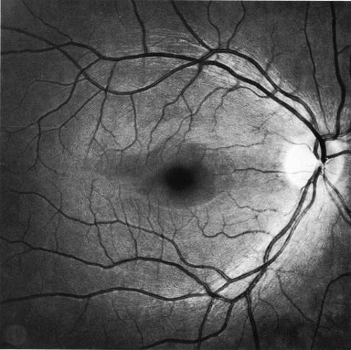 Fig. 1. Fundus photograph shows the normal pattern of the retinal nerve fiber layer. (Courtesy of P. Juhani Airaksinen, MD). |
The axons maintain an orderly grouping in the retina as bundles partitioned by sheets of Müller’s cells.1,2,3 A sheen reflected from the many bundles taking parallel courses gives the retina a striated appearance4 that is most prominent where the nerve fiber layer is thickest near the disc, especially in the arcuate bundles arriving at the poles of the disc from the temporal arcades.5,6 The attentive observer can see the striae of light reflected from these bundles and recognize their partial or total absence when bundles of axons disappear in glaucomatous or other forms of optic atrophy.4,7,8,9,10 The backscatter of light is determined by the cylindric nature and size of the light-scattering structures, possibly the axonal microtubules. They produce reflection with directional, spectral, and polarization (retardation) properties that have been studied and quantified, providing a basis for measuring the integrity of the retinal nerve fiber layer.11,12,13,14 The manner of layering of axons within intraretinal bundles2,3,6,15 may not be exactly the same in all primate species. However, a basic orderliness is present in the retina and seems to be maintained in the chiasm and beyond to the lateral geniculate body.16,17,18,19 Discrete scotomas and other defects caused by damage to nerve fiber bundles are produced by localized insults in the retinal nerve fiber layer or in the optic nerve head. Diffuse pressure on the posterior optic nerve by a mass lesion typically produces a central scotoma or diffuse visual dysfunction, but occasional instances of more localized damage in this region produce well-localized nerve fiber bundle defects, attesting to the existence of orderly bundles in this region also.12,13,14,15 On arriving at the lateral geniculate body, the axons are still organized and synapse in an orderly array.
In the retinal nerve fiber layer, axons converge from every direction toward the optic disc and turn to enter the optic nerve through an opening in the outer retina, the choroid, and the sclera. The features and anatomic variation of the normal optic nerve head, or optic disc, are illustrated in Figures 2, 3, 4, 5, 6, 7, 8, 9, 10, 11, 12, 13, 14, 15, and 16. A physiologic excavation (cup) results if the chorioscleral canal is larger than required for the approximately 1 to 1.5 million axons and the supporting glial cells and blood vessels. The size of the excavation depends on how ample is the size of the chorioscleral canal.20,21 In discs where the chorioscleral canal matches the number of axons, the chorioscleral canal itself is typically somewhat taller than it is wide (Figs. 2 and 3). However, because the number of nerve fibers entering the upper and lower poles of the disc is greater than in the temporal and nasal sectors, the boundary of the physiologic cup is more or less circular.22 When the disc is large, it may be nearly circular but the cup likewise nearly circular. However, except in discs of anomalous shape, the width of the rim of neuroretinal tissue is noted in normal, nonglaucomatous optic discs to be greatest in the inferior meridian followed by the superior meridian, and narrowest in the temporal quadrant.23
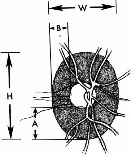 Fig. 2. Normal optic nerve configuration. Notice that the height (H) of the disc is greater than the width (W). The width of the neuroretinal tissue (A) is also greater in the vertical meridian than in the nasal and temporal meridians (B); thus, the physiologic cup is round. |
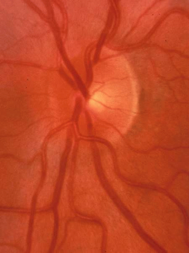 Fig. 3. Normal disc untilted, taller than it is wide. The neuroretinal tissue in the upper and lower sectors is more abundant in these sectors than others, so the central cup is round. A narrow white line marks the disc boundary and represents the lip of sclera that in humans almost universally separates the choroid from the optic disc tissue around the entire circumference. |
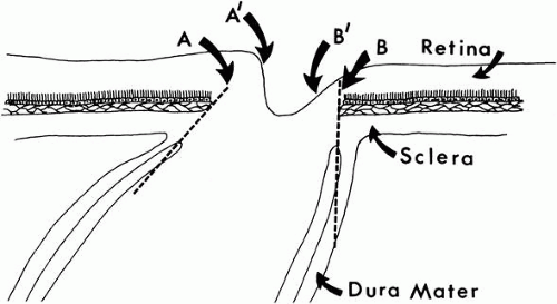 Fig. 4. Influence of the tilt of the scleral canal on the slope of the canal. An outward slope of the chorioscleral canal (A) corresponds to a steep wall of the cup (A′). A perpendicular wall of chorioscleral canal (B) corresponds to a sloping wall of the cup (B′). |
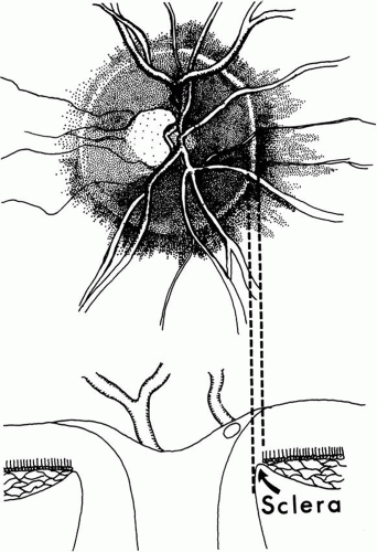 |
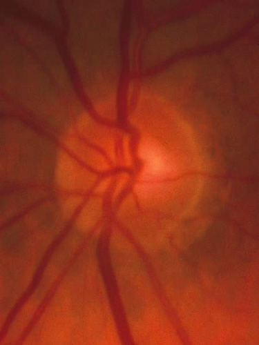 Fig. 6. Normal optic nerve head of moderate size with modest sized physiologic cup. Of note again is the white rim of sclera around the whole disc, seen most easily on the temporal side where the nerve fiber tissue crossing the disc margin is thinner and obscures the scleral rim less. There is a small peripapillary crescent (zone) of thin retinal pigment epithelium on the temporal side of the disc, as diagrammed in Figure 7. |
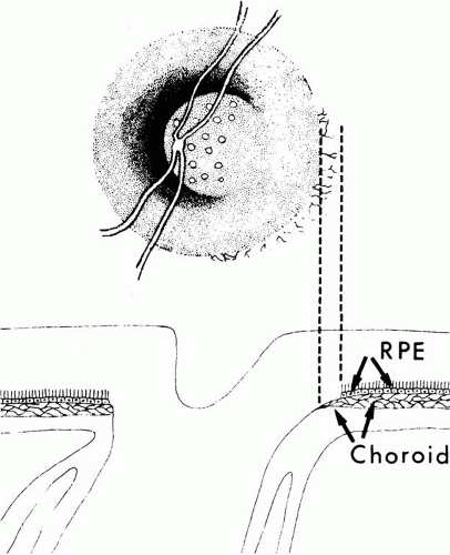 Fig. 7. Peripapillary crescent caused by misalignment of the several layers of the retina, choroid, and sclera. The choroid is not covered completely by the choriocapillaris and retinal pigment epithelium (RPE). |
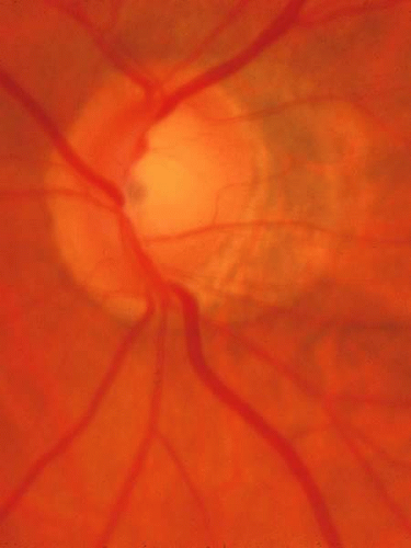 Fig. 8. Tilted disc, with vessel trunk beneath a nasal ledge, a sloping disc surface on the temporal side, and a peripapillary crescent (β zone) the edge of which envelops the upper and lower poles. Although difficult to judge at times in tilted discs, the neuroretinal tissue is thinned at the poles of the disc in a manner typical of glaucoma. |
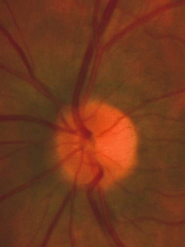 Fig. 9. Normal small disc with no cup because it is filled with neuroretinal tissue, which consists of the axons leaving the eye to form the optic nerve, along with the astroglia and the capillary bed. |
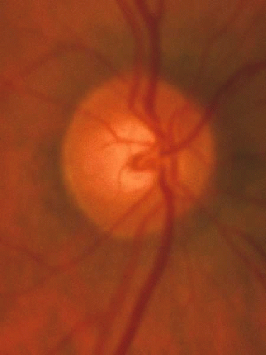 Fig. 10. Physiologic cup of approximately one third the disc diameter, resulting from a chorioscleral canal slightly larger than required to accommodate the exiting axons along with the astroglia and capillaries. The scleral lip is again present around the entire circumference, even though not conspicuous where the pink nerve fiber layer is thick enough to obscure it. There is no peripapillary zone of thin retinal pigment epithelium or choroid to form a crescent or halo. |
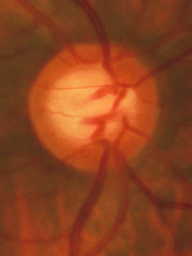 Fig. 11. Normal large disc with a large cup. The disc margin is nearly circular, and the cup has a slight horizontal oval shape, because the neuroretinal tissue is more abundant in the inferior and superior sectors than in the nasal and temporal sectors. |
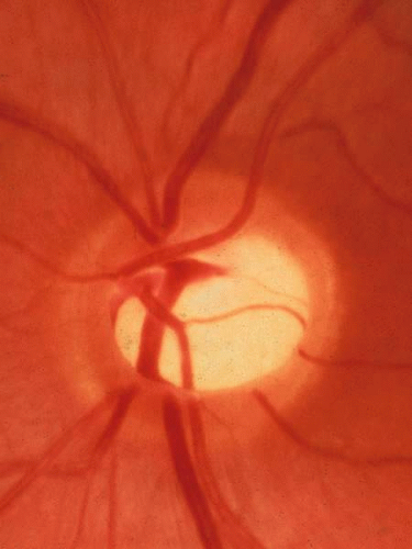 Fig. 12. Another large cup in a large disc. There is no location where the tissue is absent with the cup reaching the rim of the disc. The most typical sequence of the bulk of neuroretinal tissue, from greatest to least, is inferior, superior, nasal, temporal (ISNT). |
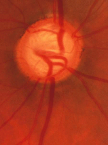 Fig. 13. Large disc that had a large physiologic cup, on which is superimposed glaucomatous tissue loss. The glaucomatous damage can be recognized by the fact that in the upper and lower sectors the tissue is thinner than nasally and temporally. In particular, careful inspection shows that at the inferior pole in a narrow region where a small venous tributary crosses the margin at the 6:30 position, the cup reaches just about to the disc margin, which is the inner edge of the white stripe that represents the scleral lip. The scleral lip sometimes has a sufficiently pink hue that is mistaken to be remaining neuroretinal tissue in a location where in fact there are not remaining axons. The ongoing glaucomatous process was signified by occasional splinter hemorrhages in this patient. Because the physiologic cup was large initially, the visual loss is strikingly moderate because despite the large cup, only a modest proportion of the original neuroretinal tissue has been lost. |
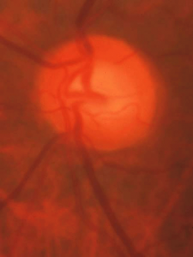 Fig. 14. Normal disc with a sloping surface of the disc surface (which is the same as a sloping margin of the cup wall). If tissue loss begins in the lower temporal sector of a disc, as is typical of glaucoma, it may be difficult to recognize the beginning of glaucomatous damage with backward displacement of the surface of “saucerization.” It would be particularly difficult in this disc, because the tissue is a homogeneous color with few vessles by which to judge the location of the tissue surface. |
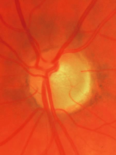 Fig. 15. Disc without glaucomatous disease, with a tilted exit of the optic nerve. The center of the optic nerve with the vascular trunk is markedly displaced toward the upper nasal edge. The bottom of the cup is represented as a small pale area just temporal to the place where the major retinal vessels penetrate the lamina cribrosa. The lower temporal disc surface, which in a sense is the lateral and temporal wall of the cup, is a broad sloped area of neuro-retinal tissue, somewhat pale because it is spread out over the scleral support of the sloped exit canal. This is another example of a normal disc within which early glaucomatous damage in the lower temporal sector might be difficult to recognize. |
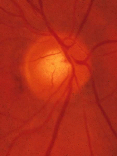 Fig. 16. Normal disc with moderate-sized cup. The vascular trunk is at the upper nasal edge of the cup, and the lower temporal sector has a sloped neuroretinal surface. This example has the fundamental anatomic features that make it difficult to recognize early glaucomatous loss in the lower temporal sector, but not to the unusually exaggerated degree of the previous two figures. |
The shape of the physiologic cup is affected by the obliquity of the wall of the canal through the choroid and sclera. The wall of the cup is steep where the wall of the canal is angled outward.24 Where the canal has a wall perpendicular to the ocular coats the cup has a sloping wall (Fig. 4). The slope of the cup may vary from one sector to another, and there is considerable individual variation in the size and shape of the chorioscleral canal and therefore of the optic cup. In many eyes, the optic nerve exits through a canal the passes nasally and upward into the orbit, so that the upper nasal neuroretinal rim is bounded by a steep wall when viewed from the ophthalmoscopic viewpoint, while the lower temporal wall is of the cup is sloped, so that ophthalmoscopically the cup wall is, in essence, being viewed from within the cup rather than from above. In other discs, particularly those with a large cup, the walls are perpendicular to the scleral wall in all meridians. There is also typically no evident slope of the exit canal in eyes with only a small dimple of a cup, resulting from a near-perfect match between the size of the chorioscleral canal and the amount of neuroretinal tissue (axons and glia) that comprise the optic disc. The normal anatomic variations were noted and classified by Eschnig,25 and the glaucomatous changes to be discussed below are superimposed on the anatomic character present natively.
The canal through which the optic nerve exits from the eye traverses several layers of peripapillary tissue: the outer retina, the pigment epithelium, Bruch’s membrane, the choriocapillaris, the outer choroid, and the sclera. In nearly all human eyes, in all or some meridians, a flange of sclera extends as far forward as the pigment epithelium, forming something of a border for the canal,25,26 and this can be seen ophthalmoscopically as a scleral rim around the disc (Figs. 5, 6, and 7). With or without such an obvious flange, the several chorioretinal layers may not be perfectly aligned with one another. Perhaps most typically, as the optic nerve exits obliquely in an upper nasal direction, the lower temporal sector of the disc viewed ophthalmoscopically is seen to have a crescent of misaligned edges of the several chorioretinal layers, such that the outer choroid and sclera may be left uncovered by the choriocapillaris and pigment epithelium (Fig. 8). Because of the tilt, the scleral flange is not seen as a narrow band; instead, by ophthalmoscopy its inner surface is viewed obliquely as it merges with the optic nerve sheath. In some myopic eyes, the crescent is most extensive on the temporal side, and in other eyes the crescent may be directly inferior, or for that matter less frequently in almost any position. In yet other variations the crescent encompasses much or all of the entire circumference of the disc, with misalignment or retraction of the various chorioretinal layers. When these eyes have glaucoma and the thin or retracted choroid surrounds the disc entirely, the area of thin choroid and absent retinal pigment epithelium has been termed a glaucomatous halo. The deformed anatomy associated with the tilted exit canal or the peripapillary crescent may mark the sector of the disc that is most susceptible to injury from elevated intraocular pressure27; for example, the lower temporal sector of the disc is most frequently the first sector affected.
PRESSURE-INDUCED DAMAGE
In the past, some believed that the damage to the optic disc in glaucoma occurred as the result of some sort of local sclerosis. Elevated intraocular pressure was not thought to be the cause, but simply to be an accompanying manifestation of a sclerotic process, that affected both the trabecular meshwork and the optic disc. Seeming evidence for this idea was a dissociation between the degree of cupping and the degree of pressure elevation,28,29,30 which is now attributed to individual variation in susceptibility to damage from intraocular pressure.31
A causative role of intraocular pressure is evident from cases of secondary glaucoma and from the fact that nerve damage is produced by elevated intraocular pressure in glaucoma produced experimentally in animals. Experienced clinicians have long agreed that progressive damage to the disc is halted when satisfactory lowering of intraocular pressure is achieved,32 and several recent clinical trials have now also shown more formally a cause-and-effect relationship between the level of intraocular pressure and damage.33,34,35 However, considerable individual variation is evident. Some individuals have intraocular pressure well above the normal range (ocular hypertension) for a long time without developing the anatomic and functional changes typical of glaucoma. Others, meanwhile, undergo progressive harm while the intraocular pressure is in the statistically normal range (normal-tension glaucoma), and the rate of harm is reduced with the intraocular pressure is lowered. Something makes the some optic nerves more susceptible to pressure-induced injury, and the gradation of susceptibility extends down into the normal range.
Factors that determine the susceptibility of the optic nerve to pressure-induced injury must exist and vary in amount to explain the individual variation in susceptibility to pressure-induced injury. Although the occurrence of damage does correlate with the level of intraocular pressure (and with its correlate, the resistance to aqueous humor outflow), the correlation with age is equally strong.28,29 This indicates that some of the susceptibility factors are age-related and that they are as important as the intraocular pressure in determining the visual loss in glaucoma. That some of the factors are genetic is evident from relationships to family history and race. Genetic makeup may impact not only the tendency to elevated intraocular pressure, but also susceptibility to harm that depends on the level of intraocular pressure. Observed relationships of particular susceptibility to female gender, migraine and other manifestations of vascular dysregulation, and clinical correlates or manifestations of vascular occlusive disease suggest the types of physiologic processes that may be altered by altered by the level of intraocular pressure.
Details of the pathophysiologic mechanism by which intraocular pressure produces optic nerve damage, exactly what the participating causative factors are, and how they interact together remain largely unknown.36 However, some clues are emerging. In some cases, inability to regulate blood flow in the optic nerve (dysregulation or inadequate autoregulation, which may be associated with migraine, with vascular overreaction to cold or stress, or with low blood pressure) is postulated by many37,38 to limit the ability of blood vessels in the optic nerve head to maintain sufficient blood flow when the intraocular pressure rises and challenges the circulation by raising venous pressure. It has been shown that individuals vary in their ability to regulate blood flow in the optic nerve,39 and potential mechanisms for local blood flow control by optic nerve capillaries have been studied in the laboratory.40 In other cases, vascular occlusive disease seems to play a role. If ischemia is a major cause for optic nerve damage in glaucoma, the different clinical presentation and appearance of a glaucomatous disc from that of a major acute vascular occlusive event (acute anterior ischemic optic neuropathy) remains a puzzle.
The are other events in the pathogenic pathways. Pressure seems to affect the astroglia, which express inducible nitric oxide synthase when the pressure is high,41 which on one hand might enhance vasodilation, but can also cause cytotoxic levels of nitric oxide. Axonal transport is impaired in the region of the lamina cribrosa, perhaps as a result of local ischemia,42 which may be reversed in time.43 With more serious or prolonged ischemia, damage to the axon membrane and entry of calcium may stimulate calcium-dependent proteases with irreversible damage to the axon segment would be expected. Any of these local events may prohibit transport of trophic factors, in particular nerve growth factor, to the retinal ganglion cells. In turn, after a time of inadequate arrival of growth factors, apoptosis is triggered with eventual death of the ganglion cell and its entire axon.44 Once triggered, apoptosis may occur even if the axon ischemia and blockage was reversed. It may be speculated that individual variation in the degree and duration of ischemia, free radical damage, and time of growth factor depravation before apoptosis is triggered might account for individual variation in susceptibility and in the rate of glaucomatous damage. Release of glutamate by dying ganglion cells has been postulated to add to the damage through excitotoxicity of adjacent retinal ganglion cells.45 While an interesting possibility, it remains uncertain whether excitotoxicity plays a major role in the pathogenic process.
Especially in cases in which the intraocular pressure is not particularly high, the collapse and stretching of the lamina cribrosa and other connective tissue in the region remains unexplained but is the histologic correlate to the clinically different appearance of the glaucomatous disc from other forms of optic atrophy (simple loss of axons). Recent evidence46 that a thin cornea places an eye at higher risk of damage when carrying an elevated intraocular pressure has suggested to some that the character of connective tissue in these individuals affects vulnerability, but there remains debate about whether the thin cornea simply produces an erroneous pressure measurement that masks the magnitude of the elevation of intraocular pressure.
Individuals vary in the rate of damage and in the level of intraocular pressure that is harmful, but perhaps also in the details of which of several root causes might participate or dominate in an individual, and which consequential events in the pathogenic sequence might be influenced by the intraocular pressure. Young individuals with secondary glaucoma and substantially elevated intraocular pressure develop cupping that is in part a diffuse stretching of the supporting tissues, and the elastic stretching may be reversible, while axon loss is not reversible. Older individuals with normal or slightly elevated intraocular pressure may develop shallower cupping with pallor thought to be related to chronic ischemia, which may involve all sectors of the disc or be quite localized. Cases may vary further depending on whether an occlusive disease or regulatory dysfunction participates in the postulated ischemic component, with the level of intraocular pressure possibly being more relevant in nonocclusive cases.47 There is more to learn about the entire pathogenic process, how to identify the dominant participating pathogenic processes in a given individual, and the pathways that are common to all cases that produce the typical clinical features of chronic glaucoma. Such information can be used in part to assess individual risk. Moreover, each of the contributing causes and participating pathophysiologic pathways involved in all cases or in particular identified individuals may be amenable to treatment that will improve the visual outcome.
We wait for the day when we will know more about the pathogenic process, know what the various susceptibility factors are, are able to measure them for the purpose of predicting how much pressure the eyes of a given person can tolerate, and can attack the susceptibility factors therapeutically. Until then, we know that a particular person is highly susceptible to future glaucomatous damage mainly when we observe that the pathogenic process is active or that the damage has begun,31 for example noting splinter optic disc hemorrhages or early glaucomatous anatomic or psychophysical (visual field) abnormality. The level of intraocular pressure is a weak predictor, but remains the focus of attention mainly because it is the only risk factor participating directly in the pathogenic mechanism that we know both how to measure and to treat.
The susceptibility factors seem in large part to be in the constitution of the person, perhaps the result of systemic influences (e.g., vascular status) or the fact that the two eyes of a person possess similar anatomic and physiologic traits on a genetic basis. Thus, if one eye has suffered damage from elevated intraocular pressure, the other eye is likely soon to follow if it too has or develops a pressure elevation.48 The two eyes may not always be damaged to the same degree. When one eye carries a somewhat higher pressure, it is usually the more damaged eye.49,50 Occasionally, a unilateral carotid artery obstruction, an anatomical difference between the two eyes (e.g., marked anisometropia) or some unidentified factor seems to make one eye more susceptible than the other, even with symmetric intraocular pressures.
It appears that the susceptibility factors are absent, or present only to a low degree, in the majority of persons, especially the young. Therefore, many people can carry a moderately elevated intraocular pressure for many years without damage, but there is a limit, and few eyes will tolerate a pressure of 50 or 60 mm Hg for very long. Approximately half of eyes have suffered harm when discovered to have an intraocular pressure of 35 mm Hg.30 It can be presumed that susceptibility factors are stronger in persons who suffer damage from mildly abnormal pressures (in the low and middle 20s) or in the normal range than in the majority of eyes, which typically tolerate such modest pressures easily for a long time. A few persons are exceptionally susceptible to damage and they suffer damage from modest pressure elevations, or in the high teens (low-tension glaucoma, or more accurately, normal-tension glaucoma). Damage while the pressure is in the low teens occurs even more rarely.
Although it is sometimes thought surprising that a pressure in the normal range can be damaging, it is not so surprising if it is kept in mind that 18 mm Hg is, as an absolute pressure level, nearly 80% of 23 mm Hg, which is readily accepted as potentially damaging to the susceptible person. If susceptibility factors lower the resistance of the disc to the point that 23 mm Hg is damaging, it is not a much further step to make the disc sensitive to 18 mm Hg. This reasoning suggests that it is arbitrary to consider low-tension glaucoma as being different than ordinary chronic open-angle glaucoma. The distinction that is made in traditional diagnostic classification results from fallaciously equating abnormal (i.e., a pressure level that is infrequent) with unsafe (i.e., a pressure that is harmful).
Indeed, there is growing sentiment that the basic nature of the disease is the same when the intraocular pressure is normal as when it is abnormally high, so that primary open angle glaucoma should no longer be distinguished from normal-tension glaucoma. While it is important to clarify our thinking by considering these diseases to have the same underlying mechanisms, there may be cases of glaucomatous damage that occurs without the intraocular pressure playing a part (and these would mainly have normal intraocular pressures), and in any event, cases that occur with normal intraocular pressure must have particularly active participation of the susceptibility factors. Noting that a case if glaucoma has a normal intraocular pressure guides the clinician to recognize that these susceptibility factors are more dominant in this patient than the intraocular pressure and one day may guide that clinician to focus attention on treatment directed at these factors in these cases and on the intraocular pressure in those cases with particularly elevation intraocular pressure.
In typical cases of chronic glaucoma, the damage occurs over a prolonged period of time. After the most susceptible axons are already damaged, it may be speculated that damage to other, previously unaffected axons may occur as a result of one or more of the following: (1) age causes a reduction in resistance to damage so that the next most susceptible axons become susceptible to the pressure that is being carried; (2) weakening of the disc by partial cupping increases the susceptibility of the remaining axons; (3) intraocular pressure progressively rises or has higher peaks of pressure as glaucoma continues; (4) damage depends not only on the pressure level but also on the time of exposure to the pressure (thus, the damage occurs as a cumulative effect of the force exacted by the intraocular pressure); or simply (5) somewhat arbitrarily a certain number of axons (or proportion of remaining axons) suffer during numerous small episodes, with cumulative damage.
Sometimes, however, in susceptible individuals glaucomatous cupping of the disc can proceed rapidly, within a month or two, even with moderate pressure elevation (25 to 30 mm Hg) if the pressure rose to this new level rather suddenly and exceeds the susceptibility level of all the axons all at once. This situation is encountered with a sudden (but perhaps mild) pressure rise in secondary glaucomas. Many cases may suffer no harm from a sudden pressure rise to 35 mm Hg from trauma or uveitis, for example, but a few eyes will suffer rapid, nearly total cupping in just a week or two with lesser elevation of pressure, say to the mid or upper 20s. Therefore, cases with recent onset of pressure rise must be monitored closely until it is clear whether this eye is being damaged or not.
In some cases of chronic glaucoma the cupping and field loss are recognized to progress in a series of small episodes a year or two apart, perhaps as a result of episodic upward swings of intraocular pressure or episodic swings of the constitutional factors that decrease the tolerance level. Even in cases that seem to be a continuous slow loss of axons, there may be smaller more frequent episodes that affect a few axons or bundles every week or two, but are each too small to be perceived as separate events. A single dramatic episode of transient vulnerability is sometimes blamed for nerve damage and field loss in patients who have had a hemodynamic crisis. Patients with such a history are less likely than others to progress51 if they have returned to good health and episodes of ill health are not recurrent. Progression may be accompanied by the occurrence of splinter hemorrhages that appear transiently on the optic disc.52,53,54,55,56,57 Although observed only occasionally, such hemorrhages may occur between ophthalmoscopic examinations in the majority of untreated patients and in patients whose glaucoma is progressing despite treatment. They are seen most often in cases of normal-tension glaucoma, perhaps because they most likely have an abundance of susceptibility factors with intraocular pressure that has not been lowered sufficiently to halt damage.
Often one region of the disc is affected earlier and more severely than another. When axon loss occurs more predominantly in certain bundles, cupping characteristically extends toward the disc rim in the sector that has lost neural tissue. If the preferential loss is in the most typical location (in the upper and lower sectors, most characteristically just temporal to the inferior pole), the cup expands vertically,22 and may form a notch at the disc rim, for example, at the inferior the pole of the optic disc. In such typical cases, the loss of axons and ganglion cells predominates in the corresponding arcuate regions of the temporal retina.
In contrast, when axon loss is evenly diffuse, the cup expands concentrically,58,59 and there is an accompanying generalized depression of the field. In the middle of the spectrum, where the majority of cases lie, there may be some widespread diffuse loss of axons but more severely near the pole of the disc. Thus, as the cup extends and deepens vertically, there is also temporal and nasal unfolding of the cup.
VISUAL EFFECTS
Loss of axons naturally affects the visual function in the regions of the retina from which the axons arose. Color sense,60,61,62,63 contrast sensitivity,63,64,65,66 and acuity67,68 are among the functions lost, in addition to the differential intensity visual threshold typically measured in classic visual-field testing.69,70,71,72,73,74,75 A certain proportion of nerve fibers must be lost in the affected retinal region before the diminished visual function is recognizably reduced from the normal range, from the previously documented visual status, or from the threshold in the surrounding unaffected (or less affected) region.76 For example, a 50% loss of axons in a bundle serving a given retinal region may produce a 0.5 log-unit (5 dB) reduction in visual sensitivity. If one or several adjacent bundles lose 50% or more of their axons, a scotoma results in which visual sensation in the corresponding region of the field will be recognizably reduced compared to that in the surrounding regions. A localized scotoma may be recognized because of major loss of axons in a confined region while no axons are lost in 95% of the disc area. However, if the axon loss is more diffuse, the resulting mild widespread depression may remain for quite a while within the range of normal visual sensation. Such diffuse axon loss is more difficult to recognize as an acquired reduction of visual sensation,58,63,66,71,77,78,79 unless comparison is made either with a previously documented baseline in that person or with the opposite, less affected eye. Fortunately, bilaterally equal diffuse loss of nerve fibers with bilaterally equal cupping and loss of visual field is infrequent. Even when axon loss is diffuse, one region is usually more affected than another and one eye more than the other. Therefore, glaucomatous nerve damage and visual-field loss can usually be recognized at an acceptably early stage by fundus examination and automated perimetry by80: (1) characteristic localized cupping and field loss, (2) by asymmetry of field in the upper and lower positions, (3) by asymmetry of cupping or field loss between the two eyes that is in keeping with asymmetry of intraocular pressure, or (4) by a change from a previous baseline disc photograph or baseline threshold perimetry. The conclusion that an acquired or asymmetric generalized depression of visual field is glaucomatous must be in keeping with other findings suggestive of glaucoma and out of keeping with any other causes of visual depression that might be present, such as amblyopia, anisometropia, alteration in media clarity, or indeed simply deviant ocular anatomy or function that may accompany high refractive error.
Stay updated, free articles. Join our Telegram channel

Full access? Get Clinical Tree


