(1)
University of Sydney, Sydney, Australia
The Iris
Overview
2.
The iris has a central aperture, the pupil, which determines the amount of light entering the eye.
3.
The iris contains two muscles: the sphincter and dilator pupillae.
These control the pupillary aperture, allowing the pupil size to vary from 1 to 9 mm.
4.
The iris consists of an anterior stromal layer and a posterior double-layered epithelium.
The sphincter and dilator muscles are located within the stroma.
Development
1.
The double iris pigment epithelium is derived from the optic cup [2].
The posterior epithelial layer is derived from the internal layer of the cup.
The anterior epithelial layer is derived from the external layer of the cup.
Both the dilator and sphincter pupillae muscles are derived from the anterior epithelial layer [3].
Structure (Fig. 6.1) [1, 5]
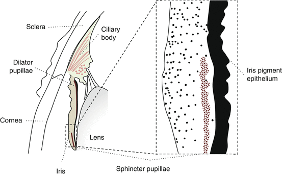
Fig. 6.1
Structure of the iris
1.
Iris stroma
The iris stroma consists of fibroblasts, melanocytes, blood vessels, and nerves in a collagen-rich extracellular matrix.
The anterior surface is velvety and lacks epithelium. It is not a barrier for solutes or fluid.
Stromal vessels have non-fenestrated endothelium that maintains the blood-aqueous barrier [6].
Stromal melanocyte pigment concentration determines iris color [7].
A lightly pigmented iris is blue/green as longer wavelengths are absorbed and shorter reflected.
2.
Sphincter pupillae
The sphincter is a 1-mm-wide ring of smooth muscle within the pupillary border.
Smooth muscle cells are connected by gap junctions and innervated by parasympathetic nerves.
Uniform pupillary constriction is achieved through simultaneous stimulation of each muscle segment and spread of current through gap junctions [8].
3.
Dilator pupillae
The dilator is a thin peripheral layer of myoepithelium extending from the iris root.
The muscle fibers are long basal processes extending from cells in the anterior iris epithelial layer.
These are interconnected by gap junctions and innervated by sympathetic nerves.
Like pupillary constriction, uniform pupillary dilation is achieved through simultaneous stimulation of each muscle segment and spread of current through gap junctions [8].
4.
Iris epithelium [9]
There are 2 iris-pigmented epithelial layers, anterior and posterior, aligned apex to apex.
The anterior is continuous with the outer pigmented ciliary body epithelial layer.
The posterior is continuous with the inner non-pigmented ciliary body epithelial layer.
The Pupil
Overview: Functions of the Pupil
1.
2.
Control of optical aberrations
3.
Depth of focus
Approaching objects remain relatively well focused as only radially oriented light enters the eye.
4.
Aqueous conduct channel
The pupil acts as a aqueous conduct channel between the posterior and anterior chambers [15].
This prevents a dangerous buildup of pressure in the posterior chamber.
Control of Pupillary Aperture
The size of the pupil aperture is influenced by the following factors:
(a)
The light reflex
(b)
The near reflex
(c)
Reflexive dilation
(d)
Other factors
The Light Reflex (Fig. 6.2)
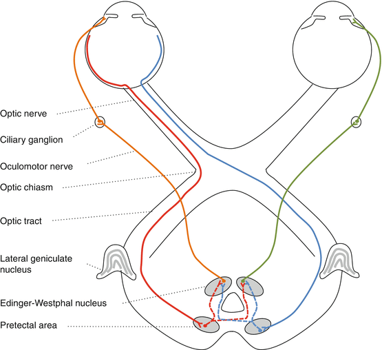
Fig. 6.2
The light reflex
The neural pathway consists of afferent, interneuron, and efferent divisions.
1.
2.
Afferent division: intrinsically photosensitive ganglion cells
A specific ganglion cell type (the γ-cell) mediates the light reflex to midbrain pretectal nuclei [19].
3.
Afferent division: pretectal input
Each pretectal nucleus receives:
(a)
Uncrossed ipsilateral temporal RGC axons
(b)
Crossed contralateral nasal axons
These travel via the optic nerve, chiasm, and then tract.
4.
Interneuron division
Pretectal nuclei neurons send equal bilateral projections to the Edinger-Westphal (E-W) nuclei.
The E-W nuclei are located in the midbrain on either side of the periaqueductal gray matter [21].
5.
Efferent division
The E-W nucleus sends preganglionic parasympathetic fibers in the oculomotor nerve to the ciliary ganglion where they synapse with postganglionic parasympathetic neurons [1, 22].
These travel to the eye via the short ciliary nerves to innervate the sphincter pupillae.
Factors affecting the light reflex
Stimulus factors (e.g., retinal adaptation, stimulus duration, light intensity, retinal location) that influence visual perception produce a comparable change in pupil responsiveness [16, 23].
With greater intensity stimuli, the amplitude of constriction increases and latency decreases [5].
With long duration stimuli, the pupil may undergo oscillations (hippus) or slow dilation (pupil escape) after initial contraction due to light adaptation [24].
The Near Reflex
The near response occurs when visual fixation shifts from far to near.
1.
Stimuli for the near reflex
2.
Efferent pathways
Each arm of the triad is conveyed by distinct oculomotor nerve fibers
(i)
Miosis
Miosis involves the same E-W nuclei and efferent pathways involved in the light reflex [28].
(ii)
(iii)
Convergence
Pupil Reflex Dilation
The dilator pupillae cannot overcome the stronger sphincter pupillae without sphincter relaxation [5].
Iris sphincter relaxation is achieved by centrally mediated E-W inhibition.
1.
Tonic Edinger-Westphal nuclei inhibition
When awake, centrally mediated sympathetic activity inhibits the E-W nuclei [30].
This inhibitory activity is overcome by the light or near reflexes [31].
Stay updated, free articles. Join our Telegram channel

Full access? Get Clinical Tree


