Fig. 1
Multi-level acquired subglottic stenosis in a 4-month-old child intubated during infection with respiratory syncytial virus
Since not all intubated neonates develop SGS, theorized risk factors for subsequent scarring include long-term intubation, traumatic intubation, excessive tube movement, intubation with an inappropriately large tube, multiple intubations, and systemic factors such as reflux and sepsis. However, the relative risk attributable to each of these factors has never been proven in a large, multicenter study. A 1996 review by da Silva found prematurity to be the only factor consistently associated with development of subglottic stenosis, while Manica et al. found length of intubation and need for rescue doses of sedation to be associated with development of SGS [6, 7].
Clinical Presentation
In the neonatal period, less severe forms of SGS may manifest as intermittent stridor or rapid fatigue with feeding. Infants under 6 months of age diagnosed with “croup,” or children under 1 year of age who have been diagnosed with multiple episodes of croup should arouse suspicion for underlying structural narrowing of the airway.
Among premature and critically ill children, SGS often presents as failure to wean from ventilatory support once associated pulmonary pathology has been effectively managed. Inability to intubate an infant with an age-appropriate endotracheal tube may also be the first sign of subglottic narrowing.
Diagnosis and Management
Initial evaluation of patients suspected of having SGS begins with a detailed history of birth, growth, development, and comorbid conditions. Patterns of exacerbation of stridor or respiratory effort should be noted. A detailed, but directed physical exam should assess the infant for signs of increased respiratory effort. The respiratory cycle creates alternating compressing and dilating forces on the intrathoracic and extrathoracic airways, and therefore the phase of respiration during which stridor is heard can help to localize the site of narrowing. Generally, supraglottic or glottic narrowing leads to inspiratory stridor, while tracheal and bronchial pathology cause expiratory stridor. SGS often presents with biphasic stridor.
Bedside flexible laryngoscopy can provide useful information about vocal fold mobility and supraglottic pathology in extubated patients, and may allow for limited examination of the subglottis. Simple neck X-rays can also detect SGS, and are often used to diagnose the acute subglottic narrowing seen in croup. Airway fluoroscopy provides a dynamic assessment of the larger airways and can aid in the diagnosis of tracheobronchomalacia or vascular compression.
The gold standard for diagnosing SGS is rigid microlaryngoscopy and bronchoscopy (MLB). Assessment during spontaneous ventilation is preferred so that laryngotracheal dynamics may also be observed. The Cotton-Myer grading system (Fig. 2) is the most widely used classification scheme for SGS, and is easily adapted to standard endotracheal tube sizes [8]. “Sizing” of the subglottic airway is typically done with uncuffed endotracheal tubes under endoscopic visualization. The patient is intubated with serially larger tubes and assessed for an air leak around the tube at normal ventilatory pressures (10–25 cm H2O). The outer diameter of the largest tube that permits a leak at appropriate pressures is considered to be the size of the airway. If a smaller-than-age-appropriate tube is needed, simple calculation can determine the percent of luminal obstruction.
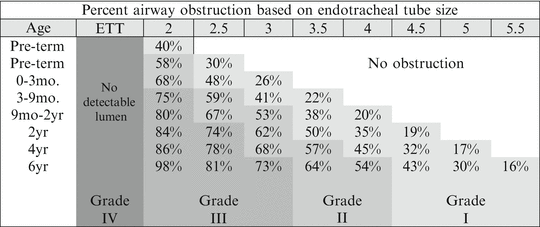
Fig. 2
Redrawn with modification from: Myer CM 3rd, O’Connor DM, Cotton RT. Proposed grading system for subglottic stenosis based on endotracheal tube sizes. Ann Otol Rhinol Laryngol. 1994 Apr;103(4 Pt 1):319–23
Medical Management
Children with grade I SGS can often be managed conservatively with observation as long as the child is able to grow and effectively participate in normal daily activities. Intermittent medical treatment with inhaled or systemic steroids with or without racemic epinephrine may be required during periods of inflammatory upper airway obstruction. A strong working relationship between the caregivers and patient is essential, and open lines of communication must be in place so that small setbacks during periods of illness do not spiral into the need for intubation. These children often benefit from thorough aerodigestive evaluation to ensure that all potentially contributing factors are appropriately managed.
Although there continues to be debate in the literature surrounding the significance of acid reflux management in SGS, animal studies have shown that acid application to the injured subglottis causes fibrosis and scar proliferation, and adult studies have shown that idiopathic SGS refractory dilation may resolve with appropriate reflux management [9–11].
Endoscopic Balloon Dilation
Dilation has been a long-standing therapeutic option for subglottic stenosis. Beginning in the 1970s, balloon dilation gradually gained favor over the use of rigid, metal dilators due to the minimization of shearing forces, and the increased precision associated with endoscopic placement of the balloons. Typically, the area of stenosis is exposed with a suspension laryngoscope and the balloon is centered at the narrowest part of the airway, often with endoscopic guidance. The balloon is then inflated to just under burst pressure either for a pre-defined time period (such as 60 s) or until the patient exhibits evidence of oxygen desaturation. Depending on the response, dilation may be repeated with the same size balloon or with one of larger diameter. Topical or injected therapies, including triamcinolone or mitomycin, may be used to augment the effects of dilation and prevent further scar formation [12].
Initial evidence that thin or evolving stenoses responded better than firm, mature scars has been borne out with wider adoption of the technique. [13] Hebra et al. reported that patients who show no initial response are unlikely to benefit from further dilations [14]. In our experience, patients with very inflammatory stenoses or stenoses extending beyond the subglottis respond poorly to dilation. The need for repeat dilation is the rule rather than the exception, although there is limited evidence to show benefit beyond three repeated procedures for an acute symptomatic exacerbation.
Adjuvant Endoscopic Therapies
Focal obstruction of the subglottis by cysts or masses may also be managed endoscopically. Microlaryngeal instruments are often appropriately sized for use in the neonatal and infant subglottis. Simple scars and webs may be sharply divided prior to dilation, and small masses may be removed with microlaryngeal biopsy forceps. A powered tissue shaver (with an appropriately sized, laryngeal blade) can also be used for management of obstructing masses, subglottic cysts, or small hemangiomas [14].
Anterior Cricoid Split
Anterior cricoid split (ACS) was introduced in the 1980s as an alternative to tracheotomy in infants with SGS, with variable success rates. Reported extubation rates range from 58 to 100 %, although rates as low as 27–35 % have been noted among extremely premature infants (gestational age less than 28 weeks) [2, 15]. The procedure can be performed in open or endoscopic fashion, and involves complete transection of the anterior cricoid ring with or without associated balloon dilation of the airway. The lower portion of the thyroid cartilage is often divided in the midline as well, although the surgeon must take care to avoid the anterior commissure. A nasotracheal tube is left in the airway to serve as a stent during the preliminary healing phase—approximately 1 week—and soft tissue fibrosis is expected to fill the gap between the cut ends of the cricoid. The posterior cricoid may also be split to provide greater expansion of the subglottic airway.
Laryngotracheal Reconstruction
The addition of autologous cartilage grafts to augment the airway expansion achieved with cricoid split alone defines procedures classified as laryngotracheal reconstructions (LTR). In infants and neonates, reconstruction with a thyroid alar cartilage graft is the most commonly performed procedure. Pathologic study has shown that the thyroid alar graft can be effectively used from the glottis to the second tracheal ring, and is best for patients with grade II and select grade III stenoses [16]. Advantages of using the thyroid ala as a graft source include use of a single incision, elimination of potential for pulmonary complications related to the rib harvest and post-operative pain/splinting, and the excellent size match between the thyroid ala and the anterior cricoid ring in infants. Several retrospective studies have found increased operation-specific extubation rates when comparing primary LTR (81–89 %) with ACS alone (27–83 %) [15, 17, 18].
Reconstruction with costal cartilage grafting is typically done outside of the neonatal/infant period, after a bridging tracheotomy has been performed (Fig. 3). Techniques have been well-described in prior literature, and involve vertical incisions in the anterior or posterior midline of the cricoid plate with interposition of autologous cartilage grafts to stent open the airway. Anterior and posterior grafts may be placed together or in isolation, depending on the pathology present. Advantages of deferring LTR until around age 2 (or later) include growth of the airway leading to more working room and better tolerance of post-operative edema, maturation of the pulmonary system so that patients are better able to tolerate the surgery and post-operative recovery, and growth of the actual rib cage so that adequate grafts can be harvested with minimal morbidity. Recent studies, however, have focused on younger patients as earlier decannulation has been associated with better speech and language development outcomes [19].
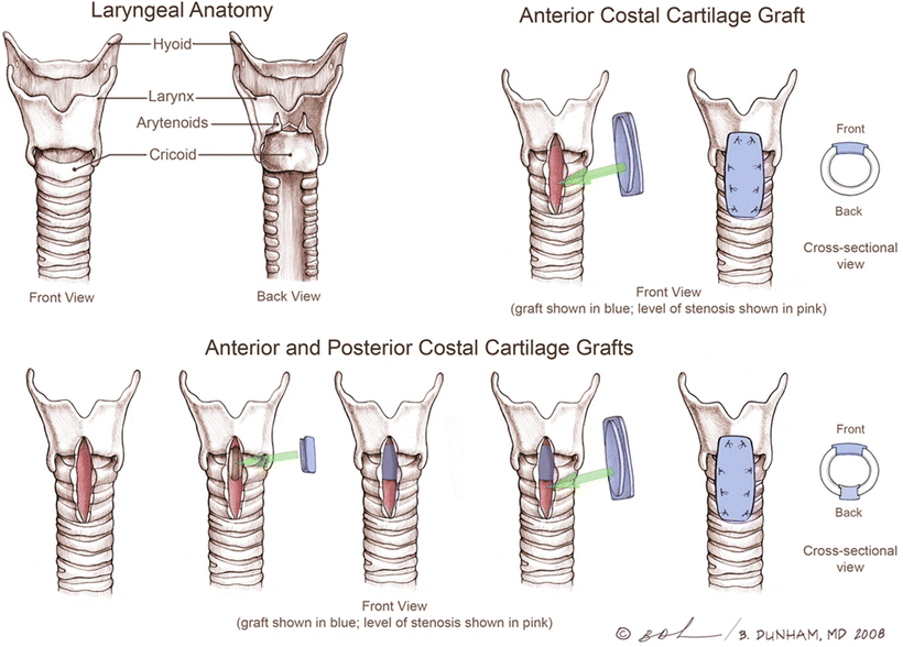
Fig. 3
Diagram of placement of anterior and posterior cartilage grafts during LTR, with resultant effect on cross-sectional airway diameter. (Permission granted for use of illustration by Brian Dunham, M.D © 2008)
Cricotracheal Resection
For children with high-grade III to grade IV lesions, cricotracheal resection (CTR) is an alternate surgical option. The anterior portion of the cricoid ring is removed with the associated stenotic segment of airway, and the distal trachea is anastomosed to the remaining cricoid posteriorly and to the inferior thyroid cartilage anteriorly. The procedure requires significantly more dissection around the airway, and therefore carries a greater risk to the recurrent laryngeal nerves than does LTR, but may offer better odds of operation-specific decannulation in selected patients [20, 21]. Ikonomidis et al. have shown CTR to be safe and effective in children weighing less than 10 kg, but did find a high rate of post-op dysphonia (71 %) [20].
Tracheostomy
In patients with high-grade stenosis, inflammatory subglottic obstruction, or severe concomitant cardiac or pulmonary disease, tracheostomy may be the best initial option. This allows time for growth and for other medical comorbidities to be brought under control. Airway expansion surgery and decannulation can then be pursued under less acute circumstances.
Tracheostomy in the neonatal period is not without risk, though. The risk of mortality directly attributable to pediatric tracheostomy status is 0.5–1 % in the United States, with as many as 43 % of patients experiencing serious complications such as tube occlusion and accidental decannulation [22, 23]. Recent research has also suggested that neurodevelopmental outcomes may be worse among preterm infants who require tracheostomies [24].
Congenital Subglottic Stenosis
Epidemiology
Congenital SGS the third most common cause of neonatal stridor, and is thought to represent between 5 and 15 % of airway malformations [25, 26]. Congenital SGS refers to abnormal development of the cricoid ring, often resulting in a thickened, irregular structure imparting an elliptical shape to the subglottic airway. The true incidence of congenital SGS is unknown, as most infants with significant levels of obstruction are intubated at birth, thus making it very difficult to differentiate between acquired and congenital pathology.
Pathogenesis
In early embryonic development, the respiratory tree and foregut freely communicate. As the embryonic trachea is separated from the esophagus by the esophago-tracheal membrane, the epithelium of the airway proliferates rapidly, resulting in complete occlusion of the larynx and trachea. Recanalization is typically complete by the 7th week of gestation. Varying degrees of pathology may result when recanalization is incomplete, ranging from laryngeal webs to mildly obstructive subglottic shelves to complete atresia.
Stay updated, free articles. Join our Telegram channel

Full access? Get Clinical Tree


