Fig. 6.1
Schematic illustration showing the anatomical relationships between the auditory nerve (AuN) and the surrounding structures. The auditory nerve is a bundle of bipolar neurons that form synaptic contacts with the hair cells peripherally and cochlear nucleus cells centrally. The cell bodies of the auditory neurons (spiral ganglion cells, SGC) are housed in the Rosenthal canal (RC). The tractus spiralis foraminosus (TSF) is an osseous canal through which the axons of the auditory nerve pass from the Rosenthal canal to the axis of the auditory nerve (modiolus). CN cochlear nucleus, CPAC cerebellopontine angle cistern, HC hair cell, TB temporal bone, TZ transitional zone. (From Sekiya et al. [9])
Auditory nerve that occupies one of the central places in hearing restorative medicine along with hair cells and other structures [1, 5, 6] (see Chap. 29) holds unique anatomical features in the scope of auditory nerve regeneration.
6.2 Transitional Zone (TZ) of the Auditory Nerve
The interface between the PNS (peripheral nervous system) and the CNS (central nervous system) portions is called the transitional zone (TZ) or the Obersteiner-Redlich zone [7, 8] (Fig. 6.1). Centrally from the TZ, myelin sheaths are formed by oligodendrocytes, and the supporting tissue is astrocytic. The peripherally convex shape of distal end of TZ is clearly visualized by an antibody to GFAP (glial fibrillary acidic protein) because of the presence of astrocytes only in the CNS portion of the nerve [9]. Peripherally, the sheaths are formed by Schwann cells that are enveloped in endoneurium [7]. The interface (TZ) is penetrated only by axons. The central portion of auditory nerve is exceptionally long among cranial nerves except olfactory and optic nerves [10]. This lengthy CNS portion of this nerve holds a crucial significance in investigations of hearing restoration [9, 11, 12]. Experimental results about cell migration trespassing TZ are diverse (see Fig. 29.2). A study demonstrated an occurrence of cell migration with peripheral to central direction trespassing TZ [13]. About central to peripheral cell migration trespassing TZ, the experimental results were conflicting [14, 15]. These discrepancies might have been due to the differences of donor cells, conditions of the host animals, and experimental settings. In the light of auditory nerve regeneration, however, it should be noted centrally growing neurites from distal side are sensitive to the repellent effects of astrocytes at TZ [16, 11], although peripherally growing neurites crossed the TZ [15].
6.3 Neurotrophins
BDNF (brain-derived neurotrophic factor) and NT-3 (neurotrophic factor-3) are synthesized in the hair cells and transported to the SGCs where their high-affinity receptors trkB and trkC are expressed and the survival and synaptogenesis of auditory neurons depend on these growth factors [17–19]. Auditory neurons receive neurotrophins from several other sources, including the cochlear nucleus, and even SGCs themselves [18, 20]. Hair cells do not necessarily need the presence of auditory neurons for survival [19, 21–23]. Hair cells may become a crucial nutrient source for transplanted cells in the attempt to regenerate auditory neurons (see Chap. 29).
6.4 Target Pathologies for Auditory Nerve Replacement
When dendrites or axons of auditory neurons are damaged initially, auditory neurons degenerate but hair cells tend to be preserved to various degrees (primary auditory nerve degeneration) [5] (Fig. 6.2). In contrast, following the inappropriate use of ototoxic drugs or acoustic overstimulation, hair cells may degenerate and subsequently auditory neurons degenerate (secondary auditory nerve degeneration) (Fig. 6.2). Collectively, the auditory neurons commonly degenerate in either of these pathological processes, indicating the auditory neurons occupy an indispensable part in reviving lost hearing.
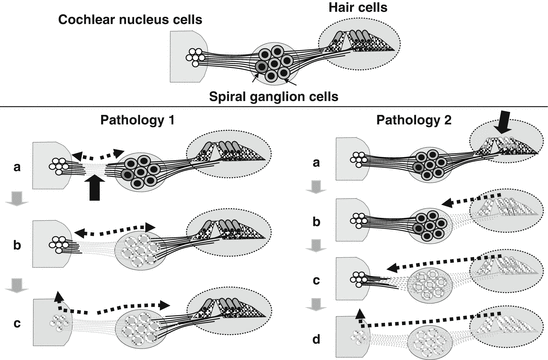
Fig. 6.2
Pathological processes occurred in deafness. In Pathology 1, the initial insult to the auditory nervous system occurs as a lesion in the dendrites or axons of the nerve (A, solid arrow). In time, the degeneration of the auditory neurons proceeds toward the cochlea and the brainstem (B and C, dotted arrows). In this pathology, the hair cells tend to be preserved to various degrees (C). In Pathology 2, the hair cells are damaged initially (A, solid arrow), and the dendrites and the auditory ganglion cells secondarily degenerate (B, C, dotted arrows). At the most advanced stage of this type of pathology, the degeneration of the cochlear nucleus cells might occur (D). The cell transplantation approach should cope with each of these pathologies properly. (From Sekiya et al. [5])
Selective auditory neuronal degeneration with sparing of hair cells was recognized as auditory neuropathy where ABR (auditory brainstem response) was absent or profoundly distorted with otoacoustic emissions (OAE) and/or cochlear microphonics (CM) preserved [24]. Auditory neuropathic type auditory nerve degeneration is observed in various clinical disorders and elderly people [25–28]. Temporary threshold shift (TTS) has been assumed to indicate reversal damage to hair cell and auditory neurons without delayed auditory dysfunction, but a recent study demonstrated noise-induced primary auditory nerve degeneration occurred in a delayed fashion without hair cell damage in situation where TTS had been observed [29]. In small vestibular schwannomas, the hair cells are assumed to be well preserved [30–32]. Hearing preservation in vestibular schwannoma treatment still remains as an unresolved problem in neurosurgery [9]. Taken together, emerging evidence indicates auditory neuropathic type auditory nerve degenerations exist much more than once thought. This pathological situation may become most suitable for cell transplantation intervention because survived hair cells can provide trophic factors to donor cells [33, 34] (see Chap. 29).
6.5 Deafness, a Neurodegenerative Disorder
Whenever insults, such as mechanical trauma, ischemia, radiation, genetic disorders, or chemical insult, are imposed to the CNS, quiescent astrocytes resume proliferation, become hypertrophic, and upregulate GFAP and finally glial scar is formed along with the progression of neuronal degeneration [35, 36]. We demonstrated that compression of CNS portion of auditory nerve induced glial scar not only in the auditory nerve but also in the cochlear nucleus [9]. One recent report demonstrated that with hair cell damage glial scar was induced in the auditory nerve [12]. From these results, it has become apparent that glial scar is induced in the auditory nerve not only in primary but also secondary auditory nerve degenerations. Probably, glial scar formation may be more severe when the site of insult is closer to the brainstem, because we observed glial scar formation even in the cochlear nucleus region in direct compression to the CNS portion of auditory nerve [9, 37]. In every effort to restore lost auditory nerve function, glial scar in the auditory nerve and cochlear nucleus has to be overcome because glial scar is believed to strongly inhibit neural regeneration [38]. In our future investigations for hearing restoration, deafness should be regarded as a neurodegenerative disorder because it is fraught with the same glial scar problem as in other neurodegenerative diseases including spinal cord injury, Parkinson’s disease, and amyotrophic lateral sclerosis (ALS).


