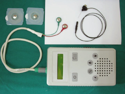
21 Spasmodic Dysphonia Spasmodic dysphonia (SD) is a focal laryngeal dystonia that is an action-induced laryngeal movement disorder. The action that triggers the spasms in the voice is speaking itself. Only the volitional cortically driven system is affected in SD. Dystonias are generally classified into two groups, generalized and focal, and may be primary (idiopathic) or secondary to birth injury, hypoxia, infection (encephalitis/meningitis), toxicity (drugs), or stroke. The most common types of focal dystonia are laryngeal dystonia, blepharospasm, torticollis, and writer’s cramp. Laryngeal dystonia, also called SD, is a focal, primary dystonia, affecting the muscles of the larynx. SD patients classically have a spasmodic speech pattern with adductor or abductor spasms. Adductor spasms give a strained and choked quality to the voice and this is referred to as adductor spasmodic dysphonia (ADSD). There is an abrupt initiation and termination of voicing, resulting in short breaks of phonation. In contrast, abductor spasms give a breathy quality to the voice referred to as abductor spasmodic dysphonia (ABSD). It is possible to have a combination of both these types of SD referred to as mixed SD. A study by Cannito and Johnson1 suggests that all patients have a mixed SD, with one type of an activity predominating the other. Approximately one-third of persons with SD also have voice tremor, which makes the pitch and loudness of the voice waver at 5 Hz during vowels and is most evident when “/a/” as in the word “all” is produced for at least 5 seconds.2 Historically, various terminologies have been used to describe SD including spastic dysphonia,3 spastic aphonia,4 and coordinated laryngeal spasm.5 It was Aronson who helped establish SD as an organic disease and also described two distinct types of SD, the adductor and the abductor varieties.6 Though SD is today thought to be a completely distinct entity to psychogenic dysphonia, there is a worsening of patients’ symptoms noticed in the presence of strangers, in crowds, and over the telephone. There is often an improvement of the SD patient’s voice with whispering, singing, and shouting or after an alcoholic beverage is consumed. This is in fact one of the mysteries of SD, that it is task specific. Spasms only occur during speaking and not during emotional expressions such as laughter, crying, and shouting. This may be explained by Kuypers’ study (1958), which indicates that only humans have a direct corticobulbar pathway from the laryngeal cortex to the nucleus ambiguus.7 Possibly, neural systems involved in learning speech are likely affected in SD, while those involved in emotional vocalization are not. To identify the neural abnormalities in SD, differences between these two neural systems (one for emotional vocalization and the other for speech) must account for symptoms being absent in the former and present in the latter.8 Occasionally, patients with ADSD compensate by producing a breathy voice referred to as compensatory abductor dysphonia. Similarly, ABSD patients may try to tightly contract the vocal folds producing compensatory adductor dysphonia, though this is more uncommon.9,10 Historical Anecdotes •Traube used the term spastic dysphonia in 1871 while describing a patient with nervous hoarseness. •Aronson in 1968 helped establish SD as an organic and not a psychiatric condition and also in later studies described the two types of SD, i.e., adductor and abductor SD. •Dr. Herbert Dedo in 1976 introduced recurrent laryngeal nerve (RLN) sectioning as a treatment for ADSD. •Dr. Andrew Blitzer performed the first laryngeal injection of botulinum toxin (BTX) for SD in 1984. •Isshiki, in 1998, reported treating a case of ADSD with type 2 thyroplasty. •In 1999, Dr. Berke described the denervation–reinnervation surgery. The basal ganglion plays an integral role in movement. Inputs from the cerebral cortex, especially the primary motor strip and primary somatosensory cortex, are received in the basal ganglia and the substantia nigra. The exact etiology of SD is as yet unknown. However, research by Simonyan and Ludlow11 has shown that the primary somatosensory cortex shows consistent abnormalities in activation extent, intensity, correlation with other brain regions, and symptom severity in SD patients and, therefore, may be involved in the pathophysiology of SD. When the brains of dystonic patients were studied pathologically, although no consistent lesions were found, most frequently mentioned lesions were in the basal ganglia.12 In ADSD, the lack of symptoms during whispering, when the vocal folds are not vibrating, suggests that changes in laryngeal sensory feedback either from the vocal fold mucosa or from subglottal pressures in the trachea may play a role in the pathophysiology of the disorder. Further research on the role of laryngeal sensory feedback in the manipulation of symptoms needs to be performed.8 A genetic component does seem to be involved in some patients as 12.1% of Blitzer and Brin’s 1991 series of laryngeal dystonia patients had a family history of dystonia.10 The DTY1 gene was first identified in one large non-Jewish family with multiple family members presenting with dystonia and has been responsible for childhood-onset dystonia in the Jewish population.13 A few cases with single mutations in THAP1, a gene involved in transcription regulation, suggest that a weak genetic predisposition may contribute to mechanisms causing a nonprogressive abnormality in laryngeal motor neuron control for speech but not for vocal emotional expression.8 Though SD may be present in both males and females, it is more prevalent among the female population, with some estimates indicating the prevalence to be as high as 80%.14 It is uncommon to find SD in children, and 45 years is the most commonage of presentation of SD. There seems to be a progression of symptoms for 2 years following which there is usually a plateau of symptoms, though the patient may complaint of a progressive increase in effort required to phonate. SD is a relatively rare disorder; however, accurate worldwide audited numbers are not available. Some estimates are as low as 1 per 100,000 cases,15 although accurate diagnosis is a significant roadblock to research. With the increasing awareness regarding SD being created, more cases are being accurately diagnosed and managed. The provisional diagnosis of SD is made more often than not within a couple of minutes of talking to the patient as the diagnosis is based primarily on auditory-perceptual features. This is possible as ADSD patients have a typical choking voice and the spasms are seen on vowels. This type affects at least 80% of persons with SD and disrupts sentences such as “we eat eggs every day.”16 This is because any sentence that has a lot of vowels dramatically worsens the voice. On asking the patient to whisper or sing, the spasms markedly decrease. In the case of ABSD patients, a typical breathy spasm is observed and is almost always accompanied by a flaring of the alae nasi. These breathy bursts take place due to prolonged voiceless consonants and while attempting to start voice after voiceless consonants such as/s/,/f/,/h/,/p/,/t/, and/k/.17 Disruption of sentences such as “he had half a head of hair”18 is observed. Flexible laryngoscopy allows an evaluation of the vocal fold movements in normal physiological phonation. The patient is asked to talk, sing, and whisper, and any change in the severity of spasms is observed. In ADSD patients, the spasms may be of only the true vocal folds or of true and false vocal folds. In severe ADSD, a complete anteroposterior and lateral compression of the supraglottis is observed. Stroboscopy may be performed using the flexible laryngoscope. In ABSD patients, a sudden abduction is observed corresponding with the breathy spasm on phonation. Stroboscopy confirms the spasms seen on flexible laryngoscopy, and the amplitude of the mucosal wave is seen to be decreased or occasionally absent. However, when stroboscopy is done using a Hopkins telescope through the mouth, the signs of SD may occasionally get masked as this test does not allow for normal physiological phonation. A team approach in the diagnosis of SD is vital. Besides the laryngologist, a speech therapist and a neurologist form the core team in the diagnosis of SD. Occasionally, the opinion of a gastroenterologist or a psychiatrist may be sought. Clinical Insights •In ADSD, voice improves with whispering, singing, or shouting and after consuming an alcoholic beverage and worsens on the phone or in the presence of strangers. •Vocal tremors may accompany spasms in almost 30% of ADSD patients. •In ABSD, flaring of the alae nasi is seen to accompany the abductor spasms. The differential diagnosis of SD includes muscle tension dysphonia (MTD), vocal tremor, Parkinson disease, Wilson disease, myasthenia gravis, motor neuron disease (MND), pseudobulbar palsy, cerebellar disorders, laryngopharyngeal reflux, laryngeal cancer, and functional voice disorders. MTD may be occasionally mistaken as SD. The cardinal differences between the two are that SD is task-specific unlike MTD and in SD the strain in the voice is spasmodic unlike MTD where the strain is continuous. MTD is often associated with tenderness over the greater cornu of the hyoid and in the thyrohyoid membrane. Obliteration and tenderness in the cricothyroid membrane may also be observed. A lidocaine block of the RLN improves the voice of both ADSD and MTD patients. However, the primary treatment of MTD is laryngeal massage and speech therapy. Vocal tremor is an involuntary, rhythmic, oscillating movement of the vocal folds. Vocal tremors are not task-specific and are present on talking, singing, and on a sustained/eee/. This vocal tremor may be part of an essential tremor known as “benign heredofamilial tremor.” This may involve only the voice or other body parts such as the head and neck also. Vocal tremor occurs in 10 to 20% of patients with essential tremor. It is essential to rule out cerebellar disease, parkinsonism, thyrotoxicosis, and drug-induced and psychogenic causes of the tremor. In 30% of SD patients, an associated vocal tremor is present. Speech therapy as a modality of treatment in ADSD patients does not meet with much success. However, it may be a viable option in patients with very early ADSD. Speech therapy is also of value in those patients who have developed wrong compensatory techniques of a breathy voice in ADSD or compensatory abductor dysphonia in ADSD. In ABSD patients, speech therapy especially with a biofeedback mechanism has shown promising results. This has a very positive bearing in the treatment of ABSD patients who typically respond to BTX poorer than their ADSD counterparts. One of the key biofeedback techniques used for the ABSD group is to ask the patient to stand in front of the mirror and talk while making a concentrated effort in controlling the flaring of the alae nasi that accompanies each abductor spasm. Medical treatment may benefit patients of essential tremor or those who besides having SD have a generalized dystonia. Patients of SD occasionally are also anxious or depressed and may need psychiatric treatment for the same. Clostridium botulinum is the bacteria that produce seven serologically distinct botulinum neurotoxins that are designated from A to G. These seven neurotoxins are antigenically distinct but do have similar molecular weight and a common subunit structure. These neurotoxins are synthesized as single-chain polypeptides. However, this single chain gets cleaved by trypsin or bacterial enzymes to form a bichained (one heavy chain and one light chain) molecule in which both the chains are linked by a disulfide bond. It is in this form that the molecule becomes potent. Clinical studies have shown that BTX does not affect the synthesis or storage of acetylcholine. The toxin probably modifies the release of synaptosomes from the microtubular subsystem. Though the clinical effects of BTX are probably due to its peripheral effects, the action of BTX on the laryngeal muscle contractions cannot be assumed to have altered only muscle spasms. There may be retrograde transport affecting input to the laryngeal motor neurons.19 In addition, as the muscle spasms are reduced not only in the laryngeal muscle injected but also in other laryngeal muscles on the opposite side of the larynx,20 the sensory feedback from the larynx is altered by less mucosal compression and lower subglottal pressures in the trachea due to reduced hyperadduction during speech. The clinical effects are seen 24 to 72 hours following the BTX injection. BTX is commercially available as a powder in vacuum-sealed vials. The quantity of active toxin is mentioned in mouse units (mu/U) and it is essential that BTX be manufactured in a standard fashion such that each subsequent vial is of an equal potency to maintain both safety and efficacy. BTX (A) is available as Botox manufactured by Allergan (Irvine, California, United States) and also as Dysport manufactured by Ipsen, Slough, United Kingdom. In India, BTX is available in 50- and 100-mu vials, where 1mu is the median lethal dose in mice. The exact lethal dose in humans is not known but is postulated to lie near approximately 2700 U. A 50-U vial of BTX when reconstituted with 2cc of preservative-free saline gives a concentration of 2.5 U BTX per 0.1mL of reconstituted solution. It has been recommended to use this reconstituted BTX within 4 hours of preparation. BTX use is currently not recommended in pregnant or lactating women. Patients with myasthenia gravis, Eaton-Lambert syndrome, and MND should be treated cautiously with BTX as the neuromuscular junction is affected. Aminoglycosides may potentiate the effect of BTX as they effect neuromuscular transmission and thus are also a contraindication to BTX use. Prolonged use of BTX may lead to antibody formation, which is seen more in the group of patients being treated by BTX for torticollis as larger doses are being used. Dezfulian et al21 have developed an enzyme-linked immunosorbent assay that appears to be sensitive and specific in the detection of antibody. Dr. Andrew Blitzer performed the first laryngeal injection of BTX for SD in 1984.22 The key to a successful BTX injection for SD lies in correct dose titration of BTX for every individual patient and an accurate placement of the BTX. To accurately inject the BTX in the affected muscle, most laryngologists use the laryngeal electromyography (LEMG) system while injecting. Some laryngologists, however, inject with the help of flexible laryngoscopic guidance, occasionally in association with LEMG. Dose titration is perfected once the response to standard doses of BTX on the patient has been audited. LEMG-controlled BTX injection in the management of SD is considered the gold standard. Electromyography (EMG) is a test measuring the micropotential reaching the muscle fibers utilizing a needle electrode placed in the muscle. When the needle is placed in the laryngeal muscle, the test is referred to as LEMG. The primary role of LEMG is in the injection of BTX in SD. However, the other therapeutic uses are in the injection of BTX in patients with cricopharyngeal spasm and contact granuloma. The diagnostic role of LEMG lies in differentiating between vocal fold paralysis and cricoarytenoid joint fixation and also in the diagnosis of various neurogenic voice disorders. It is essential that the diagnostic applications of LEMG be performed using a sophisticated, multichannel EMG system by the EMG technician in concert with the neurophysician and not by the laryngologist alone.
Etiology
Demographics
Diagnosis
Clinical
Differential Diagnosis
Muscle Tension Dysphonia
Vocal Tremor
Treatment Options
Role of Botulinum Toxin in the Management of Spasmodic Dysphonia
Pharmacology of Botulinum Toxin
Preparation of Botulinum Toxin
Procedure of Botulinum Toxin Injection in Spasmodic Dysphonia Patients
Laryngeal Electromyography in Spasmodic Dysphonia
Stay updated, free articles. Join our Telegram channel

Full access? Get Clinical Tree


