, Iacopo Dallan1 and Manfred Tschabitscher2
(1)
Department of Otorhinolaryngology, University of Insubria, Ospedale di Circolo e Fondazione Macchi, Varese, Italy
(2)
Medical University of Vienna, Vienna, Austria
Abstract
This chapter illustrates the surgical anatomy of the most challenging portion of the internal carotid artery, surgically speaking. At this level, the carotid artery runs within the skull base, and it is therefore intracranial and extradural. Helpful surgical landmarks for approaching this tract of the vessel, both from lateral and from anterior endoscopic approaches, are reported, and particular emphasis is given to the anatomical relationship of the internal carotid artery with all the bony, neural, and vascular structures of the skull base, as well as with the middle ear.
2.1 Anatomic Layout
The petrous portion of the internal carotid artery (ICA) is extradural and intraosseous. It extends from the carotid foramen to the superior margin of the petrolingual ligament (PLL). The carotid foramen is anteromedial to the styloid process, posteromedial to the temporomandibular joint. The PLL, placed between the sphenoidal lingula and the petrous apex, represents the posteromedial border of the cavernous sinus (CS). Once it enters in the petrous bone, the ICA presents a short vertical course, and then it turns in front of the cochlea, forming the posterior genu. From here, it runs anteriorly and medially. The petrous ICA then curves upward above the foramen lacerum (FL), thus giving the anterior genu. The segment above the FL is not truly intrapetrous, and it has been called the lacerum segment by some authors (Bouthillier et al. 1996). These segments, the anterior genu and the anterior vertical segment, are placed above the FL, and the artery does not cross the foramen. In this sense, it is better called the supralacerum segment (Herzallah and Casiano 2007). Anatomically, the FL is an opening in the dry skull that in life is filled by fibrocartilagineous tissue (fibrocartilago basalis). Its borders are the petrous apex, the body of the sphenoid, and medially the occipital bone. It is passed by tiny branches of the periosteal branches of the petrous carotid, the meningeal branches of the ascending pharyngeal artery, and small veins (Ziyal et al. 2005). The ICA passes above it and turns superiorly. At this level, the vidian canal is always lateral to the vertical segment. The lateral part of its terminal part is in the extradural space superior to the PLL. A little superior to the PLL, the abducens nerve crosses the lateral surface of the artery (Osawa et al. 2008). As stated, the horizontal portion of the petrous ICA runs within the temporal bone in an anteromedial direction, posterior to the tensor tympanic muscle, eustachian tube (ET) and spinosum and ovale foramina. When the artery enters into the carotid canal, the carotid sheath divides into 2 layers: 1 layer continues with the periostium of the carotid canal and the other as the periostium lining the lower surface of the skull base (Bouthillier et al. 1996). The bone above the horizontal segment thins progressively as the artery runs medially, so that, usually (80 %), only dura separates the horizontal portion of the ICA (ICAh) from the trigeminal ganglion (Osawa et al. 2008). Regarding the nervous network, the carotid branches of the cervical sympathetic ganglia form a plexus on the lateral surface of the artery. The largest bundle of this plexus usually is located anteroinferiorly and gives rise to the deep petrosal nerve (DPN), which joins the greater petrosal nerve (GPN) to become the vidian nerve. The vidian nerve passes through the pterygoid canal running from the anterior genu of the ICA to reach the pterygopalatine ganglion in the upper portion of the pterygopalatine fossa. Within the carotid canal, the horizontal portion of the ICA is surrounded by a venous plexus (Bouthillier et al. 1996; Osawa et al. 2008), in direct continuity with the CS.
2.1.1 Gross Anatomy
The tympanic cavity lies posterolaterally to the posterior vertical segment/posterior genu. Usually, a few millimetres of bone separate the carotid canal and the middle ear. The posterior genu sits directly posterior to the entrance of the ET into the middle ear (Osawa et al. 2008). The cochlea, placed within the petrous bone, is a little bit superior, posterior, and lateral to the posterior genu. The ET and the tensor tympani are located anteriorly to the ICAh. Proceeding medially, the vessel remains within its own canal, while the ET slopes gently downward and the cartilaginous portion of the tube is located within the sulcus tubae. This is located on the outer surface of the cranial base and placed anteriorly to the ICAh. The tensor tympani in most cases (>90 %) is located superiorly or anteriorly to the ET, and it is separated from the carotid canal by a thin bone.
2.1.2 Neural Structures
2.1.2.1 Greater Petrosal Nerve
The GPN originates at the level of the geniculate ganglion, exits through the hiatus Fallopii, and courses under the dura toward the gasserian ganglion. The nerve usually runs anteromedially above the horizontal portion.
2.1.2.2 Geniculate Ganglion
The geniculate ganglion is placed above and usually posterolateral to the posterior genu (Osawa et al. 2008).
2.1.2.3 Lesser Petrosal Nerve
2.1.2.4 Gasserian Ganglion
The gasserian ganglion is placed within a depression between the trigeminal impression and the foramen ovale. The stem of the trigeminal nerve is placed within the “trigeminal impression.” The inferolateral portion of the sphenopetrosal fissure (SPF) is placed above the medial third of the horizontal portion of the ICA, while the superomedial aspect rests against the ascending limb of the intracavernous segment.
2.1.3 Vascular Branches
In most cases, the skull base segment of the ICA presents branches (Tubbs et al. 2007; Osawa et al. 2008).
2.1.3.1 Caroticotympanic Artery
The caroticotympanic artery seems to arise from the proximal vertical part of the petrous ICA and supplies the tympanic cavity. At the level of promontory, it anastomoses with the tympanic branches of the middle meningeal artery (MMA) and stylomastoid and maxillary arteries. Not all authors agree with this position; Osawa et al. thought that this vessel comes from the ascending pharyngeal artery and not from the ICA (Osawa et al. 2008). Also, previously its presence has been doubted (Paullus et al. 1977).
2.1.3.2 Arteries of the Pterygoid Canal (Vidian Arteries)
The artery of the pterygoid canal (vidian artery) is not always present. Anatomically speaking we can consider an anterior (for ECA system) and a posterior (from ICA system) Vidian artery. The posterior one may arise as a single branch or from a common trunk with a periosteal artery (Osawa et al. 2008). This vessel may arise from the horizontal segment of the petrous part of the ICA and course into the pterygoid canal to anastomose with the same-named branch of the maxillary artery (Jinkins 2000). Furthermore, it is also possible that there is no counterpart from the maxillary artery. In this case, the artery (about 0.5 mm) has been described to transverse the cartilage-filled FL to enter the pterygoid canal.
2.1.3.3 Stapedial Artery
Rarely present in adults, the stapedial artery (SA) is a branch of the second branchial arch. The SA pierces the floor of the middle ear cavity and ascends along the promontory enclosed within a bony canal to pass between the crura of the stapes to enter the facial canal. At the level of the hiatus of the GPN, the vessel enters the middle cranial fossa to become the MMA. If the SA persists, the foramen spinosum is absent (Lasjaunias 1981).
2.1.3.4 Periosteal Branch(es)
The periosteal branches are not always present. When present, most of these originate from the anterior genu and the vertical segment. When it originates as a common trunk with the vidian artery, it arises from the horizontal segment. Most periosteal arteries arising from the inferior surface of the ICA send a branch through the FL (Osawa et al. 2008).
2.1.3.5 Persistent Acoustic Artery
The persistent acoustic artery is really a rare branch connecting the carotid (petrous portion) and basilar arteries (Lang 1995).
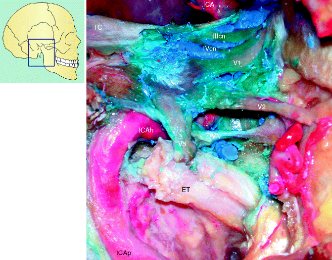
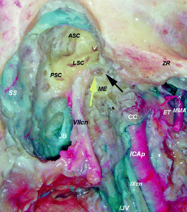
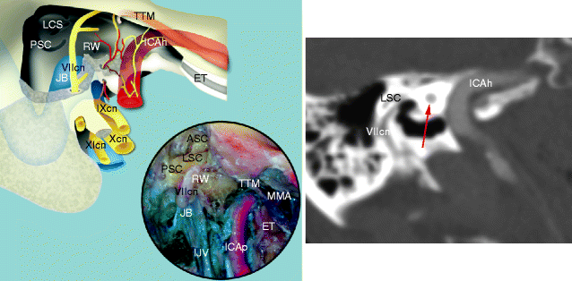
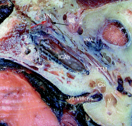
Fig. 2.1
Horizontal segment. Lateral vision of the right cavernous sinus and horizontal portion of the internal carotid artery
ET eustachian tube, ICAh horizontal portion of the internal carotid artery, ICAi intracranial portion of the internal carotid artery, ICAp parapharyngeal portion of the internal carotid artery, TC tentorium cerebelli, VN vidian nerve, V1 first branch of the trigeminal nerve, V2 second branch of the trigeminal nerve, V3 third branch of the trigeminal nerve, IIIcn oculomotor nerve, IVcn trochlear nerve
Fig. 2.2
Lateral vision of the right jugulomastoid region
ASC anterior semicircular canal, CC carotid canal, ET eustachian tube, ICAp parapharyngeal portion of the internal carotid artery, IJV internal jugular vein, JB jugular bulb, LSC lateral semicircular canal, ME middle ear, MMA middle meningeal artery, PSC posterior semicircular canal, SS sigmoid sinus, ZR zygomatic root, VIIcn facial nerve, IXcn glossopharyngeal nerve, V vestibule, yellow arrow stapes, black arrow incus
Fig. 2.3
Right exposure of the middle ear and jugular foramen. Note the intimate relationship between the eustachian tube and the carotid canal
ASC anterior semicircular canal, ET eustachian tube, ICAp parapharyngeal portion of the internal carotid artery, ICAh horizontal portion of the internal carotid artery, IJV internal jugular vein, JB jugular bulb, LSC lateral semicircular canal, MMA middle meningeal artery, PSC posterior semicircular canal, RW round window, TTM tensor tympani muscle, VIIcn facial nerve, IXcn glossopharyngeal nerve, Xcn vagus nerve, XIcn accessory nerve, red arrow cochlea
2.2 Anatomic Pictures
The posterior bend of the ICAh within the petrous bone is located just below and anterior to the cochlea. It is separated from the tympanic cavity by a thin bone that may be dehiscent.
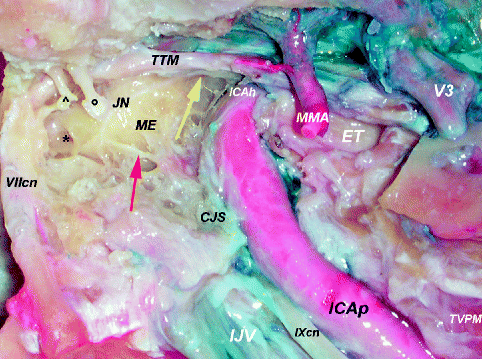
Fig. 2.4
Lateral view of the tympanojugular region
CJS carotidojugular spine, ET eustachian tube, ICAp parapharyngeal portion of the internal carotid artery, IJV internal jugular vein, JN Jacobson’s nerve, ME middle ear, MMA middle meningeal artery, TTM tensor tympani muscle, TVPM tensor veli palatini muscle, V3 third branch of the trigeminal nerve, VIIcn facial nerve, IXcn glossopharyngeal nerve, yellow arrow lesser petrosal nerve, red arrow inferior part of the inferior tympanic nerve (Jacobson’s nerve), black asterisk round window, black circle malleus, arrowhead incudostapedial joint
At the level of the jugular foramen area, the ICA moves away from the jugular bulb below the middle ear, and it is separated from it by the CJS. Jacobson’s nerve is joined by the carotidotympanic nerves from the pericarotid sympathetic plexus to form the lesser petrosal nerve (Janfaza and Nadol 2001 b).
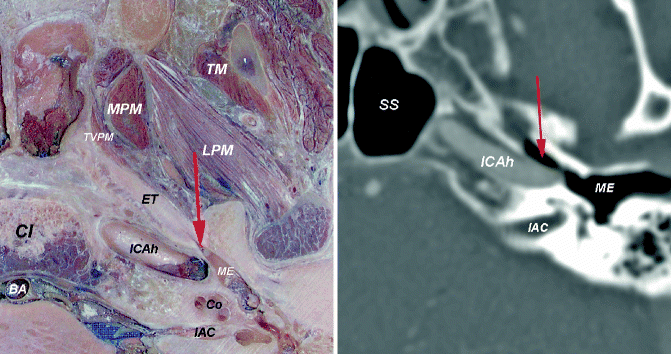
Fig. 2.5
Axial view at the level of the horizontal portion of the internal carotid artery
BA basilar artery, Cl clivus, Co cochlea, ET eustachian tube, IAC internal acoustic canal, ICAh horizontal portion of the internal carotid artery, LPM lateral pterygoid muscle, ME middle ear, MPM medial pterygoid muscle, SS sphenoid sinus, TM temporal muscle, TVPM tensor veli palatini muscle, red arrow bony segment of the eustachian tube (musculo-tubal canal)