, Iacopo Dallan1 and Manfred Tschabitscher2
(1)
Department of Otorhinolaryngology, University of Insubria, Ospedale di Circolo e Fondazione Macchi, Varese, Italy
(2)
Medical University of Vienna, Vienna, Austria
Abstract
Above the petrolingual ligament, the internal carotid artery (ICA) can be considered to be intracavernous. From an endoscopic anterior viewpoint, we can define a paraclival and a parasellar segment, especially evident in well-pneumatised sphenoid sinuses. The paraclival segment corresponds to the vertical segment of the ICAc (cavernous portion of the ICA). Endoscopically speaking, this portion of the vessel represents the lateral border of the clival window. Although some authors have divided the paraclival portion in subsegments (Herzallah and Casiano 2007), we do not consider this further classification as having surgical utility. At a variable distance from the posterior clinoid process, the vertical segment—paraclival—bends forward, forming the posterior bend of the ICAc. Then, the artery usually runs horizontally for a short distance (horizontal segment) and curves upward, thus giving the anterior bend that reaches the anterior clinoid process. For this reason, the cavernous portion in the typical neurosurgical literature has been divided into five segments: posterior vertical, posterior bend, horizontal, anterior bend, and anterior vertical (Inoue et al. 1990). It must be underlined that the configuration of the posterior bend varies significantly, and that sometimes it can bulge upward into and deform the dura of the cavernous sinus (CS) roof just lateral to the posterior clinoid process. The last segment, the anterior vertical, also named the clinoid or paraclinoid, is not completely intracavernous, but in “endoscopic” practice, it can be considered so; for this reason, we have included it in this section.
3.1 Cavernous Segment
3.2 Anatomic Layout
Above the petrolingual ligament, the internal carotid artery (ICA) can be considered to be intracavernous. From an endoscopic anterior viewpoint, we can define a paraclival and a parasellar segment, especially evident in well-pneumatised sphenoid sinuses. The paraclival segment corresponds to the vertical segment of the ICAc (cavernous portion of the ICA). Endoscopically speaking, this portion of the vessel represents the lateral border of the clival window. Although some authors have divided the paraclival portion in subsegments (Herzallah and Casiano 2007), we do not consider this further classification as having surgical utility. At a variable distance from the posterior clinoid process, the vertical segment—paraclival—bends forward, forming the posterior bend of the ICAc. Then, the artery usually runs horizontally for a short distance (horizontal segment) and curves upward, thus giving the anterior bend that reaches the anterior clinoid process. For this reason, the cavernous portion in the typical neurosurgical literature has been divided into five segments: posterior vertical, posterior bend, horizontal, anterior bend, and anterior vertical (Inoue et al. 1990). It must be underlined that the configuration of the posterior bend varies significantly, and that sometimes it can bulge upward into and deform the dura of the cavernous sinus (CS) roof just lateral to the posterior clinoid process. The last segment, the anterior vertical, also named the clinoid, infraclinoid or paraclinoid, is not completely intracavernous, but in “endoscopic” practice, it can be considered so; for this reason, we have included it in this section.
Different authors have tried to classify the ICAc, using different names but with similar conclusions (Herzallah and Casiano 2007; Jittapiromsak et al. 2010). These works both described two types of intracavernous ICA. Notwithstanding this, such differentiation appears of limited utility from an endosurgical viewpoint. Looking through a sphenoidal window, the parasellar ICA is positioned on the side of the pituitary gland, thus producing the parasellar ICA prominence on the sphenoid sinus wall. This prominence corresponds, more or less, to the distal part of the horizontal segment, the anterior bend, and the clinoid segment. The intracavernous segment of the ICA presents different branches, the most constant are the meningohypophyseal and the inferolateral trunk (ILT).
3.2.1 Gross Anatomy
This portion grossly corresponds to the sphenoid sinus. Endoscopic anatomical landmarks are identified once a wide sphenoidal window is performed: the lateral and medial opticocarotid recess (lOCR and mOCR, respectively), the parasellar and paraclival carotid canal, the sellar prominence, the clival recess, and the strut of bone over the interCS. The lOCR is given by the pneumatisation of the optic strut, thus forming a recess that extends between the optic nerve superiorly and the intracavernous carotid artery inferiorly. The mOCR corresponds, on the intracranial surface, to the lateral tubercular crest (most lateral part of the tuberculum sellae). The middle clinoid process (MCP) is placed a little bit lateral and inferior to the lateral aspect of the tuberculum sellae. It is a small eminence of the carotid sulcus at the level of the anterior part of the lateral wall of the sella turcica. The base of the MCP is just medial to the anterior genu of the parasellar ICA. And it projects laterally. The mOCR is placed at the confluence of the sella, tuberculum sellae, carotid protuberance, optic canal and planum sphenoidale. So, opening the mOCR permits visualization of the carotid canal, the optic nerve, the sella, and the medial CS. Obviously, these landmarks are easily recognizable in case of a sellar-type sphenoid sinus. In case of a presellar and conchal type, this task is respectively demanding and definitely impossible.
When looking at the lateral wall in well-pneumatised sinuses, 3 bony protrusions and 4 bony depressions can be visualized. Among the protuberances, from rostral to caudal, they correspond to the optic canal, the superior orbital fissure (SOF), and V2. The optic canal is placed above the parasellar ICA, while the SOF lies directly below the lOCR. The bony depressions include the lOCR, the mOCR, the depression between the CS apex and V2, and the depression below V2. In these cases, the vidian nerve, covered or not by bone, can be easily visualized at the level of the floor of the sphenoid sinus.
3.2.2 Neural Structures
3.2.2.1 Maxillary Nerve
The maxillary nerve corresponds to the inferior limit of the CS, where the lateral wall joins the medial wall of the CS. This nerve also can be identified anteriorly, passing through the foramen rotondum. The nerve crosses the lateral aspect of the trigeminal portion of the paraclival ICA. This landmark is superior and lateral to the vidian canal.
3.2.2.2 Oculomotor Nerve
Lateral to the intracavernous ICA, within the lateral wall of the CS, it is possible to visualize the oculomotor nerve. This nerve forms the inferior border of the optic struct triangle and runs anteriorly to the upper part of the anterior vertical portion of the parasellar ICA to reach the SOF.
3.2.2.3 Trochlear Nerve
The trochlear nerve enters the roof of the CS in the posterolateral apex of the oculomotor triangle, a little behind the entrance of the oculomotor nerve. In 20 % of cases, it enters the inferior surface of the tentorium cerebelli (Lang 1995). A small arachnoid pocket is present for a variable length inside the CS. Inside the lateral wall of the CS, it runs alone, slightly inferior to the oculomotor nerve, to gain the SOF. It is usually accompanied by the supero-proximal artery branch of the ILT. Not so rarely, bipartitions of the nerve in the lateral wall can be observed (Lang 1995).
3.2.2.4 Abducens Nerve
The abducens nerve pierces the dura of the posterior cranial fossa (PCF), on the clivus. Sheaths of dura and arachnoid follow the nerve within the basilar plexus (Lang 1995) and then through the inferior petrosal sinus. The nerve presents a short course superiorly and penetrates the CS, passing through Dorello’s canal, at the apex of the petrous bone. The nerve runs anterior and rostrally lateral to the posterior vertical segment of the intracavernous ICA and then along the inferior border of the horizontal portion of the ICAc. It courses inside the lateral part of the CS, medial to the ophthalmic nerve (Yasuda et al. 2005). Most of the time, the nerve is a single trunk, but it must be kept in mind that separated nerve bundles in the prepontine cistern or several bundles within the CS can be present. Typically, anastomoses with sympathetic fibres are present. In the prepontine cistern, in case of separated bundles, the AICA can be found between the bundles.
3.2.2.5 Ophthalmic Nerve
The ophthalmic (V1) nerve courses within the lateral wall of the CS below the trochlear nerve. It lies lateral to the abducens nerve and runs anterosuperiorly to gain the SOF. A small branch for the tentorium cerebelli has been described (Lang 1995). Close to the superior orbital fissure, it splits in three nerves: nasociliary, frontal, and lacrimal nerves.
3.2.2.6 Sympathetic Plexus
The sympathetic plexus fibres diverge from the ICAc to adhere to the abducens nerve while crossing to join the ophthalmic nerve (V1). The main target exit is V1 on the lateral wall of the CS (Jittapiromsak et al. 2010).
3.2.3 Vascular Branches (Isolan et al. 2005; Yasuda et al. 2005; Tubbs et al. 2007)
3.2.3.1 Meningohypophyseal Trunk
Also called the dorsal main stem artery (Jinkins 2000), the meningohypophyseal trunk (MHT) typically arises from the posterior bend of the ICA. This trunk originates a mean of 10.2 mm distal to the foramen lacerum (Inoue et al. 1990). Two types of the vessel are described: complete and incomplete. In the complete version, there are three branches: the tentorial artery (also called the Bernasconi-Cassinari artery), the dorsal meningeal artery (also called the dorsal clival artery) (Conti et al. 2008), and the inferior hypophyseal artery (IHA). The incomplete type demonstrates one or more of these three vessels arising directly from the cavernous ICA. More often, it is the dorsal meningeal artery. Rarely, all three vessels arise directly from the ICA. The other two tentorial branches are described by Jinkins (Jinkins 2000) as the basal meningeal and the marginal tentorial arteries that travel along the attached and free borders of the tentorium cerebelli, respectively. The IHA travels superior and medially toward the pituitary gland and primarily to its posterior lobe. In some cases, the vessel supplies the anterior lobe, mainly on its periphery (Jinkins 2000). The vessel, when approaching the pituitary gland, bifurcates and sometimes trifurcates (Parkinson 1964). The IHA and the dorsal meningeal arteries anastomose with one another on both sides, thus forming a circulus arteriosus around the root of the dorsum sellae. The dorsal meningeal artery, or dorsal clival artery, passes posterior toward Dorello’s canal and supplies the dura of the upper clivus and the proximal aspect of the abducens nerve. During its travelling, it contributes, with the artery of Bernasconi-Cassinari, to the blood supply of the proximal part of the cranial nerves inside the CS. The outer circumference of the MHT is 0.75–1 mm (Lang 1995).
3.2.3.2 Inferolateral Trunk (Artery of the Inferior Cavernous Sinus)
The ILT (also called the artery of the inferior CS) arises from the central one third of the inferior or lateral surface of the horizontal segment of the cavernous ICA, distal to the origin of the MHT (3–13 mm) (Tubbs 2007). It is also called the lateral main stem. Usually, it travels superior to the abducens nerve and then downward between the abducens nerve and V1 to reach the area around the foramina ovale and rotundum. The ILT usually divides into two branches (Krisht et al. 1994). The superior one curves posteriorly along the trochlear nerve and the edge of the tentorium. The second branch is the artery of the SOF, which runs under the first two trigeminal branches and provides vascularizaton for the foramen ovale and rotundum. The SOF artery is critical for the blood supply of the distal portion of cranial nerves III, IV, V1, and VI (Conti et al. 2008). When the MHT does not provide a tentorial branch, the artery of the inferior CS provides a marginal tentorial artery. The ILT has been described as arising from the MHT (Jinkins 2000).
3.2.3.3 McConnell’s Capsular Artery
McConnel’s capsular artery is not always present (30–50 % of the cases) (Reisch et al. 2002). The vessel(s) may come from the most superior segment of the intracavernous ICA. In less than 10 % of cases, it arises from the medial aspect of the horizontal segment of the cavernous ICA. When present, it supplies the inferior and peripheral aspect of the anterior lobe of the pituitary gland and the diaphragma sellae (Jinkins 2000). McConnell’s arteries may give branches that penetrate the sella turcica to enter the sphenoid sinus via the craniopharyngeal canal (when present).
3.2.3.4 Persistent Trigeminal Artery
When present, the persistent trigeminal artery (PTA) arises from the central middle third of the posterior bend of the ICAc. Its incidence is described as between 0.06 % and 0.6 % (Fields 1968; Silver and Wilkins 1991). In more than half of the cases, the PTA penetrates the sella turcica near the clivus to join the basilar artery. In the remaining cases, the artery travels lateral to the sella turcica (Suttner et al. 2000). There are two types of PTA, according to the relationship to the abducens nerve: a lateral or petrosal and a medial or sphenoidal. If the artery arises from the posterolateral aspect of the cavernous ICA, it runs lateral to the abducens nerve intradurally and inferior to cranial nerve VI within the CS and thus displaces the nerve superiorly. When the PTA arises from the posteromedial aspect of the intracavernous ICA, it pierces the dura over the dorsum sellae and courses medial to the abducens nerve (Salas et al. 1998).
3.2.3.5 Ophthalmic Artery
3.2.3.6 Superior Hypophyseal Artery (Arteries)
In rare cases, some perforating branches for the superior aspect of the pituitary gland (superior hypophyseal arteries, SHAs) originate from the intracavernous segment of the ICA (more commonly from the paraclinoid segment). It is also possible to observe several branches, some arising in the intracavernous segment and others coming from the cisternal one.
3.2.3.7 Recurrent Artery of the Foramen Lacerum
The recurrent artery of the foramen lacerum seems to supply the pericarotid autonomic nervous plexus (Lasjaunias 1981). It forms an anastomosis with the ascending pharyngeal artery. It can be considered a periosteal branch.
3.2.3.8 Artery of the Gasserian Ganglion
The branches of the artery of the Gasserian ganglion cross the abducens nerve within the CS as they travel laterally.
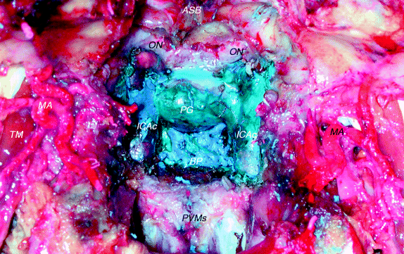
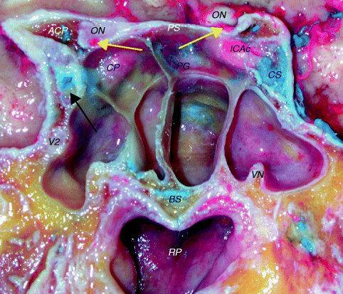
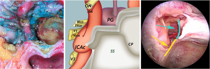
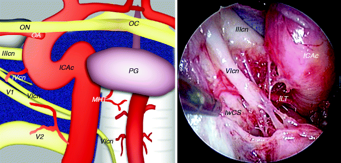
Fig. 3.1
Anterior view of the midline structures
ASB anterior skull base, BP basilar plexus, ICAc cavernous portion of the internal carotid artery (vertical segment), MA maxillary artery, ON optic nerve, PG pituitary gland, PVMs prevertebral muscles, TM temporal muscle
Fig. 3.2
Anterior view of the sphenoid sinus and the rhinopharynx
ACP anterior clinoid process, BS basisphenoid, CP carotid protuberance, CS cavernous sinus, ICAc cavernous portion of the internal carotid artery, ON optic nerve, PG pituitary gland, RP rhinopharynx, VN vidian nerve, V2 second branch of the trigeminal nerve, black arrow first branch of the trigeminal nerve, yellow arrows ophthalmic artery
Fig. 3.3
Anterior vision of the right cavernous sinus
BS basisphenoid, CP carotid protuberance, ICAc cavernous portion of the internal carotid artery (vertical segment), LRSS lateral recess of the sphenoid sinus, OA ophthalmic artery, ON optic nerve, PG pituitary gland, SS sphenoid sinus, V1 first branch of the trigeminal nerve, V2 second branch of the trigeminal nerve, IIIcn oculomotor nerve, IVcn trochlear nerve, VIcn abducens nerve, yellow arrow inferior part of the medial wall of the cavernous sinus (yellow line), blue-sky arrow superior part of the medial wall of the cavernous sinus (blue-sky line)
3.3 Anatomic Pictures
The inferior part of the medial wall of the CS faces the lateral part of the body of the sphenoid, while the upper part of the medial wall is directly related to the PG (Martins et al. 2011). In the upper part, the medial wall is given by the meningeal layer, that is a continuation of the diaphragma sellae, which surrounds the pituitary capsule inferiorly (Yasuda et al. 2005; Martins et al. 2011). In the inferior part, the medial wall is given by the endosteal layer that covers the body of the sphenoid bone. So, a sellar (superior) and a sphenoidal (inferior) part can be described. The limits of the medial wall of the cavernous sinus are the superior orbital fissure anteriorly, the dorsum sellae posteriorly, the superior margin of the maxillary nerve inferiorly and the diaphragma sellae superiorly (Yasuda 2005). The presellar segment of the ICAc corresponds to the anterior bend of the ICAc and to the clinoidal segment of the internal carotid artery.
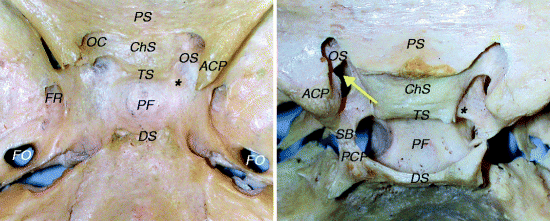
Fig. 3.4
Sphenoid sinus and bone
ACP anterior clinoid process, ChS chiasmatic sulcus, DS dorsum sellae, FO foramen ovale, FR foramen rotundum, OC optic canal, OS optic strut, PCP posterior clinoid process, PF pituitary fossa, PS planum sphenoidale, SB sellar bridge, TS tuberculum sellae, yellow arrow carotid-clinoid foramen, black asterisk middle clinoid process
ACPs project posteriorly and laterally to the optic canal and carotid sulcus. The TS separates the chiasmatic sulcus from the pituitary fossa. This is closed posteriorly by the DS. On each side of DS are the PCPs. The OS links the ACP with the sphenoid bone and separates the optic canal from the superior orbital fissure. When anterior and middle clinoid processes fuse, a “carotid-clinoid” foramen is formed. This circumstance is present in about 10 % of adults. The bony connection between anterior and posterior clinoid processes is called the “sellar bridge.” This is found in about 6 % of cases (Lang 1995). The base of the middle clinoid process is just medial to the anterior genu of the (parasellar) caverous ICA, while the apex extends in a dorsolateral direction. The middle clinoid process is, when present, a small eminence of the carotid sulcus at the level of the anterior part of the lateral wall of the sella turcica (Labib et al. 2013). The lateral tubercular crest is a bony eminence placed on the lateral part of the tuberculum sellae.
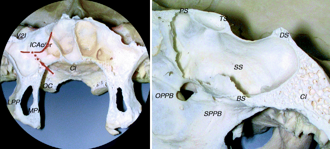
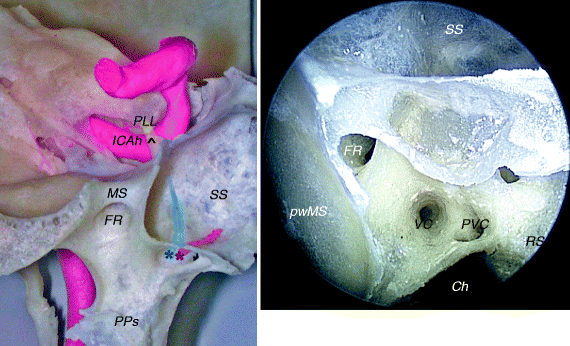
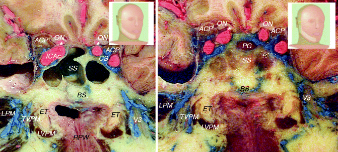
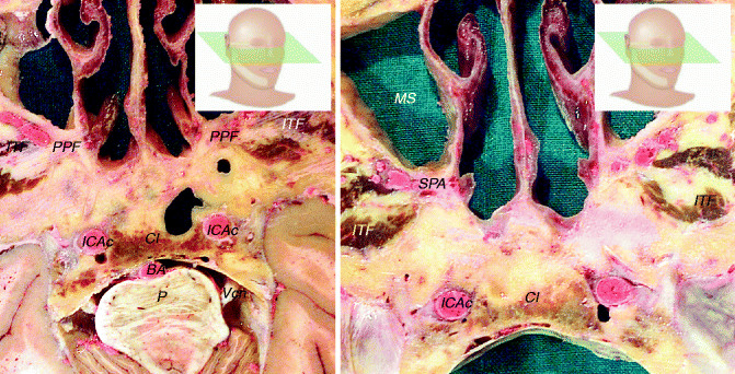
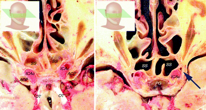
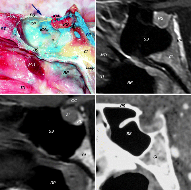
Fig. 3.5
Sphenoid sinus and bone
BS basisphenoid, Cl clivus, DS dorsum sellae, ICAc/pr protuberance of the cavernous portion of the internal carotid artery, LPP lateral pterygoid plate, MPP medial pterygoid plate, OC occipital condyle, OPPB orbital process of the palatine bone, PS planum sphenoidale, SPPB sphenoidal process of the palatine bone, SS sphenoid sinus, TS tuberculum sellae, V2I incisure of V2, red arrow opening of the vidian canal, red asterisks vidian canal, red line internal carotid artery
Fig. 3.6
Sphenoid bone and sinus with a focus on the base of the pterygoid region and the relationship with the cavernous portion of the internal carotid artery
Ch choana, FR foramen rotundum, ICAh horizontal portion of the internal carotid artery, MS maxillary strut, PLL petrolingual ligament, PPs pterygoid plates, PVC palatovaginal canal, pwMS posterior wall of the maxillary sinus, RS rostrum sphenoidale, SS sphenoid sinus, VC vidian canal, black arrowhead lingula of the sphenoid, blue-sky asterisk and line vidian (pterygoid) canal, red asterisk and line palatovaginal (pharyngeal) canal
Fig. 3.7
Coronal views at the level of the sphenoidal region. The picture on the left shows a more anterior view than the picture on the right. Note that the picture on the left is seen from posterior, while the picture on the right is seen from anterior
ACP anterior clinoid process, BS basisphenoid, CS cavernous sinus, ET eustachian tube, ICAc cavernous portion of the internal carotid artery, LPM lateral pterygoid muscle, LVPM levator veli palatini muscle, ON optic nerve, PG pituitary gland, RPW rhinopharyngeal posterior wall, SS sphenoid sinus, TVPM tensor veli palatini muscle, V3 third branch of the trigeminal nerve, white asterisk ophthalmic artery
Fig. 3.8
Axial views at the level of the clivus. The picture on the left is seen from below. The picture on the right is seen from above
BA basilar artery, Cl clivus, ICAc cavernous portion of the internal carotid artery (vertical segment), ITF infratemporal fossa, MS maxillary sinus, P pons, PPF pterygopalatine fossa, SPA sphenopalatine artery, Vcn trigeminal nerve
Fig. 3.9
Axial view at the level of the sphenoid sinus. The picture on the left is seen from below. The picture on the right is seen from above. The picture on the left shows a more cranial view
ACP anterior clinoid process, ICAc cavernous portion of the internal carotid artery, ICAi intracranial portion of the internal carotid artery, OT optic tract, PG pituitary gland, PS pituitary stalk, SS sphenoid sinus, blue arrow abducens nerve, white asterisk third ventricle
Fig. 3.10
Medial sagittal vision of the sphenoid sinus and its surrounding structures
AL anterior lobe, BP basilar plexus, BS basisphenoid, Cl clivus, ICAc cavernous portion of the internal carotid artery, ICAi intracranial portion of the internal carotid artery, ITt tail of the inferior turbinate, Lcap longus capitis, MTt tail of the middle turbinate, OC optic chiasm, OP optic protuberance, PG pituitary gland, PL posterior lobe, PS planum sphenoidale, RP rhinopharynx, ST superior turbinate, yellow arrow superior hypophyseal artery(ies), blue arrow optic nerve
The anterior lobe of the pituitary gland is mainly fed by the superior hypophyseal arteries while the posterior lobe is fed mainly by the inferior hypophyseal artery.
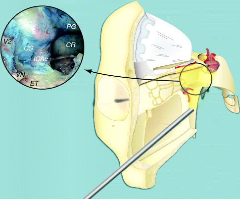
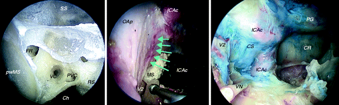
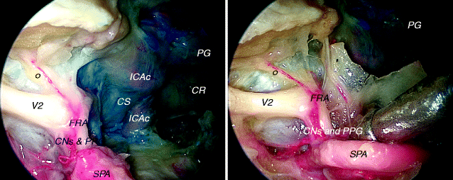
Fig. 3.11
Reconstruction showing the pterygoidal window and the region around the foramen rotundum
CR clival recess, CS cavernous sinus, ET eustachian tube, ICAc cavernous portion of the internal carotid artery, PG pituitary gland, VN vidian nerve, V2 second branch of the trigeminal nerve
Fig. 3.12
Foramen rotundum region
Ch choana, CR clival recess, CS cavernous sinus, FR foramen rotundum, ICAc cavernous portion of the internal carotid artery, MS maxillary strut, OAp orbital apex, PG pituitary gland, PVC palatovaginal canal, pwMS posterior wall of the maxillary sinus, RS rostrum sphenoidale, SS sphenoid sinus, VC vidian canal, VN vidian nerve, V2 second branch of the trigeminal nerve, blue-sky arrows superior orbital fissure
Fig. 3.13
Endoscopic views of the foramen rotundum region
CNs and PPG connecting nerves (V2 and pterygopalatine ganglion) and pterygopalatine ganglion, CR clival recess, CS cavernous sinus, FRA artery for the foramen rotondum, ICAc cavernous portion of the internal carotid artery, PG pituitary gland, SPA sphenopalatine artery, V2 second branch of the trigeminal nerve, black circle zygomatic nerve
Foramen rotundum transmits the second branch of the trigeminal nerve (maxillary nerve), arteries (foramen rodundum artery), and veins.
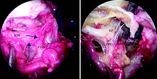
Fig. 3.14
Endoscopic vision of the pterygopalatine ganglion
ICAc cavernous portion of the internal carotid artery, ION infraorbital nerve, PPG pterygopalatine ganglion, SPA sphenopalatine artery, ZN zygomatic nerve, V2 second branch of the trigeminal nerve, blue arrows artery for the foramen rotundum
The zygomatic nerve exits the pterygopalatine fossa via the inferior orbital fissure and diverges from the ION to enter the orbit (Janfaza et al. 2001). The PPG is one of the four parasympathetic ganglia of the head (the other are the otic, the submandibular, and the ciliary). It is reached by the vidian nerve (parasympathetic and sympathetic), but only parasympathetic fibres synapse in the ganglion (Janfaza and Montgomery 2001).
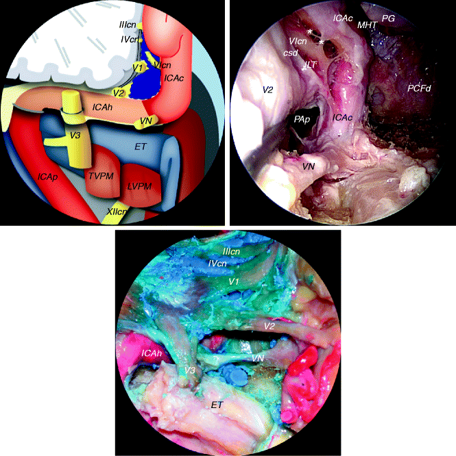
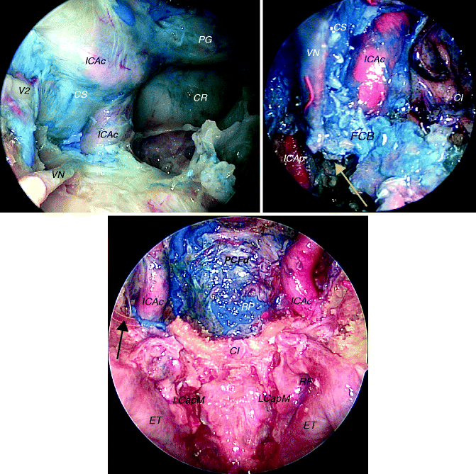
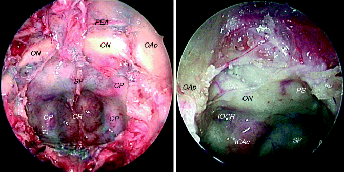
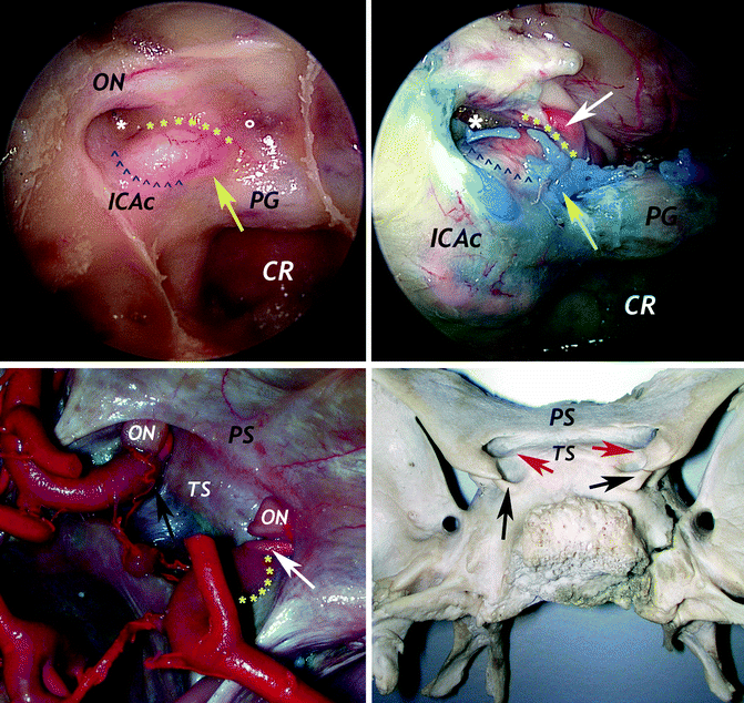
Fig. 3.15
Relationship between the vidian nerve and the anterior genu of the internal carotid artery
BS basisphenoid, CS cavernous sinus, CSd dura of the cavernous sinus, ET eustachian tube, ICAc cavernous portion of the internal carotid artery, ICAh horizontal portion of the internal carotid artery, ICAp parapharyngeal portion of the internal carotid artery, ILT inferolateral trunk, LVPM levator veli palatini muscle, MHT meningohypophyseal trunk, PAp petrous apex, PCFd posterior cranial fossa dura and periosteum, PG pituitary gland, TVPM tensor veli palatini muscle, VN vidian nerve, IIIcn oculomotor nerve, IVcn trochlear nerve, V1 first branch of the trigeminal nerve, V2 second branch of the trigeminal nerve, V3 third branch of the trigeminal nerve, VIcn abducens nerve, XIIcn hypoglossal nerve, white asterisks sympathetic fibres connecting the VIcn
Fig. 3.16
Relationship between the vidian nerve and the anterior genu of the internal carotid artery
BP basilar plexus, Cl clivus, CR clival recess, CS cavernous sinus, ET eustachian tube, FCB fibrocartilago basalis, ICAc cavernous portion of the internal carotid artery, ICAp parapharyngeal portion of the internal carotid artery, LCapM longus capitis muscle, PCFd dura and periosteum of the posterior cranial fossa, RF Rosenmuller’s fossa, VN vidian nerve, V2 second branch of the trigeminal nerve, yellow arrow petrous apex and petroclival region (bone removed), black arrow vidian nerve (bordered in yellow)
Fig. 3.17
Sphenoidal endoscopic views
CP carotid prominence, CR clival recess, ICAc cavernous portion of the internal carotid artery, lOCR lateral opticocarotid recess, OAp orbital apex, ON optic nerve, PEA posterior ethmoidal artery, PS planum sphenoidale, SP sellar prominence
Fig. 3.18
Endoscopic and external views showing the position and relationship of the middle clinoid process and medial optico-carotid recess
Cl clivus, ICAc cavernous portion of the internal carotid artery, ON optic nerve, PG pituitary gland, PS planum sphenoidale, TS tuberculum sellae, yellow asterisks upper dural ring, blue arrowheads lower dural ring, white asterisk lateral optico-carotid recess, white circle medial optico-carotid recess, white arrow ophthalmic artery, black arrows middle clinoid process, red arrows lateral tubercular crest, yellow arrows endocranial region corresponding to MCP
The lOCR corresponds to the pneumatisation of the optic strut. The clinoid segment of the ICA sits against the posterior surface of the optic strut (Yasuda et al. 2005). The endoscopic removal of the bony wall a little bit inferior and lateral to the lOCR exposes the medial aspect of the superior orbital fissure. The middle clinoid process does not corrispond to the medial optico-carotid recess. The MCP is located at the roof of the cavernous sinus, and it indicates the transition between the ICAc and the paraclinoidal ICA. It has no relationship with the optic nerve.
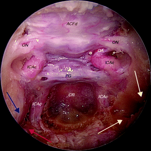
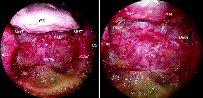
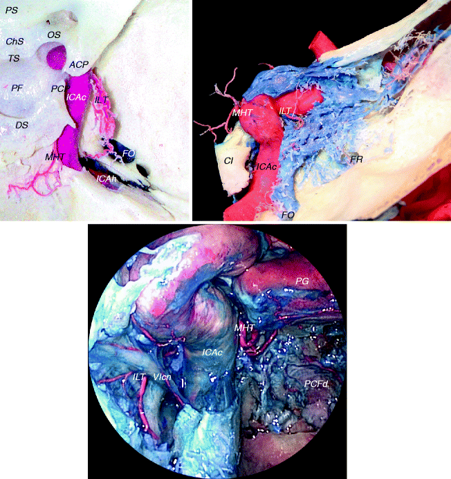
Fig. 3.19
Sphenoidal window
ACFd anterior cranial fossa dura, CR clival recess, ICAc cavernous portion of the internal carotid artery, OA ophthalmic artery, ON optic nerve, PG pituitary gland, red arrow vidian nerve, blue arrow second branch of the trigeminal nerve, white arrow maxillary prominence, yellow arrow vidian prominence, white asterisk lateral opticocarotid recess, white circle medial opticocarotid recess
Fig. 3.20
Clival region. The vascularisation of the pituitary gland related to the inferior hypophyseal arteries is clearly visible
CS cavernous sinus, DMA dorsal meningeal artery, ICAc cavernous portion of the internal carotid artery, IHA inferior hypophyseal artery, MHT meningohypophyseal trunk, PCFd dura and periosteum of the posterior cranial fossa, PG pituitary gland
Fig. 3.21
Branches of the cavernous ICA
ACP anterior clinoid process, ChS chiasmatic sulcus, Cl clivus, DS dorsum sellae, FO foramen ovale, FR foramen rotundum, ICAc cavernous portion of the internal carotid artery, ICAh horizontal portion of the internal carotid artery, ILT inferolateral trunk, MHT meningohypophyseal trunk, OS optic strut, PCFd dura and periosteum of the posterior cranial fossa, PF pituitary fossa, PG pituitary gland, PS planum sphenoidale, TS tuberculum sellae, VIcn abducens nerve
The most important branches of the cavernous carotid artery are the meningohypophyseal trunk and the inferolateral trunk. When present, capsular arteries arise from the medial wall of the ICAc (horizontal segment), run anteromedially to the hypophyseal capsule, and pass through the medial wall of the CS to join the inferior hypophyseal arteries on the floor of the PF. McConnell’s artery has been described in about one-third to one-half of the CS (Rhoton 2002).
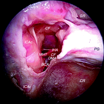
Fig. 3.22
Meningohypophyseal trunk
BCA Bernasconi-Cassinari artery, CF cavernous fat, CR clival recess, DMA dorsal meningeal artery, GR gyrus rectus, ICAc cavernous portion of the internal carotid artery, IHA inferior hypophyseal artery, MHT meningohypophyseal trunk, PG pituitary gland
By opening the dura of the medial wall of the cavernous sinus, the space between the internal carotid artery and the PG is exposed. In this space, the MHT is usually evident.
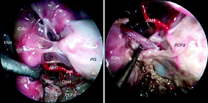
Fig. 3.23
Meningohypophyseal trunk and detail on dorsal meningeal artery
DMA dorsal meningeal artery, ICAc cavernous portion of internal carotid artery, IHA inferior hypophyseal artery, MHT meningohypophyseal trunk, PCFd dura and periosteum of the posterior cranial fossa, PG pituitary gland, VIcn abducens nerve
The MHT is traditionally described as having three branches: the inferior hypophyseal artery, the dorsal meningeal artery (also called the dorsal clival artery), and the tentorial artery (also called the Bernasconi-Cassinari artery). The DMA is in close relationship with the abducens nerve at the level of petrous apex (Cavallo et al. 2011). The DMA is the main feeder of the Dorello’s segment of VIcn (Martins et al. 2011).
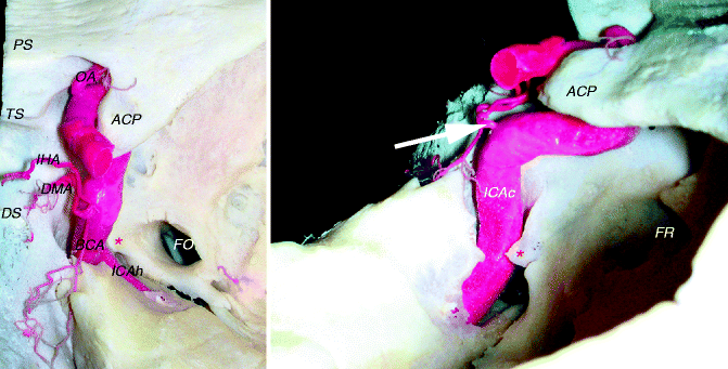
Fig. 3.24
Meningohypophyseal trunk
ACP anterior clinoid process, BCA Bernasconi-Cassinari artery, DMA dorsal meningeal artery, DS dorsum sellae, FO foramen ovale, FR foramen rotundum, ICAc cavernous portion of the internal carotid artery, ICAh horizontal portion of the internal carotid artery, IHA inferior hypophyseal artery, MHT meningohypophyseal trunk, OA ophthalmic artery, PS planum sphenoiodale, TS tuberculum sellae, white arrow MHT, red asterisk lingula of the sphenoid
The MHT is present in most cases. Not in all cases does it give off all the typical braches: dorsal meningeal artery (or dorsal clival artery), tentorial artery (or Bernasconi-Cassinari artery), and the inferior hypophyseal arteries. In about half of the cases, some branches arise directly from the ICAc (Jittapiromsak et al. 2010). The tentorial artery is the main feeder of the oculomotor nerve (d’Avella et al. 2008), and usually it is located on the inferior surface of the nerve. Moreover, it can be the feeder of the distal part of the trochlear nerve; in these cases, the vessel runs in close proximity of this nerve to the superior orbital fissure. Other authors show that BCA feeds cranial nerve IV along its course within the tentorium cerebelli (Martins et al. 2011).
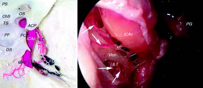
Fig. 3.25
Inferolateral trunk
ACP anterior clinoid process, ChS chiasmatic sulcus, DS dorsum sellae, FO foramen ovale, ICAc cavernous portion of the internal carotid artery, ICAh horizontal portion of the internal carotid artery, ILT inferolateral trunk, MHT meningohypophyseal trunk, OS optic strut, PCP posterior clinoid process, PF pituitary fossa, PG pituitary gland, TS tuberculum sellae, VIcn abducens nerve, white arrows branches of the ILT
The ILT is present in most cases (Krisht et al. 1994; Tran-Dinh 1987). It may arise as a common trunk with the MHT (Reisch et al. 2002). It is a single trunk in most cases. More often, it arises from the lateral aspect of the horizontal segment of the ICAc, and in most cases it passes superiorly to the abducens nerve (Inoue et al. 1990; Jittapiromsak et al. 2010). It usually gives rise to 3 or 4 branches supplying the dura and the cranial nerves within the cavernous sinus (Lasjaunias et al. 1977; Tran–Dinh 1987). The main trunk of the ILT with small secondary branches is the feeder of the ophthalmic nerve (V1). Usually, these vessels reach the inferior surface of the nerve. Obviously, the ILT also supplies the abducens nerve with several small branches.
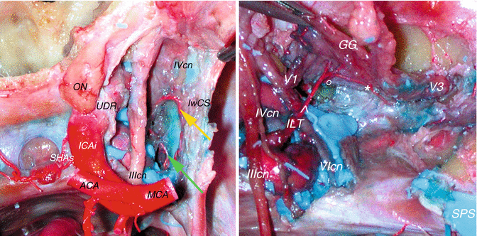
Fig. 3.26
Branches of the inferolateral trunk. In the right picture the gasserian ganglion has been lifted up
ACA anterior cerebral artery, GG gasserian ganglion, ICAi intracranial portion of the internal carotid artery, ILT inferolateral trunk, lwCS lateral wall of the cavernous sinus, MCA middle cerebral artery, ON optic nerve, SHAs superior hypophyseal arteries, SPS superior petrosal sinus, UDR upper dural ring, IIIcn oculomotor nerve, IVcn trochlear nerve, V1 first branch of the trigeminal nerve, V3 third branch of the trigeminal nerve, VIcn abducens nerve, yellow arrow superoproximal artery (branch of the ILT), green arrow indicates a tentorial branch of the ILT, white arrowhead artery of the superior orbital fissure, white circle artery for the foramen rotundum, white asterisk artery for the foramen ovale
The superoproximal artery usually curves superiorly and posteriorly, reaching the IVcn on the lateral wall of the cavernous sinus (CS) and goes to the free edge of the tentorium cerebelli (Conti et al. 2008). It is the main feeder of the proximal part of the nerve (d’Avella et al. 2008), but it can also be a supplier of the proximal part of IIIcn and V1 (Conti et al. 2008). Another constant branch is the superior orbital fissure (SOF) artery, which runs between V1 and V2 and usually splits in two branches: one for the foramen rotundum and the other for the foramen ovale (Conti et al. 2008). As a whole, the SOF artery can be considered the most important vessel for the blood supply to the distal portion of cranial nerves III, IV, VI, and V1 (Willinsky et al. 1987; Krisht et al. 1994; Tekdemir et al. 1998).
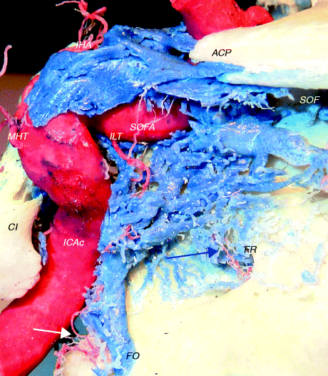
Fig. 3.27
Branches of the inferolateral trunk
ACP anterior clinoid process, Cl clivus, FO foramen ovale, FR foramen rotundum, ICAc cavernous portion of the internal carotid artery, IHA inferior hypophyseal artery, ILT inferolateral trunk, MHT meningohypophyseal trunk, SOF superior orbital fissure, SOFA superior orbital fissure artery, white arrow artery for the foramen ovale, blue arrow artery for the foramen rotundum
Obviously, the arborisation for the floor of the middle cranial fossa can be variable, and the arteries for the foramen rotundum and ovale can be given by the main trunk of the ILT. Typically, branches of the ILT anastomose with branches of the middle meningeal artery and accessory middle meningeal artery. It is also possible that tentorial branches arise from the ILT. In this case, their role in feeding neural structures is really critical.
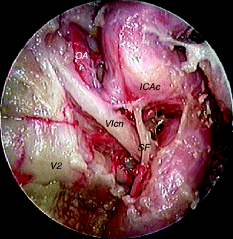
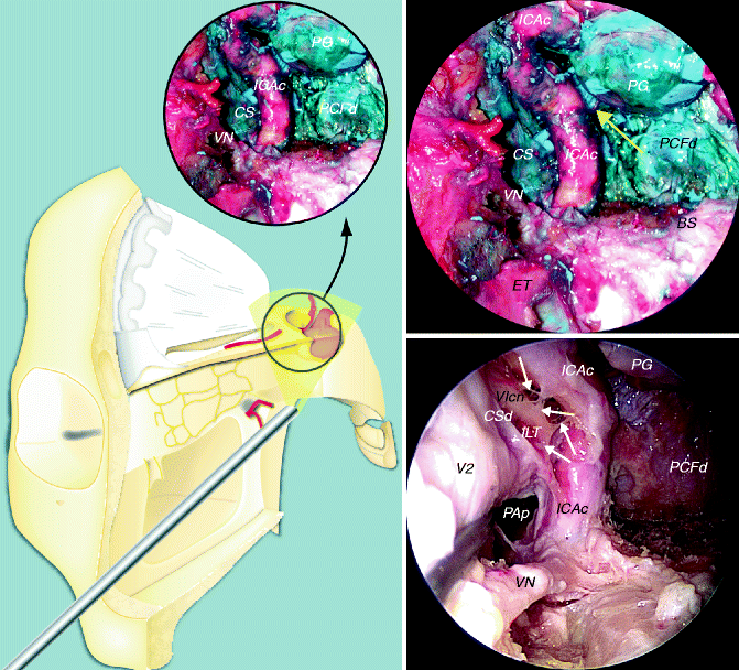
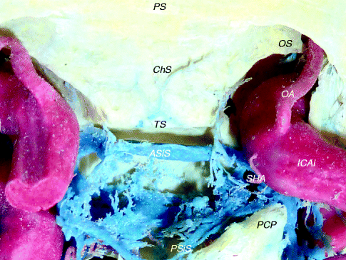
Fig. 3.28
Branches of the cavernous internal carotid artery (ICA), a rare variation: ophthalmic artery passing through the superior orbital fissure
ICAc cavernous portion of the internal carotid artery, OA ophthalmic artery, SF sympathetic fiber, V2 second branch of the maxillary nerve, VIcn abducens nerve
Fig. 3.29
Cavernous window
BS basisphenoid, CS cavernous sinus, CSd dura of the cavernous sinus, ET eustachian tube, ICAc cavernous portion of the internal carotid artery, ILT inferolateral trunk, PAp petrous apex, PCFd dura and periosteum of the posterior cranial fossa, PG pituitary gland, VN vidian nerve, V2 second branch of the trigeminal nerve, VIcn abducens nerve, yellow arrow meningohypophyseal trunk, white arrows sympathetic fibres
Fig. 3.30
Sellar region
ASiS anterior superior intercavernous sinus ChS chiasmatic sulcus, ICAi intracranial portion of the internal carotid artery, OA ophthalmic artery, OS optic strut, PCP posterior clinoid process, PSiS posterior superior intercavernous sinus, PS planum sphenoidale, SHA superior hypophyseal artery, TS tuberculum sellae
The cavernous sinuses are connected to each other via the anterior superior and posterior superior intercavernous sinuses. Together, they are called the circular sinus (Janfaza and Nadol 2001). The superior hypophyseal artery usually arises from the intracranial portion of the internal carotid artery. It supplies the optic chiasm and infundibular region of the hypothalamus. The inferior hypophyseal artery supplies the posterior lobe of the pituitary gland. The OA has been described as having an origin in one-half of the cases above the distal dural ring, in one-third of the cases at the level of the distal dural ring, and in less than 20 % of cases in the paraclinoid area (Lang 1995).
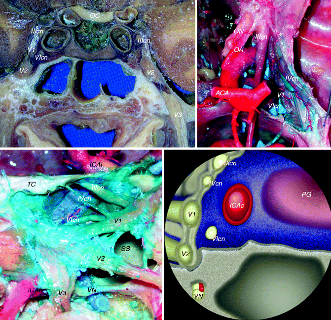
Fig. 3.31
Cavernous sinus
ACA anterior cerebral artery, ICAc cavernous portion of the internal carotid artery, ICAh horizontal portion of the internal carotid artery, ICAi intracranial portion of the internal carotid artery, OA ophthalmic artery, OC optic chiasm, ON optic nerve, PG pituitary gland, SS sphenoid sinus, TC tentorium cerebelli, VN vidian nerve, V1 first branch of the trigeminal nerve, V2 second branch of the trigeminal nerve, V3 third branch of the trigeminal nerve, IIIcn oculomotor nerve, IVcn trochlear nerve, VIcn abducens nerve
The CSs are large venous spaces located on both sides of the body of the sphenoid. Probably, the term is too restrictive, neglecting the neural and soft tissue elements. A valuable and rational name is lateral sellar compartment (Parkinson 1990). For the venous compartment, three spaces can be identified: posterosuperior, medial, and inferomedial (Reisch et al. 2002).
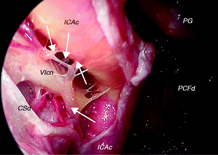
Fig. 3.32
Cavernous sinus
CSd dura of the cavernous sinus, ICAc cavernous portion of the internal carotid artery, ILT inferolateral trunk, PCFd dura and periosteum of the posterior cranial fossa, PG pituitary gland, VIcn abducens nerve, white arrows sympathetic fibres
Within the CS, the sympathetic fibres are observed mainly in the anterior part of the artery, and usually they are placed inferiorly. Most of these fibres run together with V1 (Jittapiromsak et al. 2010). The sympathetic fibres diverge from the ICAc to adhere to the abducens nerve while crossing to join the ophthalmic nerve (V1). The main target exit is V1 on the lateral wall of the CS (Jittapiromsak et al. 2010). Within the CS, the abducens nerve typically courses medially to the V1 before it exits through the superior orbital fissure.