21 Sinonasal Anatomy and Physiology • Quadrilateral cartilage • Perpendicular plate of the ethmoid • Vomer • Maxillary crest • Facial artery—labial branch • Palatine artery • Anterior ethmoid artery • Posterior ethmoid artery • Sphenopalatine artery—posterior septal branch • Anterior ethmoidal nerve • Medial superior posterior nasal (inc. nasopalatine n) • Maxilla • Nasal bones • Upper and lower lateral cartilages • Sphenoid sinuses • Cribriform plate • In continuity with MT • Overlie the superior meatus • Variable attachments posteriorly to the sphenoid face or lamina papyracea • Medial surface contains olfactory epithelium Fig. 21.1 Lateral nasal wall. I, superior meatus; ii middle meatus; iii, inferior meatus. 1, nasal vestibule; 2, opening of the nasolacrimal duct; 3, origin of the inferior turbinate; 4, semilunar hiatus; 5, insertion of the middle turbinate; 6, sphenoid sinus; 7, insertion of the superior turbinate; 8, frontal sinus; a, drainage of the antral cavity; b, drainage of the frontal sinus; c, drainage of the anterior ethmoid cells; d, drainage of the posterior ethmoid cells; e, drainage of the sphenoid sinus; f, area of infundibulum (dotted area). • Has four parts—anterior and posterior buttresses, vertical attachment, and basal lamella (horizontal attachment) • Separates ethmoidal cells into anterior and posterior • Various anomalies include pneumatization (concha bullosa) and paradoxical configurations • Overlies the middle meatus • Largest turbinate • Overlies inferior meatus (valve of Hassner) • May also exist in some people • Weeks 9–10: Formation of six ridges. • Ridges form ethmoturbinals: • Four consistent/constant landmarks: • Three inconsistent landmarks: • Hiatus semilunaris: Fig. 21.2 Four Messerklinger landmarks. Sph, sphenoid sinus; SEC(OC), spheno-ethmoidal recess (olfactory cleft); SpM, supreme meatus; SM, superior meatus; RBR, retrobullar recess; FS, frontal sinus; ANC, agger nasi cell; EB, ethmoid bulla; Max O, maxillary ostium. 1, uncinate process; 2a, anterior wall of EB; 2b, posterior wall of EB; 3, basal lamella of middle turbinate; 3a, superior turbinate; 3b, supreme turbinate; 4, anterior face of sphenoid sinus. • Ostiomeatal complex: – Uncinate process – Ethmoid infundibulum – Anterior ethmoid cells – Ostia of anterior ethmoid, maxillary, and frontal sinuses • Sinus lateralis = suprabullar and retrobullar recesses • Suprabullar recess: space between roof of ethmoid bulla and skull base • Retrobullar recess: space between posterior wall of ethmoid bulla and middle turbinate basal lamella • Crescenteric bone that forms part of the ethmoid bone • Three types of superior attachment: • With type A attachments, mucociliary outflow from the frontal sinus is directly into the middle meatus, in type B/C attachments, that mucociliary flow is into the ethmoid infundibulum • “Doorway to the sinuses” • Present at birth • Located beneath the orbit • Anterior to the infratemporal and pterygopalatine fossae • Natural maxillary ostium lies at the lateral end of the ethmoid infundibulum, behind the uncinate process; accessory ostia are often seen and mistaken for the natural ostium and can lie within the lower uncinate process or posterior to the free edge of the uncinate process • Blood supply—facial, maxillary, infra-orbital, and greater palatine arteries • Nerve supply—infra-orbital and superior alveolar branches of maxillary nerve • Ethmoid bone: • Boundaries: • Nerve supply—anterior and posterior ethmoidal nn • Blood supply—anterior and posterior ethmoidal arteries from the ophthalmic a • Anterior cells—drain into middle meatus – Most anterior of the ethmoid cells – Defines the anterior aspect of the frontal recess – Lies posterior to the uncinate process and anterior to the basal lamella – Attached to the skull base superiorly defining the posterior end of the frontal recess and demarcating the anterior ethmoidal a – Drains posteriorly into the retrobullar space – Small when a pneumatized MT present – Pneumatization along the orbital wall opposite the uncinate process—may narrow the infundibulum – Pneumatization along the anterior skull base posterior to the frontal sinus • Posterior cells—drain into superior meatus: • Occupies sphenoid bone • Intersinus septum not usually midline • Post-ethmoid a and n give vascular and sensory supplies, respectively • Ostium ~1 cm above choana • Main development occurs after puberty • Relations: • Frontal recess: • Frontal recess cells: • Occasionally absent; underdeveloped in CF patients • Variable in size • Bounded by anterior cranial fossa and orbits • Start to form after age 2 years • Blood supply = supraorbital and anterior ethmoid arteries • Nerve supply = supraorbital nn • Ventilatory: • Phonetic: • Immunological—humoral and cellular: • Olfactory • Mechanical: • Speech of particle movement = 3 to 25 mm/min at ~12 Hz • Low levels of nitric oxide related to ciliary dysfunction • Mucous secretions (from goblet cells) trap particulate matter, which is then propelled to nasopharynx • Watery secretions from serous glands evaporate to moisten the inspired air • Sinus cilial train beats towards natural ostium • Ostial patency required for adequate mucociliary clearance and gas exchange (nitric oxide) • Frontal sinus outflow along medial wall and into either infundibulum or middle meatus direct—depending on uncinate attachment
21.1 Nasal Septum
21.1.1 Structure
21.1.2 Blood Supply
21.1.3 Innervation
21.1.4 Adjacent Structures
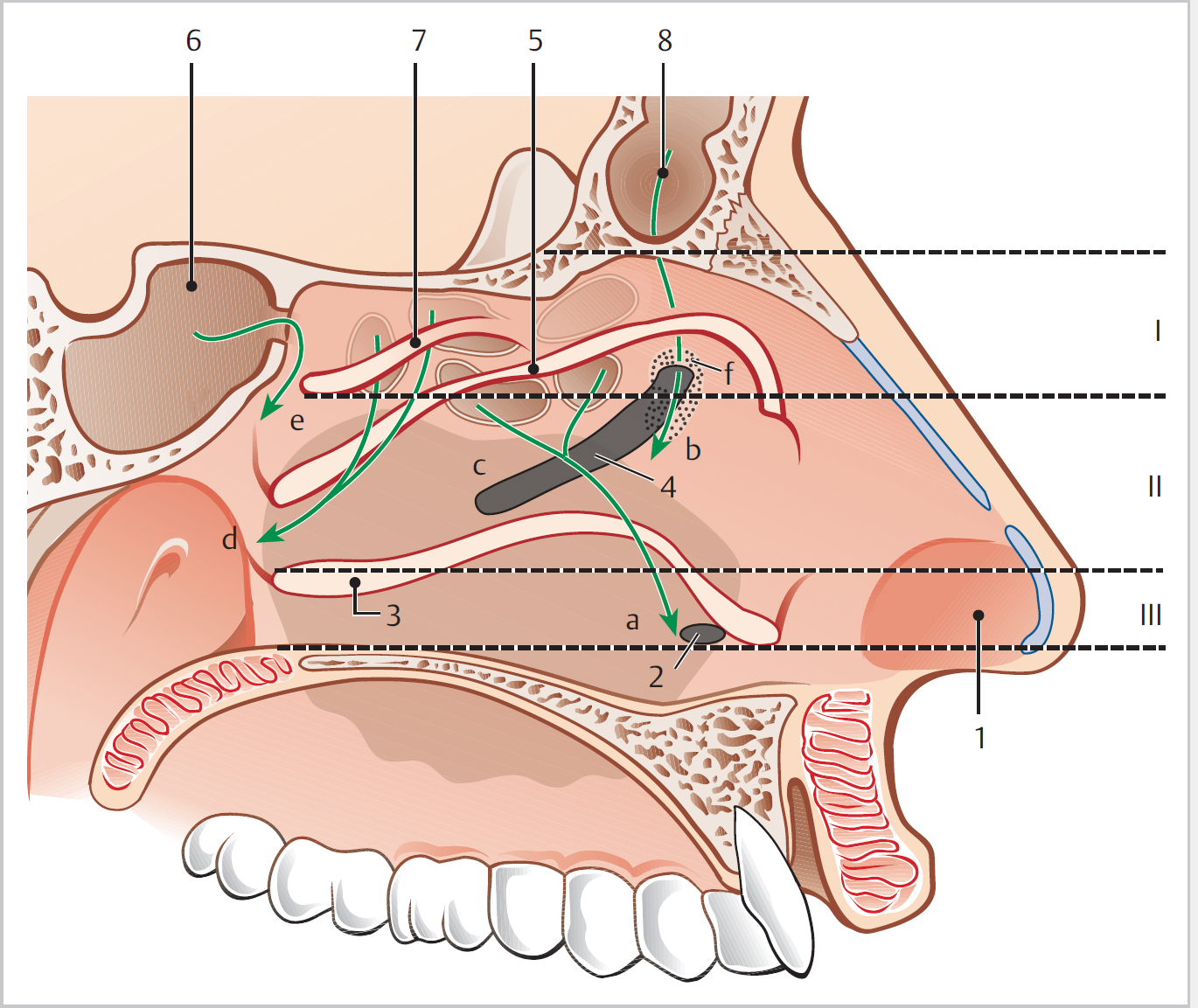
21.2 Nasal Conchae (Turbinates) (Fig. 21.1)
21.2.1 Superior
21.2.2 Middle
21.2.3 Inferior
21.2.4 Supreme
21.3 Paranasal Sinuses, Ostia, and Ostiomeatal Complex
21.3.1 Embryology
 Pars ascendens and pars descendens
 Middle turbinate: third ethmoturbinal
 Superior turbinate: fourth ethmoturbinal
 Supreme turbinate: fifth ethmoturbinal
 Inferior turbinate: maxilloturbinal
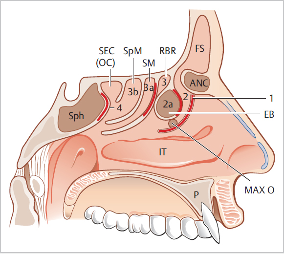
21.3.2 Messerklinger Landmarks (Fig. 21.2)
 Uncinate process
 Ethmoid bulla: anterior wall
 Middle turbinate basal lamella
 Sphenoid face
 Ethmoid bulla: posterior wall
 Superior turbinate basal lamella
 Supreme turbinate basal lamella
21.3.3 Middle Meatus
 H. semilunaris inferior: shortest distance between free posterior margin of uncinate process and anterior face of ethmoid bulla
 H. semilunaris superior: crescent-shaped cleft between ethmoid bulla and middle turbinate
 Final common drain pathway for anterior sinuses—a functional unit
 Includes:
21.3.4 Potential Spaces
21.3.5 Uncinate Process
 To lamina papyracea (A) forming a recessus terminalis
 To the skull base (B)
 To the middle turbinate (C)
21.3.6 Maxillary Sinus
21.3.7 Ethmoidal Cells
 Paired bony scaffolds, connected by cribriform plate
 Lamina papyracea of orbit
 Orbital process of the frontal bone
 (Fovea ethmoidalis1 orbital plate of the frontal bone)
 Middle and superior turbinates medially
 Lateral cribriform plate lamella (also medially)
 Sphenoid sinus
 Agger nasi:
 Ethmoid bulla:
 Infra-orbital (Haller) ethmoidal cells:
 Supraorbital ethmoid cells
 Between one and nine in number
 No middle cells
 A variant may pneumatize superior and lateral to the sphenoid sinus (sphenoethmoidal [Onodi] cell)
21.3.8 Sphenoid Sinus
 Pituitary fossa and middle cranial fossa superiorly
 Cavernous sinus and ICA laterally
 Pons and posterior cranial fossa posteriorly
 Forms roof of nasopharynx
 Optic nerve lies next to or even within the sinus
 Pterygoid canal with nerve inferiorly
21.3.9 Frontal Sinus
 Nasofrontal duct—incorrect
 Inverted funnel shape
 Anterior skull base is posterior boundary
 Anterior ethmoid artery close
 Agger nasi cell
 Frontal cells (types I–III/IV)
 Supraorbital ethmoid cell
 Frontal bullar cell
 Suprabullar cell
 Frontal intersinus septal cell
21.4 Sinonasal Physiology
21.4.1 True or Perceived Sinonasal Physiological Functions
 Humidification
 Filtration
 Airway
 Buffer pressure changes
 Voice resonance
 Reduces bone conduction of own speech
 Reduce skull weight
 Heat insulation
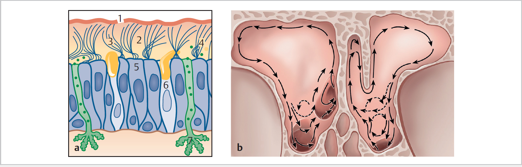
21.4.2 Mucociliary Clearance (Fig. 21.3)
![]()
Stay updated, free articles. Join our Telegram channel
 Agger nasi cell and frontal cells may occupy space in funnel—make identification and drainage difficult
Full access? Get Clinical Tree


