Acquired pediatric laryngotracheal stenosis almost always results from prolonged intubation for prematurity. An understanding of the process by which this occurs helps in prevention and treatment. Before deciding to perform cartilage augmentation procedures, more limited techniques such as medical therapy or endoscopic surgery need to be considered. Careful assessment of the patient and the stenosis aid the decision-making process for the right operation at the right time. Despite this assessment, patients with a severe or complete stenosis have a poorer prognosis, and cricotracheal resection may be a better option.
Laryngotracheal reconstruction (LTR) now is accepted as the standard of care for established pediatric laryngotracheal stenosis. It can be adapted to address almost all laryngeal stenoses including grade IV lesions, although it may not always be the most appropriate treatment. Cartilage graft augmentation, the tried and tested technique of airway reconstruction, now has been used for more than 30 years. In most pediatric otolaryngology departments the bulk of the experience is with this technique. As in all airway surgery, decision making is at least as important as the actual surgery. This article therefore covers the etiology and prevention of acquired laryngotracheal stenosis and discusses cartilage augmentation in classical (multistage) LTR with a covering tracheostomy and the more recently developed single-stage LTR. The reader is reminded that early lesions may be addressed endoscopically, thus avoiding cartilage grafting altogether.
Acquired laryngotracheal stenosis
Etiology
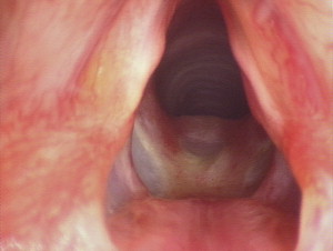
Pediatric laryngotracheal stenosis may occur in patients who have required intubation, often following premature birth. Ninety percent of acquired subglottic stenoses have a history of intubation , but stenosis also may develop after other forms of internal or external airway injury ( Box 1 ). The endotracheal tube causes pressure necrosis and mucociliary stasis in the subglottic tissues leading to mucosal edema and ulceration. The ulcer then deepens, giving rise to exposed cartilage ( Fig. 1 ), with subsequent infection and perichondritis that may progress to chondritis and cartilaginous necrosis. Granulation tissue typically forms in the areas of ulceration, and fibrous tissue is deposited in the submucosa .
Internal trauma
Intubation injury
External trauma
Blunt
Penetrating
Other
Trauma
After laryngeal surgery: high tracheostomy, glottic web, supraglottic collapse
Burns
Chronic infection (eg, tuberculosis, syphilis)
Chronic inflammation
Systemic: sarcoidosis, systemic lupus erythematosus, pemphigus, Wegener’s reflux
Laryngeal neoplasm
Primary lesion: chondroma, fibroma, carcinoma
Secondary involvement: tumor infiltration, radionecrosis, postoperative scarring
Histologic studies have shown acute mucosal injury invariably occurs after intubation of the infantile larynx, although injury progression is transient, with healing commencing within a few days and with rapid improvement and completion of healing by 30 days in most cases . The underlying etiology of laryngotracheal stenosis is multifactorial, and endoscopic studies have not identified any consistent factors in the development of postintubation subglottic stenosis in neonates . Airway trauma also may be caused by tube movement in a restless patient, by orotracheal tube placement, or by instrumentation from repeated intubations. Other sources of airway inflammation including nasogastric tubes or local bacterial infection may compound the inflammatory response and subsequent fibrosis. Gastroesophageal reflux and systemic factors including chronic illness, immunosuppression, and dehydration also increase the susceptibility of the laryngeal mucosa to injury. A congenital element is present in some patients as a result of their anatomy. Infants who have a smaller-sized cricoid may have a tendency to develop stenosis following intubation.
Prevention
Neonatal care has improved significantly since the early 1970s, but laryngotracheal stenosis continues to occur in approximately 1% of pediatric patients after intubation. Low-irritant endotracheal tubes now are used; the safest materials are polymeric silicone and polyvinyl chloride. Nasal endotracheal intubation can help minimize tube movement. The parallel-sided straight tubes generally are preferred for long-term neonatal intubation. It is important to avoid trauma during airway instrumentation by using gentle tissue handling and preparation of the patient to provide relaxation at intubation. It is essential to use an appropriately sized tube that allows a leak at 20 cm H 2 O pressure. Although pediatric patients seem to tolerate longer periods of intubation than adults, airway injury and stenosis still are more likely after longer periods of intubation. A careful approach to surgery on the pediatric larynx is paramount. For benign lesions (eg, pathology involving the anterior commissure), aggressive endoscopic interventions should be avoided with the use of staged procedures if necessary. Unavoidable high tracheostomies should be revised as soon as practical, and tracheostomy should allow maximal preservation of native tracheal cartilage by using the smallest tube possible to establish a safe, stable airway.
Treatment options for laryngotracheal stenosis
Early lesions may be treated by medical therapy, endoscopic surgery, or anterior cricoid split ( Table 1 ). Medical therapy includes treatment of any underlying conditions, including infection or gastroesophageal reflux, that would hinder laryngeal recovery. Oral, intravenous, or inhaled steroids and adrenaline nebulizers can help optimize the airway. Endoscopic treatment is being used increasingly and is beneficial in addressing soft, immature, and mild forms of stenosis. Techniques include cold steel, carbon dioxide, potassium-titanyl-phosphate laser, and balloon dilatation. Mitomycin C may be used as an adjunct. Established laryngotracheal stenosis is treated best with cartilage augmentation LTR, with cricotracheal resection being reserved for the most severe cases of airway stenosis.
| Treatment modality | Criteria/patient factors |
|---|---|
| Laryngeal rest/elective period of intubation |
|
| Endoscopic treatment of granulations and early stenosis: cold steel rather than CO 2 laser, balloon dilatation, mitomycin C, endoscopic cricoid split |
|
| Anterior cricoid split |
|
| Laryngotracheal reconstruction with cartilage augmentation |
|
| Cricotracheal resection |
|
| Tracheostomy |
|
Multistage versus single-stage laryngotracheal reconstruction
The single-stage procedure has attracted substantial interest because it has several potential benefits over the traditional multistage LTR . Tracheostomy insertion may be avoided or, if present, the tube may be removed during the reconstruction. Tracheostomy closure with the reconstruction eliminates a potential source of infection and addresses the stoma site in the same procedure. The stages of airway stabilization, healing, and decannulation, which typically take several months in the traditional procedure, are compressed into a short period of postoperative endotracheal intubation, typically lasting approximately 7 days. The problems of longer-term stenting are avoided.
It must be acknowledged that, although there is only a single open procedure, multiple subsequent endoscopic procedures often are required to monitor postoperative progress and to optimize the airway further while healing occurs. Perioperative airway complications may occur, and there is potential for unplanned extubation. There also is an inherent earlier reliance on the newly reconstructed airway at planned extubation. If respiratory compromise develops after extubation, the endotracheal tube may need to be replaced. Tracheostomy reinsertion may be required. Reintubation of these children is not always straightforward and may damage the cartilage reconstruction if performed by inexperienced staff. Postoperative care must be undertaken in a pediatric ICU, with associated issues of bed availability, cost, and the potential for complications resulting from the required prolonged intubation.
Principles of cartilage augmentation and stenting
Animal studies have confirmed the presence of viable cartilage following LTR with respiratory epithelium found lining the graft in most cases . Histologic examination after LTR with anterior cartilage grafting in rabbits also showed rapid epithelialization of the graft. Although the original graft underwent progressive necrosis and resorption, neochondrification occurred rapidly, providing the graft with excellent structural support . The cartilage used in LTR is incorporated successfully into the laryngeal framework and grows with the child. No adverse effects on laryngeal growth have been reported .
LTR traditionally is performed as a multistage procedure with a tracheostomy in situ. This surgery aims to create safely a stable airway that is age-appropriate in size and preserves or restores normal laryngeal function. Cartilage grafts are used to augment the airway during LTR and thus address the underlying stenosis. Stents stabilize and support the reconstruction while healing occurs. The single-stage procedure was developed as an extension of the experience gained from anterior cricoid split surgery, where it became apparent an endotracheal tube can be used as a short-term airway stent . Numerous techniques may be used to expand the airway in LTR, including anterior and/or posterior cricoid splits, each of which may be grafted. Grafts most commonly are fashioned using autogenous costal cartilage, which is a robust and reliable grafting material. Auricular and thyroid alar cartilage may also be used.
Assessment and decision-making factors
Each pediatric patient must undergo a comprehensive assessment to formulate an individualized treatment plan. Specific details of the history and examination are not covered in this article, but assessment should document the effect of the airway stenosis on the child, the degree of respiratory compromise, the general condition of the child, and any coexisting diagnoses that may affect the airway at other levels (eg, Pierre Robin sequence, craniofacial anomalies, choanal atresia, or chronic lung disease).
A rigid microlaryngoscopy and bronchoscopy during spontaneous ventilation remains the reference standard for endoscopic evaluation. This examination allows close, systematic inspection of the airway at all levels using an appropriately sized laryngoscope with a telescope or operating microscope, under laryngeal suspension. The cricoarytenoid joints are palpated to test passive mobility and the presence of interarytenoid scarring. The Hopkins rod telescope allows accurate assessment of the subglottis and tracheobronchial airway in addition to the remainder of the larynx. Photographic documentation and/or video recording are useful ( Fig. 2 ). Features of the stenosis, including the anatomic level, length and consistency (soft or firm), site (anterior, posterior, or circumferential), maturity (active inflammation or edema), and presence of granulations, fibrosis, or scarring, must be established carefully. The suprastomal area also should be examined in patients who have a pre-existing tracheostomy. The airway is sized formally, and the stenosis is graded using the Myer-Cotton grading system to assist in treatment planning ( Fig. 3 ). A dynamic assessment of the airway ensures that any co-existing tracheomalacia, vocal cord immobility, or laryngomalacia is detected.
The single-stage procedure has the potential to provide a decannulated stable airway sooner than the traditional multistage procedure and is thus an appealing option. Treatment options for each child must be considered on a case-by-case basis. Single-stage procedures ideally are considered when an anterior graft is performed with or without a posterior cricoid split, although posterior grafts have been performed in single-stage procedures. LTRs with a combination of anterior and posterior grafts have been reported to have a higher reintubation rate, although their subsequent outcomes are comparable with the multistage procedure . Single-stage procedures have been shown to have poorer outcomes in patients who have tracheal obstruction or tracheomalacia, particularly in those younger than 4 years . The procedure also is contraindicated in patients whose airway anatomy makes reintubation technically difficult, particularly in the emergency situation (eg, craniofacial or vertebral anomalies) and in children who have ongoing neurologic deficits or chronic lung disease that preclude decannulation.
Babies weighing more than 4 kg or with gestational ages of greater than 30 weeks have been found to have a greater chance of successful extubation and eventual airway patency . Very small babies have more complications at extubation, which may occur as a result of the comorbidities frequently present in these infants. In addition the physical dimensions of their airways leave little margin for any degree of airway compromise. These patients may be managed better by an initially conservative course while they grow or by a multistage reconstruction, ensuring a stable airway throughout the lengthy healing process.
Multistage LTR is more suitable for the severe grades of stenoses. A trend toward higher reintubation rates and tracheostomy insertions after single-stage reconstructions has been reported for severe grade III or IV stenoses . It also is preferable for patients who have poor respiratory reserve or multilevel stenoses and for those who have underlying factors that may compromise normal healing (eg, ongoing reflux or systemic problems) and who thus would require long-term stenting.
Classical laryngotracheal reconstruction using costal cartilage graft with a tracheostomy
The patient is positioned supine with the neck extended. The tracheostomy tube is replaced with an appropriately sized armored cuffed endotracheal tube. The skin is prepared, and the patient is draped, keeping the donor site separate from the neck. The senior author prefers to harvest the costal cartilage graft first, to minimize contamination of the clean donor site. By convention, the graft is taken from the right submammary region adjacent to the bony-cartilaginous junction. A transverse incision is made, and the rib margins are identified. The bony-cartilaginous junction is visualized, and the adjacent cartilaginous portion of the rib is transected carefully. A 3- to 4-cm segment of cartilage is elevated in a subperichondrial plane, taking care to avoid injury to the underlying pleura. Hemostasis is achieved, and a leak test performed. Although uncommon, any pleural defect thus can be identified and repaired immediately. The wound is closed in layers with a subcuticular skin suture.
A transverse skin incision is made superior to the pre-existing stoma site, and subplatysmal flaps are elevated from the superior margin of the thyroid cartilage to the tracheostomy site. A midline laryngofissure is performed ( Fig. 4 A). The extent of this incision depends on the site and extent of the airway pathology. For isolated subglottic stenosis, the airway is opened from just below the vocal cords (the level of the cords correlates with the midpoint between thyroid notch and cricoid in pediatric patients) through the anterior cricoid and upper tracheal rings to release the stenotic segment. At this stage a posterior cricoid split may be performed, if indicated ( Fig. 4 B). Posterior glottic stenosis is addressed by carefully dividing the glottic scar via the posterior laryngofissure. The incision is extended superiorly into the interarytenoid region through the fibrosed interarytenoid musculature and inferiorly for 0∼1 cm into the tracheoesophageal septum. The split should be splayed open with scissors to prove that the division is complete.


