Subglottic hemangioma is a rare condition that can be potentially life threatening because of airway obstruction. It is common for subglottic hemangioma to be misdiagnosed as croup initially. Infants with a subglottic hemangioma and cutaneous facial hemangiomas in a “beard” distribution should be evaluated for PHACE syndrome. Endoscopic laser resection is effective for subglottic hemangioma but carries a chance of subglottic stenosis, up to 25%. Open excision of subglottic hemangioma is an excellent option, particularly in patients with bilateral or circumferential subglottic hemangioma. It is a more extensive surgery when compared with endoscopic laser resection. Surgeons who do not have access to a pediatric intensive care unit staffed by experienced pediatric intensivists should not use this procedure.
Infantile hemangiomas, a type of vascular lesion, undergo rapid growth in the first months after birth. They are the most common tumors of the head and neck in pediatric patients . A subglottic hemangioma (SGH) is, however, a rare condition that can be potentially life threatening because of airway obstruction. SGH was first described by Morrell Mackenzie in 1864 and accounts for approximately 1.5% of all congenital laryngeal anomalies . Infants with this condition are typically asymptomatic during the newborn period . The proliferation phase begins at approximately 1 to 2 months of age, leading to symptoms of biphasic stridor, respiratory distress, and feeding difficulties. Heightened suspicion for this lesion in the first months of life, together with an endoscopic airway examination, is required to accurately diagnose a SGH. Numerous modalities have been used to treat SGH, including intralesional and systemic steroids, systemic interferon, endoscopic laser resection, and open excision. Open excision with anterior laryngotracheal reconstruction has gained attention and popularity in recent years. Increasing evidence indicates that open excision is superior to other modalities in its ability to achieve decannulation or avoid tracheostomy altogether, often with fewer complications and fewer total procedures .
Clinical presentation
Patients with an SGH are typically asymptomatic during the first several weeks of life. Once in the proliferative phase—approximately 6 to 12 weeks of age—symptoms of biphasic (inspiratory and expiratory) stridor and respiratory distress become noticeable. An infant’s voice is typically normal and there is no difficulty in swallowing, but there is frequently difficulty in feeding as the infant struggles to breathe and suck at the same time. Patients are commonly misdiagnosed as having croup, a much more common pediatric condition that causes edema in the subglottic region. Often a child has a cough that mimics the barking cough of croup. Two differentiating features between croup and SGH are the lack of fever and rhinorrhea in children with SGH. Because hemangiomas decrease in size when treated with agents typically used for croup, such as nebulized epinephrine, inhaled steroids, and systemic steroids, the clinical picture initially can be misleading. Because a short duration of therapy used for the treatment of croup is insufficient to yield lasting results in SGH, recurrence and gradual worsening of stridor and respiratory distress typically occur. Recurrent croup in the neonatal period is a “red flag” for the presence of SGH.
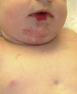
Cutaneous hemangiomas can be associated with SGH, particularly if the cutaneous lesions are found in a “beard” distribution, including the preauricular areas, chin, anterior neck, and lower lip ( Fig. 1 ). The biologic basis for this association is unknown. It seems that the more extensive the “beard” cutaneous hemangiomas, the higher the likelihood of a symptomatic airway hemangioma. In one study, 1 of l1 (9%) patients who had cutaneous hemangiomas present in only one or two of the five examined areas (right and left preauricular areas, chin, anterior neck, lower lip) was found to have an SGH, compared with 10 of 16 (63%) patients who had hemangiomas in at least four of the five examined regions . For patients with SGH, up to 50% may have cutaneous lesions .
If left untreated, SGH, like other hemangiomas, undergoes proliferation for approximately 1 year, followed by slow, spontaneous involution in most cases. Complete resolution is seen in approximately 50% of children by age 5 years and more than 70% by age 7 years. Continued improvement is observed in the remaining children until 10 to 12 years of age .
Scattered reports of various associations with SGH have long existed. Frieden and colleagues unified the association of posterior fossa malformations (P), segmental facial hemangiomas (H), arterial anomalies (A), cardiac defects (C), eye abnormalities (E), and sternal defects (S) with the umbrella term “PHACE(S) syndrome” ( Fig. 1 ). All affected patients have a segmental hemangioma (typically a plaque-like hemangioma over a specific cutaneous territory), but only one extracutaneous manifestation is needed to qualify for the diagnosis . The prevalence of PHACES syndrome as a subset of all patients with segmental facial hemangiomas is not known. Intracranial arterial anomalies are currently well recognized as a relatively frequent manifestation of the syndrome, resulting in possible arterial occlusion and infarction . Patients who have SGH and facial hemangiomas should undergo screening with MRI of the brain to rule out intracranial posterior fossa malformations and arterial anomalies.
Pathology
SGHs, like other infantile hemangiomas, are primarily composed of endothelial cells but also contain pericytes, fibroblasts, interstitial cells, and mast cells . GLUT1 is an erythrocyte-type glucose transporter that is expressed in the endothelia of blood-tissue barriers. North and colleagues discovered that GLUT1 expression was marked in infantile hemangioma, regardless of age, whereas its expression was negative in other vascular tumors and malformations, which made it a specific and useful immunohistochemical marker for infantile hemangiomas. North and colleagues further recognized that infantile hemangiomas and placenta express GLUT1, which led to speculation that infantile hemangiomas may result from angioblasts that differentiate toward a placental phenotype or, alternatively, derive from embolized placental cells.
Management
Steroid therapy
Over the years, multiple modalities have been used in the treatment of SGH. Systemic steroids have a long history of popularity and success. Although it is efficacious in the reduction of tumor size, only approximately 25% of cases resolve completely with this treatment alone . Intralesional steroids have a much higher percentage of cure, reported to be 77% in the study by Hoeve and colleagues . It often requires 30 to 50 days of intubation time while the patient is undergoing serial injections, thus increasing intubation-related morbidity and cost.
Interferon alpha-2a
Interferon is an antiproliferative agent that inhibits angiogenesis by lowering the concentration of angiogenic factors. The empiric dose for interferon is 2 to 3 million units/m 2 injected subcutaneously every day. Duration of treatment is often 6 to 12 months . Although its efficacy for hemangioma is well reported, with up to 71% showing clinical regression, substantial side effects are associated with this therapy, including fever, myalgia, transient elevation of hepatic transaminase levels, transient neutropenia, anemia, and spastic diplegia . Spastic diplegia occurs with a frequency of 5% to 20% and its mechanism is unknown, but if interferon is stopped quickly, it may be potentially reversible . The myriad possible complications make interferon therapy less than ideal for most patients; it should be reserved as a therapy of last resort.
Tracheostomy
Tracheostomy and observation were once considered the standard treatment for infants with SGH and still remains a viable alternative to tumor resection. Because of the self-limiting nature of SGH and the high percentage of spontaneous involution (>90%), the success rate for observation is high . The down side of this treatment is related to the morbidity of prolonged tracheostomy. A child with a tracheostomy has an overall mortality rate of approximately 1% and often presents challenges to parents and caregivers in the time and labor required to care for the tracheotomy . The natural course of the disease may take years to resolve, which adds a substantial burden of care to an otherwise healthy child.
Endoscopic laser resection
Endoscopic resection of SGH with a carbon dioxide (CO 2 ) laser has been the workhorse in the treatment of these lesions for the past 20 years . Although it is effective, with an overall success rate of 89%, it has some undesirable characteristics. A high recurrence rate after this procedure often requires multiple reoperations . This modality also carries a high rate of subglottic stenosis, between 6% and 25%. The greatest risk of stenosis is associated with bilateral or circumferential disease, which sometimes is not amenable to laser resection . It is also not unusual for patients to require temporary tracheostomy while undergoing serial endoscopic treatments; therefore, additional treatment options have been proposed.
Alternative lasers to CO 2 have been tried with some success. The potassium titanyl phosphate (KTP) laser is a solid-state laser that is delivered via a small fiberoptic cable. Its light is preferentially absorbed by hemoglobin and is thought to be more effective in ablating the hemangioma with less destruction of overlying mucosa . Several small series have stated that the modality is effective with low rates of complications . Most patients need one to two treatments before stabilization of the airway. The potassium titanyl phosphate laser has higher tissue absorption when compared with the CO 2 laser and potential for deep penetrating damage to the airway frame work . The possibility of subglottic stenosis development is not negligible. Larger series of study are still needed before final declaration about the safety of this modality can be made.
Some of the less commonly used lasers include neodymium: yttrium-aluminum-garnet (Nd:YAG) and pulse dye laser. Nd:YAG is a green laser with wavelength of 1064 nm. It is considered a coagulating laser with deep penetration. Although some authors have used it with success in the treatment of SGH , many consider the risk for transmural injury to be significant and are wary of recurrent subglottic stenosis . Pulse dye laser has dramatically changed the treatment of cutaneous hemangiomas since its introduction in the 1980s. With a wavelength of 585 to 595 nm, pulse dye laser is able to preserve the epidermis and target hemoglobin more precisely. The penetrating ability of the pulse dye laser is limited to 1 to 2 mm, however, and is generally less effective in the treatment of thick hemangiomas, such as the ones that can potentially obstruct the airway .
Microdebrider
Endoscopic microdebrider resection was first introduced by Pransky and Canto in 2004, but a detailed report is not yet available. With its theoretic ability to precisely excise tissue in small, confined spaces, the microdebrider has the promise for mucosal and perichondrial preservation. Hemostasis within the airway has not been problematic in early reports. The potential for decreased postoperative complications, together with minimal invasive surgery, makes this technique a viable option.
Open excision
Open excision of SGH is a more extensive surgery when compared with endoscopic treatment options. All excisions start with microlaryngoscopy and bronchoscopy to ascertain the diagnosis of SGH and assess the extent of disease ( Fig. 2 ). Once the diagnosis is confirmed, the patient is orally intubated and placed supine with the neck extended ( Fig. 3 ). The skin incision is made over the cricoid cartilage. The laryngeal framework is isolated from the surrounding tissue; thyroid ala cartilage is harvested to later augment the laryngeal framework.


