Scar Camouflage


Scar Camouflage


Shawn M. Stevens

Krishna G. Patel


Scar camouflage constitutes one of the primary roles of the facial plastic surgeon, and in no other location of the body does this bear such aesthetic importance. The surgeon must possess a sound understanding of prevention and tissue handling techniques, as well as a fundamental understanding of wound healing and patient-specific risk factors in scar formation. Additionally, a wide array of techniques must exist within the surgeon’s armamentarium, both surgical and nonsurgical, in order to appropriately manage the high variability of scar presentations (Table 176.1).

The surgeon’s goal should be to select the best technique( s) for the individual patient and scar. This constitutes a challenging decision backed by a relative paucity of highlevel evidence. Well-designed, comparison-controlled trials of various interventions are lacking for reasons including the following: (a) objective markers of scar improvement (color, elevation, texture) are inconsistent between studies; (b) interpatient scar variability makes adequate controls impossible; and (c) subjective scales such as patient and physician satisfaction scores are prone to significant inter- and intragroup variability (1). Despite these difficulties, strong trends do exist within the literature regarding surgical approaches to scar camouflage. Thus, the goal of this chapter is to provide the reader with a sound, evidence-based review of the available interventions for scar camouflage.


BASIC SCIENCE


Wound Healing

The wound healing process can effectively be broken into three distinct but overlapping phases: the inflammatory phase, the proliferative phase, and the remodeling phase.

The inflammatory phase (immediate onset lasting 2 to 5 days): This initially begins with hemostasis over the first 5 to 10 minutes via vasoconstriction and activation of the clotting cascade. This includes platelet aggregation and deposition of thrombin and fibrin. Vasodilation follows and peaks at approximately 20 minutes before vascular tone returns to its baseline. Also beginning immediately, aggregating platelets and local phagocytic cells (predominantly neutrophils for the first 24 to 48 hours) release a variety of proinflammatory and chemotactic cytokines. This in turn leads to an influx of macrophages. These are the essential mediators of early wound healing and predominant cell type from 48 hours through the remainder of the inflammatory phase. The macrophages for their part phagocytize bacteria and damaged tissue and release their own milieu of cytokines, growth factors, and chemotactic agents. This subsequently promotes the arrival of fibroblasts, endothelial and epithelial cell migration, and wound contraction by myofibroblasts.

The proliferative phase (day 2 through 3 to 4 weeks): Fibroblasts enter the wound around day 2, which signifies an overlap period of the inflammatory and proliferative phases. As the inflammatory phase ends and macrophages decline in number, fibroblasts take over as the predominant cell type. Through the resulting fibroplasia, granulation tissue forms, and extracellular matrix (ECM) is deposited consisting of disorganized collagen (mostly type III), elastin, and fibronectin. Angiogenesis and wound contraction (both begin around day 4) follow and are mediated by endothelial cell migration and wound margin myofibroblasts, respectively. Wound contraction is maximal around days 12 to 15. Reepithelialization also occurs during this phase through migration of epithelial keratinocytes from the wound margins and the bases of pilosebaceous subunits preserved in the wound at the level of the dermis. The process occurs best over a moist surface of viable tissue. This underscores the importance of a moist wound environment that not only aids migration but also prevents excess scabbing that acts as a barrier to migration. Reepithelialization ceases when the opposing sheets of migrating epithelial cells meet and reestablish contact inhibition.


TABLE 176.1 UNDESIRABLE SCAR TYPES


Atrophic scars (depressed)


Hsc (protrusive)


Irregularly shaped scars


Scars with color mismatch


Scars that cross RSTL/aesthetic subunits


Contracted scars that distort surrounding tissues


Keloids


RSTL, relaxed skin tension lines.


The remodeling phase (3 weeks to 2 years): Collagen remodeling occurs with fibers becoming aligned in a more parallel fashion and type I collagen being deposited in greater proportion. Excess cells are removed via apoptosis and phagocytosis. Also during this phase, the wound’s tensile strength will increase roughly according to the following progression: 15% of original strength by 3 weeks, 60% by 6 weeks, and 80% by 3 to 6 months, which is the maximum achieved (2).

Deregulation or interruptions of this wound healing process, including interventions by the surgeon, can lead to alterations in the scar that ultimately forms. Other systemic and/or external processes including malnutrition, diabetes mellitus, smoking, excessive sun exposure, and immunosuppression can severely impede this process and lead to poor wound healing and worsened outcomes.
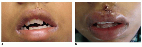

Scar Types

Scars can be classified broadly into hypertrophic scars (Hsc)/keloids and non-Hsc (1). Hsc and keloids are quite similar in nature and represent an aberration in the wound healing process. Patients prone to these types of scars have been shown to have down-regulation of various apoptotic genes and overexpression of signaling factors such as IL-6 and TGFβ1 and β2. Resulting histologic findings include ECM overproduction and deposition of highly disorganized collagen by hypermitotic fibroblasts (2, 3). Keloids, by definition, are elevated fibrous scars that extend beyond the borders of the original wound, do not regress, and usually recur after excision. They can take between 3 months and years to develop. Hsc develop quicker (within 8 weeks), are confined to the wound borders, and usually will regress over a period of 12 to 18 months (2, 4). Risks for keloid formation include darkly pigmented skin (15- to 20-fold increase), familial predisposition, and high wound tension. Hsc are less likely to be related to skin pigmentation. Care must be taken by the surgeon when working with patients at risk for keloid formation as there is a very high rate of recurrence, especially when surgical excision is used alone (5).

Non-Hsc can be divided into either atrophic (depressed) scars or scars that are flat or protrude minimally above the level of the adjacent normal skin (1). While less prone to recurrence and complications than keloids and Hsc, these scars can nonetheless be disfiguring and challenging to treat. The pathophysiology of this scar type is likely related to a hypoxic wound environment, abnormal levels of cellular apoptosis, and limited angiogenesis. The ultimate result is reduced deposition of ECM and collagen and a depression of the wound bed with regard to surrounding normal skin. Similar to keloid and Hsc, excessive tension on wound edges increases risk of atrophic scar formation.


SURGICAL TECHNIQUES


Surgical Planning and Scar Relocation

Four main factors exist that cause unsatisfactory scarring. These are (a) the direction of the scar with respect to relaxed skin tension lines (RSTLs) and if there is disruption or distortion of aesthetic units; (b) the uninterrupted length of a straight scar especially if greater than 2 cm; (c) an irregular texture to the scar surface; and (d) a color mismatch between the scar and surrounding normal tissues. Many techniques exist to treat these problems. In general, the goal is to achieve relocation, excision, irregularization, and/or disruption of a scar (Table 176.2). When planning to camouflage a scar, it is of critical importance to examine the scar’s relationship to the facial aesthetic units, their borders, and the general locations of RSTLs (Fig. 176.1). Facial aesthetic units include the forehead, eyelids, nose, cheeks, lips, and chin. Aesthetic borders between or at the margins of these units are excellent locations for incision placement or scar relocation and include the hairline, infraorbital rims, nasofacial grooves, melolabial folds, vermilion borders, and preauricular sulci. Careful acknowledgment of RSTLs is important, as an incision or closure that crosses these at an angle greater than 30 to 40 degrees will produce an aesthetically displeasing result. With careful planning, many scars can be excised via fusiform incision/primary closure and achieve excellent results. Thus, one of the ideal camouflage techniques is achieved by placement of incisions within existing skin folds produced by RSTLs (6, 7, 8, 9, 10).

Scars or defects near the center of an aesthetic unit, resting over a convexity or concavity, or of great length make simple excision less effective. If extensive undermining of surrounding tissue is necessary for tension-free closure or the anticipated closure will distort a free margin of
the face (lid, lip, nose) and/or cross an RSTL, alternative interventions should also be considered, such as local flap reconstruction (11).


TABLE 176.2 CHARACTERISTICS OF AN IDEAL SCAR


Thin width


Surface flush with surrounding skin


Color matches surrounding skin


Runs parallel to RSTL or along aesthetic subunits


Irregularized if long length


RSTL, relaxed skin tension lines.


Figure 176.1 Schematic illustrating the RSTLs of the face. (Modified illustration from Larrabee WF, Sherris DA. Soft tissue biomechanics and physiology. In: Larrabee WF, Sherris DA, eds. Principles of facial reconstruction, Chapter 1. New York: Lippincott-Raven, 1995:3.)


TABLE 176.3 FLAP CLASSIFICATION SCHEMES


Classification


Subclassification


Subtypes


Examples


Vascular supply


I. Random supply


The majority of local flaps


II. Axial supply (by named vessel)


A. Local


a. Paramedian forehead (supratrochlear artery)


B. Regional


b. Platysmal (branch of facial artery)


C. Free tissue transfer


c. Pectoralis major (thoracoacromial artery)


Composition


I. Cutaneous


II. Fasciocutaneous


III. Musculocutaneous


IV. Osteomusculocutaneous


Method of transfer/design


A. Advancement flap


a. Primary fusiform closure, W-plasty, GBLC, V to Y, island, etc.


B. Rotation flap


b. Cervicofacial, O to Z, Karapandzic, etc.


C. Transposition flap


c. Z-plasty, bilobed, rhombic, etc.


D. Interpolated


d. Paramedian forehead, melolabial, nasofacial, etc.


E. Free tissue transfer


e. Latissimus, scapular, radial forearm, etc.


Flaps

Flaps can be classified in a number of ways. Classification can be based on vascular supply, composition, and/or design and method of transfer (Table 176.3). Practical use often employs a combination of the above. Transposition flaps and local advancement flaps constitute the principle techniques utilized for scar camouflage. When performed correctly, these procedures can achieve excellent irregularization or disruption of a scar and provide near perfect color and texture match to surrounding healthy skin (8). The most commonly used techniques include the Z-plasty, W-plasty, and geometric broken line closure (GBLC) and will be discussed in detail below. Multiple variations of these techniques also exist and can be tailored to the individual patient and scar.


Z-plasty

One of the most common and important techniques in a surgeons’ armamentarium is the Z-plasty transposition flap (Fig. 176.2). The basic Z-plasty can provide excellent cosmesis for a variety of scar revision purposes and is chiefly used to (a) lengthen a scar, (b) release a contracture, (c) disrupt a scar, or (d) realign a scar within an RSTL (Fig. 176.3). However, performing a Z-plasty also has its drawbacks. Lengthening the scar in one direction will result in shortening of the perpendicular direction, and a total of three scars must be made in the place of one.


Figure 176.2 Classic design of the Z-plasty with 60 degree limbs. (From Hochman M. Scar Camouflage. In: Bailey BJ, Calhoun KH, eds. Bailey-head and neck surgery—otolaryngology, Chapter 166, Figure 166.3, 3rd ed., Vol. 2. New York: Lippincott Williams & Wilkins, 2001:2115.)

Technique—The traditional Z-plasty has two constant features. First, three incisions of equal length are made, one central and two limbs. The central incision, ideally, is designed to run parallel to the scar’s long axis. Upon closure, the central component of the incision will achieve the greatest directional change, which makes careful planning of the central limb vital when attempting to realign the scar with an RSTL (Fig. 176.4). Most often, the defect left by a fusiform excision of the entire scar itself (or other defects such as that left by Mohs surgery) forms the central limb of the Z-plasty (11). The second feature is the two angles made by the limb incisions. In the traditional Z-plasty, these angles are equal and 60 degrees from the center. This results in the formation of two triangular skin flaps. When raised in the subcutaneous plain, the triangle tips are transposed such that the shared sides of the triangles approximate the skin side of the opposing limb incisions.


Figure 176.3 Patient with a history of cleft lip repair, who demonstrated a vermilion-cutaneous step-off deformity. Z-plasty technique was utilized to correct the step-off deformity and to realign the vermilion along the natural aesthetic border. A: Preoperative photo demonstrating the misalignment of the vermilion and skin of the upper lip disrupting the normal border of the lip vermilion subunit. Lines mark the planned limbs for the Z-plasty transposition flap. B: Immediate postoperative photo demonstrating the correction of the vermilion border and restoration of the natural cupids bow contour.

The effect achieved is a disruption of the scar itself and reorientation along the lines of the limb incisions, which do not move appreciably and should ideally be drawn within RSTLs. It is critical to note that if the initial scar was already oriented with an RSTL, the central incision will fall perpendicular to the same RSTL. Scars angling less than 40 degrees from an RSTL are often better managed with simple excision. A final important feature of the Z-plasty is that intelligent angle design allows the surgeon to anticipate the new length and degree of reorientation achieved after closure. The simple rule of thumb is that the 60-degree Z-plasty will produce a 75% increase in length and 90 degrees of rotation (Fig. 176.5). A change in angle produces predictable change of these variables (Table 176.4). If further lengthening is desired, serial Z-plasty can be performed (6, 11). Of note, angles should never be less than 30 degrees as the flap tips will risk ischemia and necrosis.

A number of variations of the traditional Z-plasty exist including double-opposing Z-plasty, unequal triangle Z-plasty, four-flap Z-plasty, compound Z-plasty, and planimetric Z-plasty (6).


W-plasty

One of two common advancement flaps used for scar irregularization, the W-plasty produces a regularly irregular incision and should be used on long straight scars. Such scars reflect light homogenously and are easy for the eye to follow. Irregularization scatters that reflected light making it less visible to the observer. W-plasty is best suited for scars angling greater than 35 degrees from RSTLs or for scars that cross unforgiving areas of convexity or concavity (12). Unlike the Z-plasty, this technique does not increase the length of the scar. W-plasty is faster to perform than GBLC but forms a more predictable and conspicuous scar. As in GBLC, some normal tissue is removed, which increases overall tension on the wound.


Get Clinical Tree app for offline access