Purpose
To assess the results of a single eye bank preparing a high volume of Descemet membrane endothelial keratoplasty (DMEK) tissues using multiple technicians to provide an overview of the experience and to identify possible risk factors for DMEK preparation failure.
Design
Cross-sectional study.
Methods
setting : Lions VisionGift and Wilmer Eye Institute at Johns Hopkins Hospital. study population : All 563 corneal tissues processed by technicians at Lions VisionGift for DMEK between October 2011 and May 2014 inclusive. observation procedures : Tissues were divided into 2 groups: DMEK preparation success and DMEK preparation failure. main outcome measures : We compared donor characteristics, including past medical history.
Results
The overall tissue preparation failure rate was 5.2%. Univariate analysis showed diabetes mellitus ( P = .000028) and its duration ( P = .023), hypertension ( P = .021), and hyperlipidemia or obesity ( P = .0004) were more common in the failure group. Multivariate analysis showed diabetes mellitus ( P = .0001) and hyperlipidemia or obesity ( P = .0142) were more common in the failure group. Elimination of tissues from donors either with diabetes or with hyperlipidemia or obesity reduced the failure rate from 5.2% to 2.2%. Trends toward lower failure rates occurring with increased technician experience also were found.
Conclusions
Our work showed that tissues from donors with diabetes mellitus (especially with longer disease duration) and hyperlipidemia or obesity were associated with higher failure rates in DMEK preparation. Elimination of tissues from donors either with diabetes mellitus or with hyperlipidemia or obesity reduced the failure rate. In addition, our data may provide useful initial guidelines and benchmark values for eye banks seeking to establish and maintain DMEK programs.
Descemet stripping automated endothelial keratoplasty (DSAEK) and Descemet membrane endothelial keratoplasty (DMEK) have gained popularity over penetrating keratoplasty for the treatment of corneal endothelial diseases because of numerous inherent advantages. Thus, the demand for eye bank precut corneas for endothelial keratoplasty has increased dramatically in the United States. In 2013, the Eye Bank Association of America reported that 37% of corneas distributed for keratoplasty were for endothelial keratoplasty, representing a 12.4% increase from 2012. Despite studies showing better results over Descemet stripping endothelial keratoplasty or DSAEK, DMEK accounted for only about 6% of the endothelial tissues provided. Possible reasons include the challenges associated with preparing and handling the delicate graft tissue and lack of standardization of DMEK graft preparation by both surgeons and eye banks. Regarding the latter issue, knowledge of possible risk factors for failure in DMEK donor tissue preparation could provide both eye banks and corneal surgeons with information that would allow better selection of corneas to use for this procedure.
Donor factors such as age and endothelial cell density have been shown to influence the properties of DMEK grafts, and thereby the duration of the surgical procedure. In a recent publication, Gorovoy and associates studied some risk factors for DMEK preparation, including donor age, gender, postmortem tissue time interval, contralateral eye data, peel time, and peel complications, in 116 consecutive DMEK donor tissues that were prepared by a single surgeon. They proposed that the major risk factor for DMEK preparation failure in the fellow eye is complications during peeling the first eye. Another risk factor proposed was donor age younger than 50 years. Recently, Greiner and associates also related diabetes mellitus as a risk factor for preparation failure.
Given that eye banks are beginning to provide tissues prepared for DMEK and that interest in the procedure is evolving, it is likely that the volume of eye bank prepared tissue for this surgery will increase. Thus, the purpose of this study is to assess the results of a single eye bank preparing a high volume of DMEK tissues using multiple technicians to provide an overview of the experience and to identify possible risk factors for DMEK preparation failure. Such information may be important for eye banks seeking to develop DMEK preparation programs and for surgeons and eye bank personnel involved in the selection of donor tissues for this procedure.
Methods
The Institutional Review Board at Legacy Health Systems, (Portland, Oregon, USA) determined that approval was not required for cross-sectional data queries. This was a retrospective, cross-sectional study of electronic eye bank database records of tissue from donors prepared for DMEK at Lions VisionGift (Portland, Oregon, USA).
Protocol
We retrospectively studied all 563 corneal tissues that were processed by trained technicians at Lions VisionGift for DMEK between October 2011 and May 2014 inclusive. The tissues were divided into 2 groups: DMEK preparation success and DMEK preparation failure (see “Tissue Preparation,” below). The following parameters were assessed: endothelial cell count before and after cutting; death to preservation time; donor age; donor past medical history (including diabetes mellitus, and its duration when available; hyperlipidemia, obesity, or both; hypertension; history of cancer; and tobacco and alcohol use); past ocular history, including superficial surgeries (eg, pterygium, refractive surgery), intraocular surgeries, and any other clinical ocular history (corneal or retinal diseases and glaucoma); technician experience; duration of tissue preparation (minutes); and days from death to tissue preparation. The influence of the learning curve for technicians was determined by the number of failures per each 50 consecutive procedures. We also reported the number of previous DSAEK tissues prepared by each technician (Galloway JS).
Tissue preparation
Corneas were prepared for DMEK per standard protocol of the eye bank. In brief, Descemet membranes were peeled under Optisol GS Bausch and Lomb (Rochester, New York) from the underlying stroma over 80% to 90% of their surface area, leaving a small hinge of residual attachment that facilitated storage and transportation in situ against the host donor corneoscleral tissue. Failure was defined as a tear that occurred while separating Descemet membrane from stroma that rendered the tissue unusable for transplantation. Endothelial cell counts before and after tissue preparation were performed using a standard protocol with an EB-10 eye bank specular microscope (Konan, Irvine, California, USA).
Technician training
The Lions VisionGift training program consists of 4 phases: observation, hands-on mentored training for basic competence, solo practice to solidify the new skill set, and finally a sequence of at least 10 consecutively observed and documented tissue preparation procedures in which the trainee is expected to perform at a level acceptable to the medical director of the eye bank or at a level comparable with that of previously approved technicians. Tissues included in this study were prepared by 3 technicians who had completed all 4 training stages.
Statistical Analyses
Data are reported as mean ± standard deviation unless otherwise indicated. Welch 2-sample t tests were used to test for association between outcome and individual continuous variables such as age and duration of diabetes mellitus, whereas Fisher exact tests were used for factorial variables such as diabetes status and history of tobacco use. Logistic regression with the logit link function was used to construct a multivariate prediction model, evaluated by receiver operating characteristic (ROC) analysis to select optimal decision rules. To obtain unbiased estimates of predictive performance, logistic regression was applied within a leave-1-out cross-validation design wherein the model parameters were re-estimated within each iteration. Statistical analyses were carried out in the R program ( www.cran.us.r-project.org ) using standard functions for tests and logistic regression and customized functions for plotting ROC curves and calculating area under the curve. All tests were 2-tailed and P values of less than .05 were considered significant.
Results
A total of 563 tissue reports were analyzed, which included 534 successful and 29 failed procedures, representing a 5.2% failure rate for all attempts. The mean donor age (± standard deviation) was 64.0 ± 6.7 years (range, 43 to 73 years) in the failure group and 65.0 ± 6.8 years (range, 35 to 79 years) in the success group ( P = .44).
The donor past medical and ocular history could be assessed in 488 cases, including 462 (87%) in the success group and 26 (90%) in the failure group. Diabetes mellitus was more than 3-fold more common in the failure group than in the success group (69% vs 24%; P = .000028). It was possible to assess diabetes mellitus duration in 16 donors (89% of the diabetics) in the failure group and in 85 donors (77% of the diabetics) in the success group, and the mean ± standard deviation durations were 13.9 ± 15.5 years in the failure group and 6.5 ± 8.4 years in the success group ( P = .023). Higher rates of hypertension (85% vs 62%; P = .021) and hyperlipidemia, obesity, or both (85% vs 48%; P = .0004) also were found among the failure group. Tobacco use was nearly 2-fold more common (19% vs 10%) in the failure group ( P = .175). The other clinical and ophthalmologic parameters studied were not significantly different and can be seen in Table 1 . After Bonferroni multiple-test correction, only diabetes mellitus and hyperlipidemia, obesity, or both were significantly associated with donor preparation failure.
| Outcome Group | Diabetes | Diabetes Duration (y) | Hypertension | Hyperlipidemia, Obesity, or Both | Tobacco | Cancer | Alcohol | Intraocular Surgery | Superficial Surgery | Other Ocular Disorders |
|---|---|---|---|---|---|---|---|---|---|---|
| Failure (n = 26) | 18 (69) | 13.9 ± 15.5 | 22 (85) | 22 (85) | 5 (19) | 11 (42) | 3 (12) | 1 (4) | 1 (4) | 2 (8) |
| Success (n = 462) | 110 (24) | 6.5 ± 8.4 | 287 (62) | 224 (48) | 46 (10) | 181 (39) | 75 (16) | 24 (5) | 28 (6) | 43 (9) |
| P value | .000028 | .023 | .021 | .0004 | .175 | .84 | .78 | 1.00 | 1.00 | 1.00 |
Before attempting to peel Descemet membrane, the mean endothelial cell count among success and failure groups were, respectively, 2763 ± 293 cells/mm 2 and 2753 ± 318 cells/mm 2 ( P = .86). After preparation, it was 2768 ± 248 cells/mm 2 in the success group and could not be determined in the failure group.
The mean death to preservation time was 09 hours and 58 minutes in the success group and 10 hours and 29 minutes in the failure group ( P = .61). The mean duration of tissue preparation was 25 ± 7 minutes in the success group and 25 ± 6 minutes in the failure group ( P = 1.0). The mean death to tissue preparation time was 3.6 ± 1.7 days in the success group and 4.2 ± 2.0 in the failure group ( P = .058).
Next, we fit a multivariate logistic regression model to the data using the logit link function. Results are shown in Table 2 . In addition to diabetes mellitus and hyperlipidemia, obesity, or both, which were the only significant predictors of failure in univariate analysis after Bonferroni multiple test correction, we also included donor age because of a previously published association, with success ( Table 2 ). Diabetes mellitus remained the most significant risk factor in the multivariate model, with diabetic donors having nearly 6 times the risk of failure of nondiabetic individuals. Hyperlipidemia, obesity, or both likewise was statistically significant in the multivariate model, with an odds ratio of 4, suggesting that both diabetes mellitus and hyperlipidemia, obesity or both have independent predictive value after adjusting for the effects of each other. Although donor age was not statistically significant, the expected trend, decreased risk of failure in older patients, was observed with an approximate 5% reduction in risk per year over the age range of donors included in this study.
| Variable | Odds Ratio for Failure | 95% CI | P Value |
|---|---|---|---|
| Diabetes mellitus | 5.80 | 2.35 to 14.35 | .0001 |
| Hyperlipidemia, obesity, or both | 4.07 | 1.32 to 12.49 | .0142 |
| Age | 0.94 | 0.89 to 1.00 | .0588 |
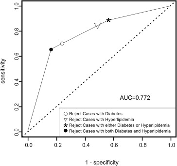
We used the logistic regression model to predict which cases were most likely to fail, using ROC analysis to help select optimal prediction rules and evaluate performance. The area under the ROC curve was 0.772 (95% confidence interval, 0.68 to 0.86; Figure ), with maximum predictive power at a sensitivity of 65% and specificity of 84%. Because both of the risk factors in the model are binary, the selection rule corresponding to this decision point is simple to describe: potential donors may have either (1) diabetes mellitus or (2) hyperlipidemia, obesity, or both, but not both (1) and (2). Applying this rule to the full donor pool (n = 563) and assuming the full donor pool has the same rates of diabetes mellitus and hyperlipidemia, obesity, or both as in the evaluable samples (n = 488), 82% (460/563) of donors included in this study would have been eligible, with a failure rate of only 2.2% (10/460), compared with 5.2% in the full donor group (29/563). Eliminating all cases of diabetes mellitus (with or without hyperlipidemia, obesity, or both) resulted in a similar failure rate (2.2%), but further diminished the donor pool so that only 74% of those included in this study were eligible. The lowest failure rate was achieved by eliminating all donors with either diabetes mellitus or hyperlipidemia, obesity, or both (1.5%), but at great cost to the available donor pool: only 236 in 563 (42%) subjects in this study would have been eligible donors under these criteria. Eliminating hyperlipidemic or obese patients (with or without diabetes mellitus; failure rate, 1.7%; eligible donor pool, 50%) gave comparable results to eliminating donors with either diabetes mellitus or hyperlipidemia, obesity, or both.