Chapter 50 Retrosigmoid Approach to Tumors of the Cerebellopontine Angle
 Videos corresponding to this chapter are available online at www.expertconsult.com.
SURGICAL ANATOMY
Historically, the earliest approach to the posterior fossa was undertaken through the suboccipital convexity. Krause1 first employed this technique during the latter portion of the 19th century. Until the 1970s, the technique in widespread use was the so-called suboccipital approach. In this procedure, a large bone window is removed, and the anterior limit of the craniectomy is the first mastoid air cell encountered. Curtailment of the anterior opening at the first contact with pneumatization was predicated on the assumption that the mastoid was bacterially contaminated, and that opening its air cell tracts created an increased risk of meningitis. Because of its more posterior angle of view, the suboccipital approach required a greater degree of cerebellar retraction, and sometimes necessitated a partial cerebellar resection.
In recent years, as a result of increased experience with CPA surgery, the classic suboccipital approach has been modified to become the retrosigmoid approach, which is now the preferred method for exposing the CPA behind the sigmoid sinus. In this technique, bone is removed anteriorly up to the level of the posterior border of the sigmoid sinus and superiorly to the inferior margin of the transverse sinus (Fig. 50-1). Although mastoid air cells are frequently transected during this maneuver, experience has not shown an increased incidence of postoperative infection. The slightly higher risk of CSF leak associated with this more anterior exposure is more than offset by its more favorable angle of view into the CPA and the markedly reduced need for cerebellar retraction with this approach.
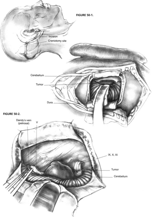
The anatomic exposure of the posterior fossa provided by the retrosigmoid approach is bounded superiorly by the tentorium cerebelli and inferiorly by the jugular foramen and foramen magnum (Fig. 50-2).2–5 Access to the central nervous system includes the lateral cerebellar hemisphere and the lateral surface of the pons and upper medulla. CN V through XI are visible at their root entry zones and over their cisternal courses. Although the theoretical anterior limit of exposure is the clivus and the apical portion of the petrous pyramid, in practice, access to these ventral structures is usually limited by CN VII and VIII superiorly and CN IX through XI inferiorly, which bridge across the CPA, restricting ventral access to narrow intervals. Exposure of the prepontine cistern is largely obstructed by the lateral aspect of the pons, which does not tolerate medial retraction well.
Anatomic variations may affect the CPA exposure provided by the retrosigmoid approach. A posteriorly placed sigmoid sinus course results in the anterior edge of the craniectomy being placed more posteriorly. This placement creates a deeper field of action and a less favorable angle of view with the consequent need for more cerebellar retraction. This disadvantageous exposure may be compromised further by a low transverse sinus course, particularly if the patient also has a short neck and a prominent shoulder. This problem of restricted exposure may be overcome by combining the retrosigmoid approach with an anterosigmoid, retrolabyrinthine decompression to allow anterior retraction of the sigmoid sinus.6 A highly placed jugular bulb restricts access to the internal auditory canal (IAC), and can make the dissection of the inferior bony trough between the canal and the bulb difficult. Occasionally, the bulb may extend superiorly to overlap the IAC, partially obscuring access to the medial aspect of the canal.7
PREOPERATIVE EVALUATION AND PATIENT COUNSELING
The minimal preoperative evaluation for a patient with a CPA tumor comprises a clinical history, a physical examination, pure tone and speech audiometry, and an imaging study (preferably, gadolinium-enhanced magnetic resonance imaging [MRI]). In nonacoustic tumors, computed tomography (CT) scanning for evaluation of the osseous characteristics of the cranial base and angiography to address vascular anatomy and possibly to perform embolization are occasionally indicated. Neither vestibular diagnostic testing nor auditory evoked responses are routinely obtained in patients already diagnosed with an acoustic neuroma.8
Numerous factors affect the selection of posterior fossa craniotomy for tumors of the CPA.7,9,10 As advocates of selective management of these lesions according to the unique attributes of each tumor and the potential surgical options, we involve the patient in the discussion of the relative advantages and disadvantages of each technique. In most cases, an obvious choice can be made, whereas in others, patient preference is important. Our customary preoperative counseling includes the anticipated and potential risks to hearing, balance, and facial motor function. Less common complications that are discussed include CSF leak, meningitis, cerebrovascular accident, and death.11 Although blood transfusion is seldom required, we encourage the patient to donate 1 U of autologous blood.
PATIENT SELECTION
Common Indications in Neurotology
Hearing Preservation
The primary aim of acoustic neuroma management is removing the threat of progressive tumor growth, while avoiding injury to the central nervous system. Preservation of cranial nerve function (facial movement, facial sensation, and hearing), which has become the primary focus of acoustic neuroma surgery in recent years, is a secondary goal. Patients with acoustic tumors can be classified into three groups in terms of potential for hearing preservation. Patients for whom hearing preservation is highly improbable generally undergo translabyrinthine removal. Criteria that place a patient into this group include poor hearing (<30% speech discrimination, >70 dB speech reception threshold), large CPA component (>3 cm), and deep penetration of the IAC. Conversely, patients with good hearing (>70% speech discrimination, <30 dB speech reception threshold), small CPA component (<1 cm), and shallow IAC involvement are considered excellent candidates for a hearing conservation approach.7 It is difficult to codify a set of rules concerning selection of a hearing conservation approach for the numerous patients who lie between these parameters. Each surgical team must rely on its own criteria, based on experience, together with the patient’s wishes in coming to a selection of surgical approach. Neurotologists would always favor undertaking a hearing conservation approach, even when the chances of success were remote, were there not potential adverse consequences from the endeavor. The lower morbidity of the translabyrinthine approach, especially in terms of persistent headache and CSF leak, leads the clinician away from the retrosigmoid hearing conservation approach when the chances of success are limited.
The concept of useful hearing is context dependent. In a patient with a normal contralateral ear, imperfect residual hearing in the tumor ear is often of little practical benefit. When hearing in the contralateral ear is impaired or threatened, such as in cases of bilateral acoustic neuromas associated with neurofibromatosis type 2, a conservative approach to hearing conservation is prudent, occasionally even at the expense of complete tumor excision.12
Hearing preservation is seldom achieved when tumors with a CPA component exceeding 2 cm in diameter are removed.13,14 This rule should not be applied in nonacoustic CPA tumors (e.g., meningiomas), however, because hearing preservation is frequently achieved even with large tumors.15
The retrosigmoid approach exposes a variable amount of the IAC without violating the inner ear while the canal is being drilled open. Two factors should be considered in the decision of whether hearing conservation via the retrosigmoid approach is feasible: the depth to which the tumor penetrates the IAC, and the degree of IAC exposable in that patient. The relationship between the inner ear and the lateralmost extension of the tumor into the IAC may be predicated by preoperative gadolinium-enhanced MRI.16
Use of Retrosigmoid Approach in Combined Therapy of Acoustic Neuroma
Numerous studies have shown that functional outcome after conventional microsurgery is substantially poorer in patients with acoustic neuroma larger than approximately 3 cm. In these patients, the incidence of persistent facial dysfunction is high. There is also an increased risk of persistent balance dysfunction because of infarction of the middle cerebellar peduncle.17 In an effort to improve functional outcome, some centers have begun approaching larger tumors with subtotal resection leaving a rind of tumor on the pons and along the course of the facial nerve. When the patient has serviceable hearing, the retrosigmoid approach is typically used. To reduce the risk of recurrence, it is essential to remove the IAC component. Such surgical remnants resume growth in approximately one third of cases.18 If the remnant grows on serial imaging, it may be treated with stereotactic radiation with a greater than 90% probability of halting its growth.
Acoustic Neuroma in a Patient with Chronic Otitis Media
Although patients with acoustic neuromas rarely have concomitant chronic middle ear infection, in patients who do the translabyrinthine approach for acoustic neuroma resection is contraindicated. The retrosigmoid approach may also open into potentially contaminated mastoid air cells lying behind the sigmoid sinus, and into air cells that may surround the IAC. To avoid potential intracranial infection, chronic middle ear infection should be controlled with tympanoplasty, antibiotics, or both, before tumor surgery whenever possible.7
Tumors with Limited Extension into Meckel’s Cave
The retrosigmoid approach is also useful in approaching extra-axial posterior fossa tumors that possess minor extensions into Meckel’s cave (cavum trigeminale). Most such tumors are trigeminal schwannomas and petroclival meningiomas. Added exposure is obtained by removing the apical petrous bone between the IAC and the tentorium. This maneuver provides access to approximately 1 to 2 cm of the posterior aspect of Meckel’s cave for tumor removal.19–21
Relative Contraindications
Deep Extension into Internal Auditory Canal
Generally, tumors extending into the lateral one third of the IAC are not resectable by the retrosigmoid approach without destroying hearing. In such cases, the translabyrinthine approach ensures complete resection and reduces operative morbidity.7
Large Tumors
Although large CPA tumors (>3 cm) may be approached through either the retrosigmoid or the translabyrinthine technique, when we intend radical removal we prefer to use the latter because it provides ample exposure and minimizes the need for cerebellar retraction. In addition, when the pons and the cerebellar peduncle are substantially displaced medially and posteriorly, the translabyrinthine approach, by virtue of its more anterior placement, provides a more favorable angle of view posteriorly toward the brainstem interface. In cases of facial nerve disruption, which is more common in large tumors, the translabyrinthine approach affords more reconstructive options through mastoid meatal rerouting.7
PATIENT PREPARATION AND POSITIONING
At most centers, the operation is performed by a multidisciplinary team consisting of a neurotologist, neurosurgeon, neuroanesthesiologist, neurophysiologist, and specialized operating room nurses. The operation is done with the patient under general anesthesia. A short-duration muscle relaxant is used to facilitate endotracheal intubation. Thereafter, anesthesia is maintained with inhalational agents alone, avoiding the use of muscle relaxants, which would prevent effective intraoperative cranial nerve electrophysiologic monitoring. In addition to the routine neuroanesthesia monitoring equipment, antithrombotic stockings and a urinary catheter are used. The retrosigmoid approach may be carried out in one of three surgical positions: supine, lateral supine (park bench position), and sitting.10 Supine is the favored position because it affords excellent exposure and carries the lowest risk of complication (discussed later).
The patient is secured in the optimal operating position by means of a head holder attached to the bed frame (e.g., Mayfield). This apparatus facilitates exposure of the suboccipital region while the patient is in the supine position. Optimal surgical field exposure is obtained by a combination of head rotation, neck flexion, and ipsilateral shoulder elevation. Excessive neck torsion should be avoided to prevent cervical injury and to reduce the risk of cerebellar swelling secondary to compromised flow through the vertebral venous system. The cranial nerve–monitoring electromyographic electrodes are placed into the muscles supplied by CN V, VII, and XI. When intraoperative auditory brainstem monitoring is indicated, scalp electrodes are placed, and an earphone is inserted into the ipsilateral external auditory canal.22
SPECIAL INSTRUMENTS
Operating room electric circuitry and neuroanesthesia electric monitoring equipment should be grounded and electronically quiet to minimize 60 Hz noise production, which interferes with the cranial nerve electrophysiologic monitoring setup. The specialized equipment for intraoperative cranial nerve monitoring used in our institution has been described elsewhere in detail.22


