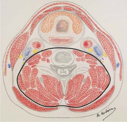
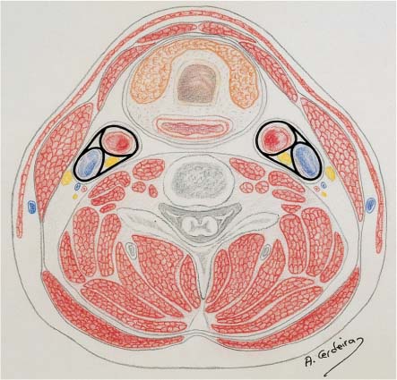
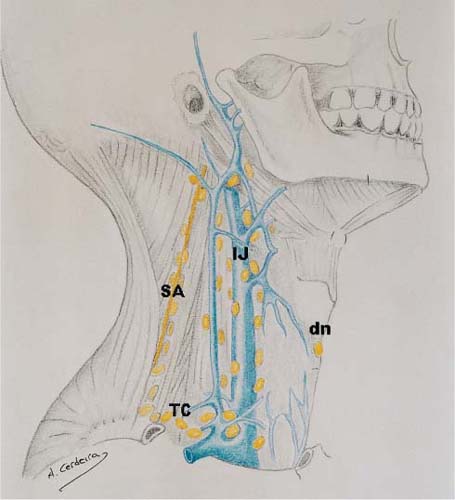
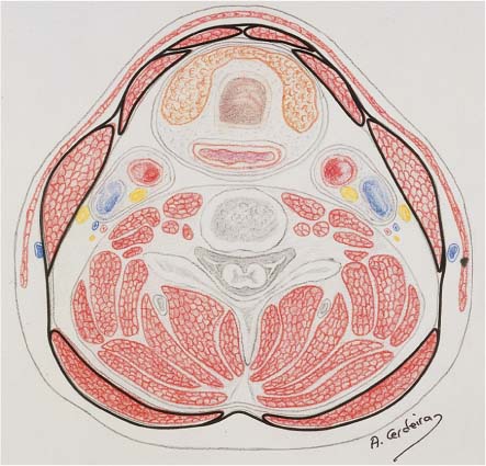
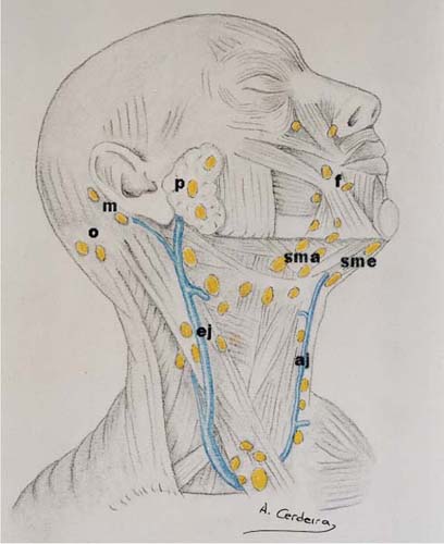
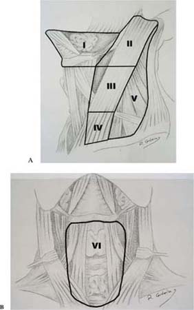
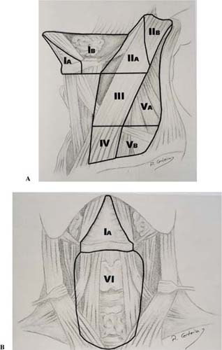
CHAPTER 2 Functional neck dissection, as described by Osvaldo Suárez, is based on the existence of a fascial barrier between the lymphatic tissue and the muscular, glandular, neural, and vascular structures of the neck. This anatomical separation allows the creation of a surgical plane of dissection. The fascial layer invests muscles and organs in the neck, forming planes and spaces where many important structures are crowded together. This fact, known as fascial compartmentalization, holds the rationale for functional neck dissection. This chapter describes the anatomical bases of functional neck dissection from a practical and surgical standpoint. The anatomical description of the fascial layers of the neck has suffered a number of different descriptions. For practical reasons we will consider two distinct fascial layers in the neck, the superficial cervical fascia and the deep cervical fascia. The superficial cervical fascia corresponds to the subcutaneous tissue. The deep cervical fascia is the key element for functional and selective neck dissection. The deep cervical fascia surrounds the neck, enveloping its different structures. For teaching purposes, two different layers are considered within the deep cervical fascia: a superficial and a deep or prevertebral layer. The carotid sheath, which is an important structure from the surgical standpoint, is located between the two layers of the deep cervical fascia. The superficial layer of the deep cervical fascia, also known as investing or anterior fascia, completely envelops the neck with the exception of the skin, platysma muscle, and superficial fascia (Fig. 2-1). It is attached to the occipital protuberance, mastoid process, capsule of the parotid gland, angle of the jaw, and body of the mandible to the symphysis, where it proceeds around the opposite side in a similar manner. It then goes posteriorly across the spinal process of the cervical vertebrae and the ligamentum nuchae. Anteriorly, it passes from the mandible to the hyoid bone and from here down to the sternum. Inferiorly, it attaches to the sternum, upper edge of the clavicle, acromion, and spine of the scapula. At the inferior border, in the midline, the superficial layer splits in two different layers just superior to the manubrium of the sternum. The space between these two layers is known as the suprasternal space ofBurns. From posterior to anterior, the superficial layer splits to enclose the trapezius, the portion of the omohyoid muscle that crosses the posterior triangle of the neck, and the sternocleidomastoid muscle. In a similar way it envelops the strap muscles, before ending in the midline. The superficial veins of the neck lie on or within this superficial layer of the deep cervical fascia. The deep or prevertebral layer, like the superficial layer, attaches posteriorly at the spinous process of the cervical vertebrae and ligamentum nuchae (Fig. 2-2). At its upper limit, it goes to the skull base at the jugular foramen and carotid canal, then passes across the basilar process to the opposite side. This fascial layer covers the muscles of the back that enter into the neck immediately deep to the trapezius muscle (splenius and levator scapula). At this level there is a potential space between both layers. At the upper limit of the posterior triangle, this space is almost virtual. The spinal accessory nerve crosses the posterior triangle at this level, along with some lymph nodes. At the lower end, both fascial layers further separate, the deep layer covering the scalene muscles, whereas the superficial layer remains attached to the trapezius muscle and the clavicle. The phrenic nerve runs inferiorly on the anterior aspect of the scalene group, covered by this fascial layer. As it proceeds medially, the deep layer attaches to the anterior tubercles of the transverse process of the cervical vertebrae. From here it crosses the midline where it attaches to the transverse process of the cervical vertebrae of the opposite side, passing posterior to the esophagus and anterior to the spine. It is this prevertebral part that gives its name to this fascial layer. The carotid sheath or vascular sheath lies between the superficial and the prevertebral layers of the deep cervical fascia (Fig. 2-3). It may be regarded as a cylinder-like structure made of fascial laminae connecting the superficial and deep layers. This vascular sheath runs from the base of the skull to the root of the neck. It has independent compartments for the internal jugular vein, the carotid artery, the vagus nerve, and the ansa cervicalis. It attaches to the prevertebral layer at the level of the anterior scalene muscle. The cervical portion of the sympathetic trunk runs posterior to the carotid sheath. Fascial compartmentalization allows the removal of cervical lymphatic tissue by separating and removing the fascial walls of these “containers” along with their contents from the underlying vascular, glandular, neural, and muscular structures. Figure 2-1 Horizontal cross section of the neck at the level of the sixth cervical vertebra showing the superficial layer of the deep cervical fascia. Figure 2-2 Horizontal cross section of the neck at the level of the sixth cervical vertebra showing the prevertebral layer of the deep cervical fascia. Figure 2-3 Horizontal cross section of the neck at the level of the sixth cervical vertebra showing the carotid sheath. The lymphatic system of the neck consists of a network of lymph nodes intimately connected by lymphatic channels. For teaching purposes, two major lymphatic networks may be considered in the neck, a superficial and a deep web. The superficial lymphatics of the head and neck drain the skin into the superficial lymph nodes located around the neck and along the external and anterior jugular veins. Superficial lymphatics include the submental, submandibular and facial, external jugular, anterior jugular, occipital, mastoid, and parotid groups (Fig. 2-4). The submental nodes, usually two or three in number, lie in a midline triangular space bounded by the anterior bellies of the digastric muscles and the hyoid bone. They drain the skin of the chin, the skin and mucous membrane of the central portion of the lower lip and jaw, the floor of the mouth, and the tip of the tongue. These nodes drain into the submandibular chain or directly into the deep cervical chains. The submandibular nodes are located along the inferior border of the horizontal ramus of the mandible. They usually lie over the submandibular gland although intracapsular nodes are also possible. The submandibular chain, along with some inconstant small facial nodes, drain the skin and mucous membrane of the nose, medial portion of the eyelid, cheek, upper lip, lateral part of the lower lip, gums, and anterior third of the lateral border of the tongue. These nodes drain into the transverse cervical and deep cervical chains. The external jugular nodes are located between the lower parotid nodes and the midportion of the sternocleidomastoid muscle, along the external jugular vein. They drain the lower part of the ear and the parotid gland into the superior deep cervical chain. The anterior jugular nodes are located on the anteroinferior portion of the neck, parallel to the anterior jugular vein. They drain the infrahyoid area toward the inferior deep jugular chain. The occipital nodes drain the skin of the occipital region and part of the superficial and deep lymphatics of the nape. The mastoid nodes are located over the mastoid process and drain the ear, external auditory canal, and skin of the temporal region. The parotid group includes both superficial and deep nodes. The superficial nodes are located over the external surface of the parotid gland, whereas the deep nodes are intraglandular and accompany the intraparotid course of the retromandibular and external jugular. The parotid nodes drain the skin of the temporal and frontal area, eyelid, auricle, middle ear, parotid, and the mucous surface of the nasal cavity. The deep lymphatics drain the mucous membranes of the upper aerodigestive tract, along with organs such as the thyroid and larynx, into the deep cervical lymph node chains. These include the internal jugular, spinal accessory, transverse cervical, retropharyngeal, and deep anterior lymphatic chains (Fig. 2-5). The internal jugular chain is formed by a variable number of lymph nodes—between 30 and 60—located along the internal jugular vein. The most posterior and smaller nodes are located over the splenius, levator scapulae, and scalene muscles, whereas the anterior nodes are in close relation with the anterior wall of the internal jugular vein. The posterior nodes drain the skin of the back of the head and receive efferent vessels from the occipital and mastoid nodes, as well as cutaneous and muscular tributaries from the neck. The anterior nodes drain the superficial and deep structures of the anterior part of the head and neck, both directly and indirectly. Figure 2-4 Superficial lymphatics of the neck. sme, submental; sma, submandibular; f, facial; ej, external jugular; aj, anterior jugular; o, occipital; m, mastoid; p, parotid. Figure 2-5 Deep lymphatics of the neck. IJ, internal jugular chain; SA, spinal accessory chain; TC, transverse cervical chain; dn, Delphian node. For practical purposes, the internal jugular chain may be divided into an upper and a lower part, with the dividing line located at the crossing of the omohyoid muscle with the carotid sheath. The spinal accessory chain follows the spinal accessory nerve in the upper part of the posterior triangle and merges with the transverse cervical chain beneath the trapezius muscle. It receives lymph from the occipital and mastoid areas. The transverse cervical chain runs along the transverse cervical vessels. It receives efferent vessels from the spinal accessory chain and from the lateral part of the neck. The retropharyngeal nodes are located at the lateral portion of the parapharyngeal space. They drain the nasal cavity, soft palate, paranasal sinuses, middle ear, nasopharynx, and oropharynx. The deep anterior chain includes the prelaryngeal (Delphian) node, the pretracheal, and the paratracheal nodes. They drain the subglottis, the trachea, and the thyroid gland. This chain is connected with the internal jugular chain. Both, the superficial and the deep lymphatic system initially drain in the nearest lymph nodes and then proceed to more central nodes. At the base of the right side of the neck, the jugular trunk (which collects all the lymph from one side of the head and neck), the transverse cervical trunk, and the subclavian trunk frequently join to form the right lymphatic duct. This large collector courses along the medial border of the scalene muscle and empties into the venous system at the junction of the right internal jugular vein and the right subclavian vein. The left thoracic duct begins in the abdomen, passes through the thoracic region, and emerges in the root of the left side of the neck between the common carotid and subclavian arteries. It then arches above the subclavian artery and in front of the vertebral artery and thyrocervical trunk, to pass behind the carotid sheath between the internal jugular vein and the anterior scalene muscle. The thoracic duct empties laterally into the venous system at the junction of the left subclavian and internal jugular veins. For practical reasons, the neck may be artificially divided into different lymph node regions. This does not mean that there is a true anatomical or physiological separation within the lymphatic system of the neck. Not only is there no physical separation within the lymphatic system of the neck, but a widespread interconnection exists between the different nodal chains, as already described. Thus, the regional lymph node classification should be regarded only as a schematic representation of the lymphatic system of the neck, and not as an anatomical transcription of the reality. As often happens in medicine, nature is much more complex than we would like it to be. The most popular terminology for subdividing the lymph node groups was proposed in 1991 by the Committee for Head and Neck Surgery and Oncology of the American Academy of Otolaryngology—Head and Neck Surgery. This committee worked to define the anatomical boundaries of lymph node groups to offer fundamental principles for a classification of neck dissection procedures. The neck is divided into six different levels (Figs. 2-6A, 2-6B). • Level I: Submental and submandibular nodes. This group includes the lymph nodes located within the submental triangle, bounded by the anterior belly of the digastric muscle and the hyoid bone. The submandibular group includes the lymph nodes located within the boundaries of the anterior and posterior bellies of the digastric muscle, the stylohyoid muscle, and the body of the mandible. The submandibular gland, which is located within this cervical space, should be removed when this nodal group is included in the resection. Figure 2-6 Regional division of the lymphatic system of the neck according to the classification of the American Academy of Otolaryngology—Head and Neck Surgery. (A) Lateral view. (B) Anterior view. Level I, submental and submandibular region; Level II, upper jugular region; Level III, middle jugular region; Level IV, lower jugular region; Level V, posterior triangle; Level VI, anterior compartment. • Level III: Middle jugular nodes. This group includes the lymph nodes located around the middle third of the internal jugular vein. The boundaries of this space are the inferior border of the hyoid bone and the carotid bifurcation superiorly, the inferior border of the cricoid cartilage and the junction of the omohyoid muscle with the internal jugular vein inferiorly, the posterior border of the sternocleidomastoid muscle posteriorly, and the lateral border of the sternohyoid muscle anteriorly. • Level IV: Lower jugular nodes. This nodal group contains the lymphatic structures located around the lower third of the internal jugular vein. Its boundaries are the inferior border of the cricoid cartilage and the omohyoid muscle superiorly, the clavicle inferiorly, the posterior border of the sternocleidomastoid muscle posteriorly, and the lateral border of the sternohyoid muscle anteriorly. • Level V: Posterior triangle. This group includes the lymph nodes located along the transverse cervical artery and lower half of the spinal accessory nerve as well as the supraclavicular lymph nodes. The boundaries are the anterior border of the trapezius muscle posteriorly, the posterior border of the sternocleidomastoid muscle anteriorly, the clavicle inferiorly, and the convergence of the sternocleidomastoid and trapezius muscles superiorly. • Level VI: Anterior compartment. This level contains the pre- and paratracheal nodes, precricoid (Delphian) node, perithyroidal nodes, and the lymph nodes along the recurrent laryngeal nerves. The boundaries are the hyoid bone superiorly, the suprasternal notch inferiorly, and the carotid arteries laterally. • Level VII. Some authors consider this an additional area. It includes the upper mediastinal lymph nodes located below the suprasternal notch. One of the main theoretical advantages of the nodal group classification is that every group of nodes may be related to different head and neck structures in order to assess the potential risk for metastasis for every primary location. However, the predictable lymph flow pattern that occurs under normal conditions may be modified by factors related to the tumor itself, the anatomical characteristics of the patient, and the influence of external factors such as previous treatment. Thus, reducing the field of dissection should be carefully planned according to the personal experience of the surgeon and the clinical features of the patient. Table 2-1 shows the relationship between the location of the primary tumor and the nodal groups at greatest risk for harboring metastases. A new modification of the nodal group classification has been recently proposed. This update of the original classification includes a subzone separation for some of the original levels and uses anatomical structures depicted radiologically to define boundaries between various neck levels to accurately designate image-depicted nodes (Figs. 2-7A, 2-7B). Level I is subdivided into • IA: submental lymph nodes • IB: submandibular lymph nodes Figure 2-7 Division of nodal groups by subzones. (A) Lateral view. (B) Anterior view. IA, submental nodes; IB, submandibular nodes; IIA, upper jugular nodes anterior to the eleventh nerve; IIB, upper jugular nodes posterior to the eleventh nerve; VA, lymph nodes in the posterior triangle located above the level of the inferior border of the cricoid cartilage; VB, lymph nodes in the posterior triangle located below the level of the inferior border of the cricoid cartilage.
Rationale and Anatomical Basis for Functional and Selective Neck Dissection
RATIONALE FOR FUNCTIONAL AND SELECTIVE NECK DISSECTION
Fascial Anatomy of the Neck
Lymph Node Distribution: Lymphatic Chains
Superficial Lymphatics
Deep Lymphatics
Major Lymph Ducts
Lymph Node Distribution: Nodal Groups
Division of Nodal Groups by Subzones
Nodal Region | Location of the Primary Tumor |
|---|---|
Area I: |
|
Submental nodes | Floor of mouth, anterior oral tongue, anterior mandibular alveolar ridge, lower lip |
Submandibular nodes | Oral cavity, anterior nasal cavity, soft tissue structures of the midface, submandibular gland |
Area II: Upper jugular nodes | Oral cavity, nasal cavity, nasopharynx, oropharynx, hypopharynx, larynx, parotid gland |
Area III: Middle jugular nodes | Oral cavity, nasopharynx, oropharynx, hypopharynx, larynx |
Area IV: Lower jugular nodes | Hypopharynx, larynx, cervical esophagus |
Area V: Posterior triangle | Nasopharynx, oropharynx |
Area VI: Anterior compartment | Thyroid gland, larynx (glottic and subglottic), apex of the piriform sinus, cervical esophagus |
Area VII: Upper mediastinal group | Thyroid, larynx (glottic and subglottic), lung |
Level II is subdivided into
• Level IIA: lymph nodes located anterior to the vertical plane defined by the spinal accessory nerve
• Level IIB: lymph nodes located posterior to the vertical plane defined by the spinal accessory nerve
Level V is subdivided into
• Level VA: lymph nodes located above the horizontal plane defined by the inferior border of the cricoid cartilage. This subzone includes part of the lymph nodes of the spinal accessory chain.
• Level VB: lymph nodes located below the horizontal plane defined by the inferior border of the cricoid cartilage. This subzone includes the lymph nodes of the transverse cervical chain.
With this modification, each side of the neck actually has nine different lymphatic regions. Their combination with the various T and N stages results in a huge number of different possibilities, which seem impractical, at least, for teaching purposes.
A Final Comment on the Nodal Group Classification
The main use of the nodal group classification is to support the worthiness of selective neck dissections. However, the artificial nature of the division creates some inconsistencies that must be kept in mind to avoid falling into a “nodal group fundamentalism,” which often happens nowadays. In our opinion, the following are the main weak points of the artificial division of the neck into nodal regions.
1. There is a notorious lack of anatomical landmarks to identify most boundaries of the proposed regions. This makes it very difficult to compare results, even if we all use the same classification. This situation has been aggravated by the recent introduction of subzones whose boundaries are especially difficult to delineate at surgery and for pathological analysis. The well-defined theoretical, anatomical, and radiological boundaries of some of the various levels and subzones are distorted at surgery by the operative maneuvers. It is not unusual to decide to stop the dissection at a given point to find later that more tissue than desired has been removed because too much traction has been used during the dissection. On the other hand, even in the ideal situation, one person’s upper level IV lymph node may easily be another’s lower level III node. This is even more probable with the subzone division of the new classification.
2. Under normal conditions the lymph flow follows a rather predictable course that is used as an argument to support the oncological safety of selective dissections. However, head and neck cancer patients do not fully satisfy the criteria of “normal conditions,” thus making selective neck dissections a controversial issue. Some operations have stood the test of time and can be considered positively safe from an oncological standpoint. Others still need documented proof of efficacy. Meanwhile, the use of selective operations should be cautiously recommended. Experience is the key factor to successful selective neck dissection.
Stay updated, free articles. Join our Telegram channel

Full access? Get Clinical Tree


