Chapter 8 This chapter can be used to diagnose a patient with positive visual phenomena. Lesions that affect the visual pathways can cause deficits in the visual field. These deficits are often described by patients as gray or dark areas where no vision is present and are called negative visual phenomena. If this is the case, refer to Chapter 7. Positive visual phenomena are simple or complex unformed or formed images perceived by the patient. Depending on the type of positive image, its etiology may be associated with an abnormality of the eye (e.g., metamorphopsia) or may be caused by abnormalities in the visual cortex or other parts of the brain. In the formulation of a differential diagnosis and workup for positive visual phenomena, it is imperative to determine whether systemic disease, neurologic abnormalities, or abnormalities of the afferent visual pathway are present. Attention should be given to visual acuity, color vision, pupillary reactivity, funduscopic examination, and visual field testing. Diagnosis of the various types of positive visual phenomena often depends on the history, especially detailed descriptions of the visual symptoms. The examiner should ask the patient to describe the details of the visual phenomena including content, complexity, static or dynamic features, frequency, duration, and repetitiveness of what is perceived. It is helpful to have the patient draw the image that is perceived. It should be established whether or not the patient knows if the perception is a hallucination. Before using Table 8–1, you should establish the following: • Does the patient see multiple images of real objects? • Does the patient see images that are not actually there, that is, hallucinations? • Does the patient see distorted images of real objects? 1. Visual perseveration can be defined as the persistence, recurrence, or duplication of a visual image in the absence of either uncorrected refractive error or misalignment of the visual axes. Examples include palinopsia and cerebral polyopia. 2. Visual perseveration may be associated with occipital or parietal lobe lesions. Visual field testing and computed tomography (CT) or magnetic resonance imaging (MRI) of the brain always should be obtained as part of the patient’s workup. 3. Is there a history of seizure disorder and/or electroencephalogram (EEG) abnormalities? 4. Is there a recent or remote history of illicit drug use? • Persistence of an entire image or parts of an image in time after the object is removed from the patient’s visual field (Fig. 8–1) • The image can persist for minutes, hours, days, or longer. • The image may persist at fixation and move as the eyes move, or appear in a simultaneously existing visual field defect. • The image can be multiplied across an intact visual field, creating a strobe-like effect (see cerebral polyopia, below). • Palinopic images are distinguishable from physiologic afterimages that are produced by retinal overstimulation by a bright light. Afterimages are often perceived in a complementary color to that of the object stimulus and last for shorter periods of time, whereas palinopic images are identical to the original stimulus. • A homonymous visual field defect, typically caused by a lesion in the parieto-occipital region, is often associated with palinopsia. • Etiologies include drugs, psychiatric conditions, seizures, metabolic disorders, focal brain lesions, migraine, and multiple sclerosis. • Preservation in space of an image with the affected individual seeing two or more images of the object that are equal in clarity • Occurs with monocular viewing • Multiple images do not resolve when looking through a pinhole. • Associated with homonymous visual field defects, occipital lobe injury, encephalitis, seizure, multiple sclerosis, migraine, and tumors 1. Visual hallucinations are defined as perceptions that occur in the absence of a corresponding real external visual sensory stimulus. There are several types of isolated visual hallucinations: release hallucinations, visual seizures, migraines, and hallucinations caused by retinal or optic nerve phenomena.
POSITIVE VISUAL PHENOMENA
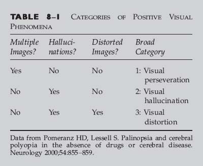
BROAD CATEGORY 1: VISUAL PERSEVERATION
QUESTIONS TO ASK AND POINTS TO KEEP IN MIND
PALINOPSIA
Classic Presentation
Red Flags
 Misalignment of the visual axes due to cranial nerve palsy, strabismus, or orbital abnormality: these result in binocular diplopia, in which the second image disappears if either eye is covered. See Chapter 7.
 Uncorrected refractive error that may lead to perception of overlapping shadow images can be the cause of monocular diplopia.
CEREBRAL POLYOPIA
Classic Presentation
Red Flags
 Misalignment of the visual axes due to cranial nerve palsy, strabismus, or orbital abnormality: these result in binocular diplopia, in which the second image disappears if either eye is covered. See Chapter 7.
 Uncorrected refractive error that may lead to perception of overlapping shadow images
BROAD CATEGORY 2: VISUAL HALLUCINATIONS
QUESTIONS TO ASK AND POINTS TO KEEP IN MIND
![]()
Stay updated, free articles. Join our Telegram channel

Full access? Get Clinical Tree


Ento Key
Fastest Otolaryngology & Ophthalmology Insight Engine
