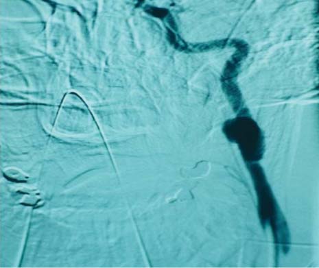
81 A 16-year-old female passenger was broadsided and ejected from her car. A sharp metallic fence rod impaled her left upper cervical region (zone II). She was seen emergently at an outside hospital and was found to be hemodynamically stable (blood pressure 170/84, pulse rate 107) with spontaneous respirations, 14 per minute. She is unconscious. Her cervical spine is clear. No active bleeding was present from the 4-cm left neck wound. On physical examination, she was in no respiratory distress and had no stridor. Only a right carotid pulse could be palpated. Two 14-gauge intravenous (IV) lines were started. No other injuries were present. The wound was not probed. At the outside facility, an emergent tracheotomy was done and the carotid artery appeared not to be exposed in this wound (Fig. 81.1). Fig. 81.1 Penetrating neck wound entering zone II. Any patient who has sustained trauma initially should be evaluated according to the Advanced Life Support System protocol. The airway is managed first and may require tracheotomy with the patient under local anesthesia to establish a stable airway for laryngotracheal injuries. Large-bore IVs are then started and the patient’s fluid resuscitated as needed. The neck is divided into three zones for trauma evaluation. Zone I is from the clavicle to the cricoid cartilage; zone II is from the cricoid to the angle of mandible; and zone III is from the mandible angle to the skull base. If any immediate life-threatening signs or symptoms from penetrating neck trauma (i.e., massive bleeding, expanding hematoma, nonexpanding hematoma in the presence of hemodynamic instability, hemomediastinum, hemothorax, systemic shock not responding to fluid resuscitation, increasing subcutaneous emphysema, major hemoptysis) are evident, emergent neck exploration is warranted. In the stable patient, the treatment choices remain controversial. Important parameters to consider are the zone that is involved (I, II, or III), mechanism of injury, and velocity of the projectile. These parameters are helpful to determine whether injury to vital structures has occurred. Currently many trauma centers manage stable patients with non–life-threatening penetrating neck trauma with selective exploration if the following is available: on-site angiography, flexible and rigid endoscopy, and close physical examining monitoring. High-velocity projectiles (>610 m/s) cause a significantly greater amount of damage than low-velocity projectiles. Zone I and zone III injuries usually require arteriography on all stable patents. Stable patients with zone II injuries may require angiography. Because hemorrhage is the leading cause of death for penetrating neck injuries, hemodynamic and neurologic status should always be monitored closely for at least 48 to 72 hours. For blunt neck trauma, the laryngeal, cervical, vascular, and digestive structures can be injured. Symptoms can manifest in a delayed fashion and are easily underdiagnosed. Thus, in patients who have had significant blunt neck trauma, careful observation should be undertaken to monitor for progressive signs and symptoms of laryngeal, digestive, cervical, and vascular injury. In Zone III injuries, exposure can be exceedingly difficult because the carotid follows its course medial to the mandible toward the skull base. Fig. 81.2 Angiogram showing a 3-cm traumatic aneurysm of the internal carotid several centimeters from the wound site at the level of C1. It was not actively bleeding on angiogram. Angiogram had been performed before surgery and revealed a 3-cm traumatic aneurysm of the internal carotid several centimeters from the wound site at the level of C1. It was not actively bleeding on the angiogram. Computed tomography (CT) scan did not show any larynx or tracheal fracture or foreign bodies. Barium swallow was normal (Fig. 81.2). 1. Zone II penetrating neck injury 2. Traumatic aneurysm of left internal carotid at level of C1 Initial management of patients with penetrating neck injuries should follow the basic tenents of trauma care. This emergent management requires (1) establishing an airway, (2) maintaining blood perfusion, and (3) defining the severity of the wound. In the emergency department, the airway can be established by intubation, cricothyroidotomy, or tracheotomy. Direct transcervical tracheal intubation is safer than oral or nasal intubation when the oral cavity, pharynx, or larynx are traumatized and filled with blood. In the setting of a gunshot wound, it may be difficult to evaluate the cervical spine fully until the airway is controlled. Multiple blind intubation attempts will risk enlarging a lacerated piriform sinus wound and extending it iatrogenically into the mediastinum. Similarly extending the neck in an injured trachea can distract apart the proximal and distal segments, thus enlarging a tracheal tear. The airway must be established and the hemodynamic status stabilized before transporting the patient to the angiography suite. Large-bore IV lines are placed, even when the patient is not hypotensive, so that fluids can be rapidly introduced if needed. The IV lines should be placed on the opposite side of the injury because of the possibility of venous lacerations proximal to the IV. The patient can be placed in the Trendelenburg position to minimize the potential of air embolism. Under no circumstances should a penetrating neck wound be probed because clot dislodgement and uncontrollable bleeding can occur.
Penetrating Neck Trauma
History

Differential Diagnosis—Key Points
Test Interpretation
 Hemoglobin 10 g/dL/hematocrit 31%. Chemistry panel, prothrombin time, partial thromboplastin time were within normal limits
 Chest radiography—no pneumothorax
Diagnosis
Medical Management
Stay updated, free articles. Join our Telegram channel

Full access? Get Clinical Tree


