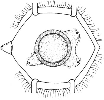
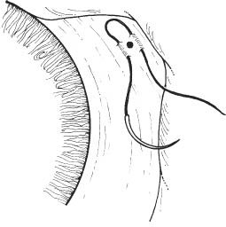
66 See Chapter 3. 1. Complete retinal evaluation. 2. Ultrasound examination of retina and vitreous if cataract precludes direct visualization. 3. Dilate pupil. a. Cyclopentolate 1% and phenylephrine 2.5% every 10 minutes × 3, starting 1 hour before surgery. b. Can use flexible iris retractors intraoperatively if iris dilates poorly (e.g., posterior synechiae). 4. Optional: Topical nonsteroidal anti-inflammatory agent (e.g., flurbiprofen) every 30 minutes starting 2 hours before surgery (to minimize intraoperative miosis). Note: In this chapter, the Alcon Accurus vitrectomy machine’s phacofragmentation unit is used for illustrative purposes. The authors recognize that other excellent machines are available and settings may differ based on machine. The authors have no financial interest in the Accurus system. 1. Anesthesia: General or retrobulbar ± lid block. 2. Prep and drape eye. 3. Place lid speculum. Figure 66.1 4. Perform conjunctival peritomy at limbus 4 clock hours temporally and superonasally in area of planned entry sites (Westcott scissors, toothed forceps) (Fig. 66.1). 5. Secure hemostasis with cautery (optional). 6. Place infusion contraindicated, inferotemporally (unless e.g., due to choroidal detachment) and two sites for bimanual manipulation superotemporally and supero-nasally at 10 and 2 o’clock, respectively. a. Sclerotomies should be 3.0 mm from limbus (measure with calipers). b. Sclerotomy sites should be parallel to the limbus. c. Mark location of sites with cautery (optional). d. Prepare infusion site. Figure 66.2 i. Preplace 4–0 white silk (or 6–0 Vicryl) mattress suture through partial-thickness sclera spanning the sclerotomy site (Fig. 66.2).
Pars Plana Lensectomy
Indications
 Select cases of traumatic, subluxed, and/or dislocated cataractous lenses. A pars plana approach is particularly useful in cases where zonular rupture with vitreous pro-lapse into the anterior chamber precludes a safe phacoemulsification, extracapsular, or intracapsular cataract extraction approach.
 Select congenital cataracts.
 Other cases in which lens removal combined with posterior vitrectomy is indicated.
Preoperative Procedure
Instrumentation
 Lid speculum
 Fine-toothed tissue forceps (e.g., 0.12 mm straight Castroviejo and/or Colibri)
 Westcott scissors
 Cautery, bipolar or disposable (optional)
 Castroviejo calipers
 Marking pen (optional)
 20-gauge microvitreoretinal (MVR) blade
 Needle holder
 4 mm infusion cannula (6 mm cannula in cases with choroidal detachment, 2.5 mm cannula in pediatric cases)
 Sutures (4–0 silk or 6–0 Vicryl, 7–0 Vicryl, and 8–0 Vicryl or 6–0 plain gut)
 Cotton-tipped applicators
 23 gauge butterfly needle
 Ultrasonic phacofragmentation unit with 20 gauge tip
 Vitrectomy suction/cutting instrument
 Intraocular forceps
 Fiber optic illuminator
 Corneal ring
 Contact lenses
 Traditional lens system (TLS): 20 degrees and 30 degrees prism, wide angle (48 degrees), macula (34 degrees), and biconcave (90 D) lenses.
 Wide angle lens system (WAS): wide-angle lens (Ocular 155D or Volk’s Mini Quad/Quad XL), equator lens (Ocular 91 D lens or Volk’s Central Retinal lens), macula lens (Ocular 66D or Volk’s Super Macula).
 Scleral plugs
 Indirect ophthalmoscope
 20 D and/or 28 D lens
 Fluted needle (Charles) with or without silicone tip
 Retinal cryopexy unit and/or indirect laser ophthalmo-scope
 Heavier than water liquid (e.g., perfluoro-octane), optional
Operative Procedure
Stay updated, free articles. Join our Telegram channel

Full access? Get Clinical Tree


