FIGURE 8.1 The Development of the Immune Cells.
BFU, Burst-forming unit; BM, basophil mast cell; CFU, colony-forming unit; E, erythroid; Eo, eosinophil; GM, granulocyte-macrophage; Ig, immunoglobulin; MEG, megakaryocyte; NK, natural killer; TCR, T-cell receptor. (From Flint PW, Haughey BH, Lund VJ, et al. Cummings Otolaryngology—Head and Neck Surgery. 6th ed. Philadelphia, PA: Saunders; 2015, fig. 38-1.)
• Class II: immunocompetent antigen-presenting cells
• HLA-DR
• HLA-DQ
• HLA-DP
Major Histocompatibility Complex (Fig. 8.3)
• Class I: all nucleated cells
• Class II: antigen-presenting cells
• Macrophage
• Dendritic cells
• B cells
Complement System (Fig. 8.4)
• Classic pathway
• Lectin pathway
• Alternative pathway
Cytokines (Table 8.1)
• Broad category of small proteins important in cell signaling
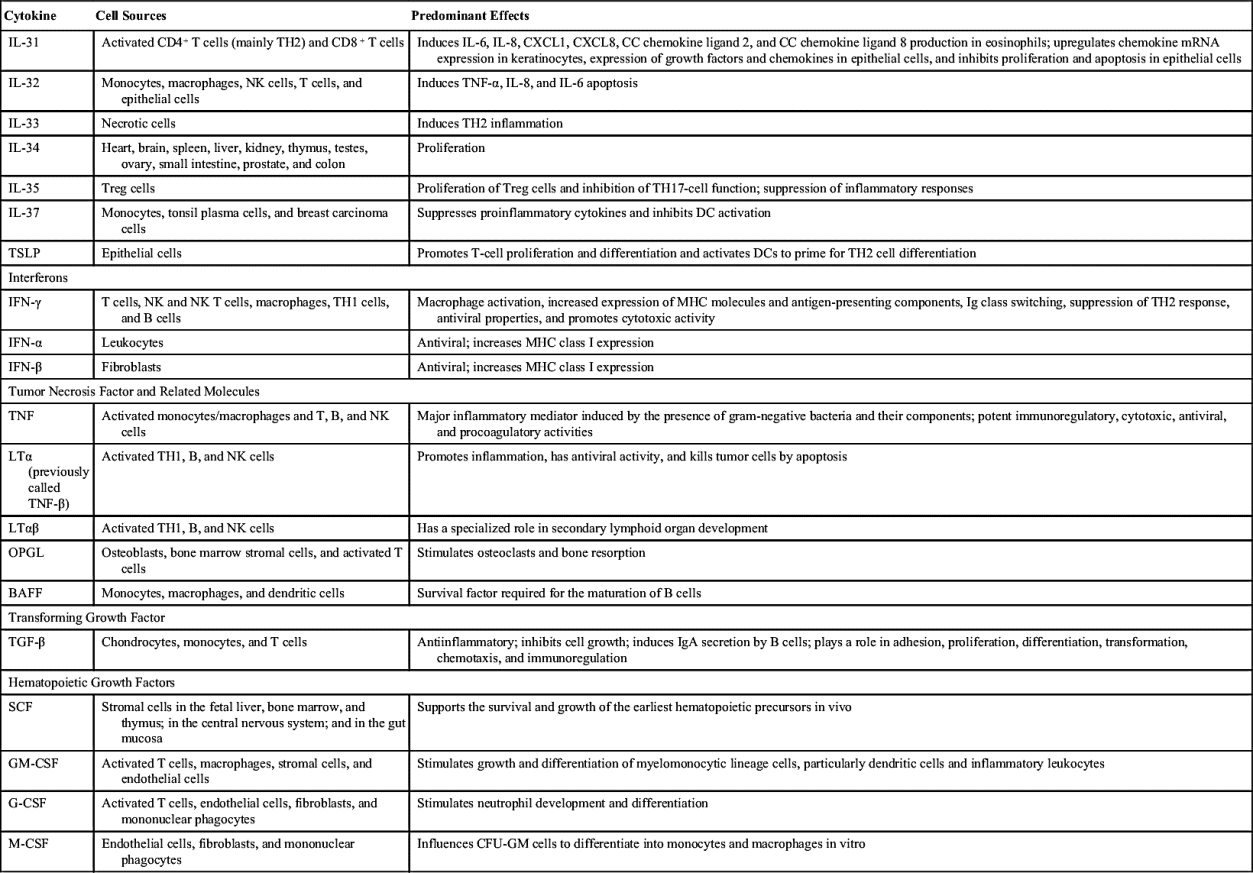
Chemokines (Table 8.2)
• Small proteins important in signaling chemotaxis
• Four categories: CXC, CC, CX3C, and CX
Cell Adhesion (Fig. 8.5)
• Intercellular adhesion molecule (ICAM)-1
• ICAM-2
• E-selectin
• P-selectin
• Vascular cellular adhesion molecule (VCAM)
Allergic Triggers: Categories
1. Inhalants
2. Ingestions
3. Injectables
4. Contactants
Stages of Development of an Allergy
• Early response: minutes after exposure antigen
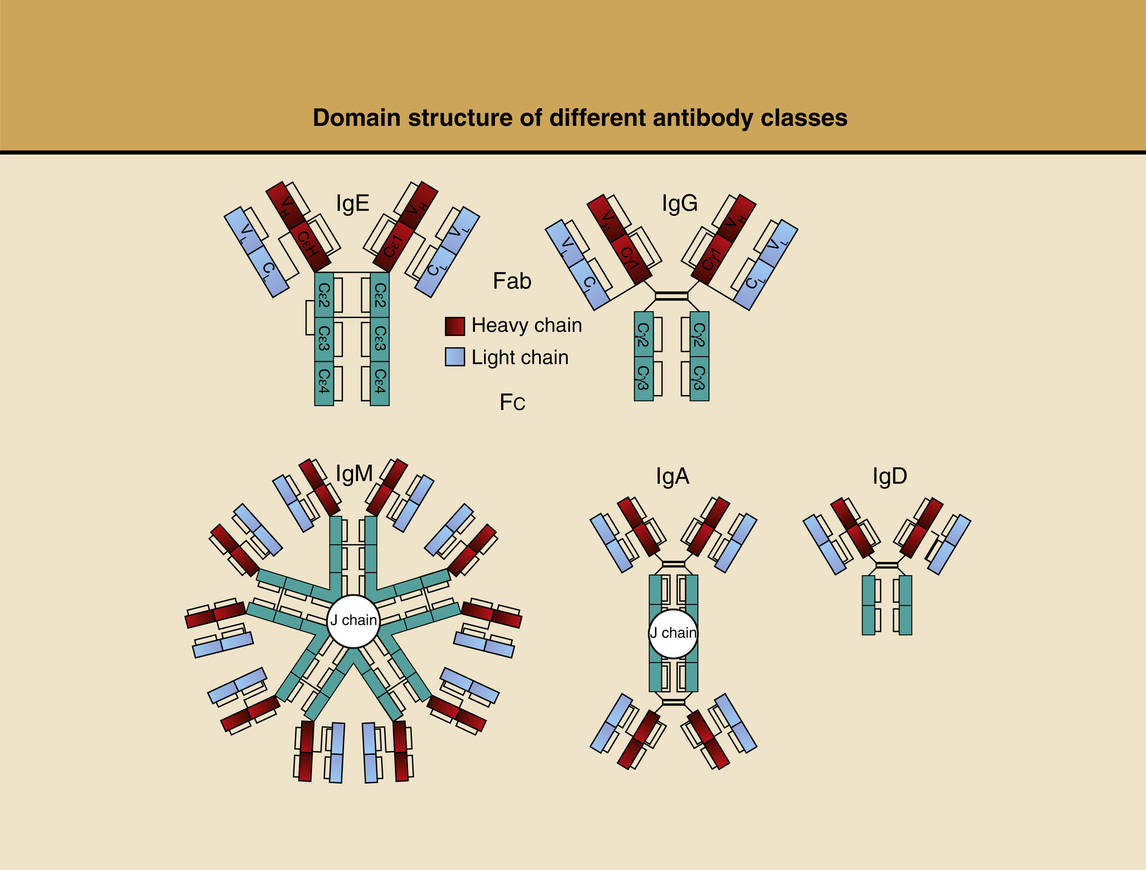
FIGURE 8.2 Domain Structure of Various Antibody Classes.
(From Krouse JH. Introduction to allergy. In: Krouse JH, Derebery MJ, Chadwick SJ, eds. Managing the Allergic Patient. 1st ed. Philadelphia, PA: Saunders Elsevier; 2008. Originally reproduced from Holgate ST. Allergy. 2nd ed. London: Mosby/Elsevier; 2001:245, fig. 16.4.)
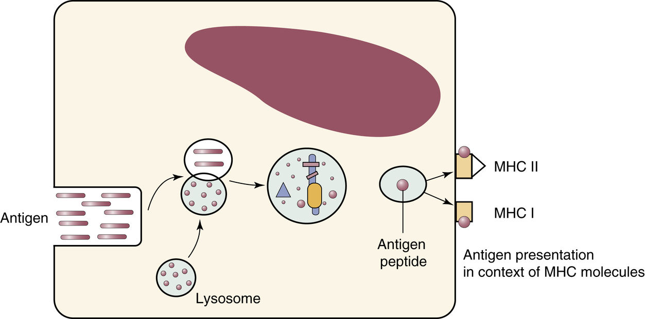
FIGURE 8.3 Antigen Processing and Presentation.
The antigen undergoes hydrolytic cleavage within antigen-presenting cells, and the resultant oligopeptides are loaded on the antigen-binding grooves of major histocompatibility complex (MHC) molecules and are expressed at the cell surface. (From Flint PW, Haughey BH, Lund VJ, et al. Cummings Otolaryngology—Head and Neck Surgery. 6th ed. Philadelphia, PA: Saunders; 2015, fig. 38-2.)
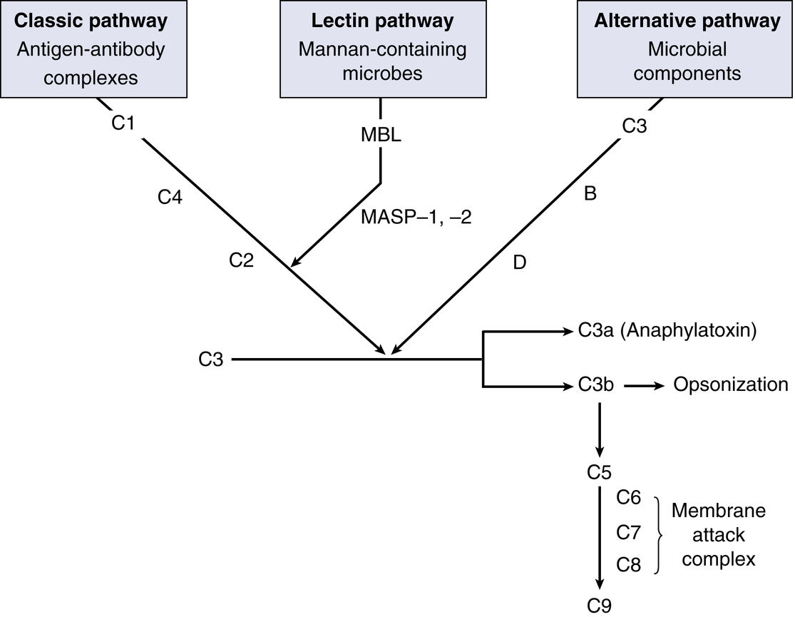
FIGURE 8.4 Complement Pathways.
Three pathways of the complement system. B, Factor B; D, factor D; MASP, MBL-associated serine proteases; MBL, mannan-binding lectin. (From Flint PW, Haughey BH, Lund VJ, et al. Cummings Otolaryngology—Head and Neck Surgery. 6th ed. Philadelphia, PA: Saunders; 2015.)
Table 8.1
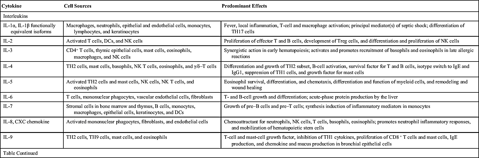
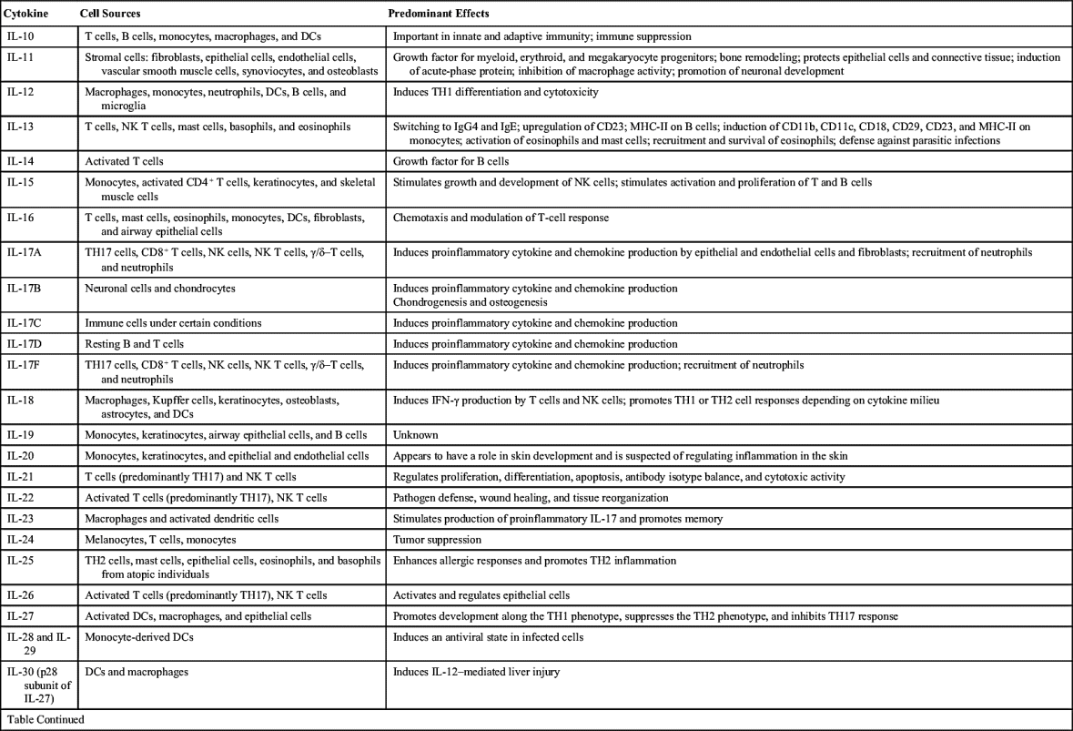
From Flint PW, Haughey BH, Lund VJ, et al. Cummings Otolaryngology—Head and Neck Surgery. 6th ed. Philadelphia, PA: Saunders; 2015, table 38-1.
Table 8.2
From Flint PW, Haughey BH, Lund VJ, et al. Cummings Otolaryngology—Head and Neck Surgery. 6th ed. Philadelphia, PA: Saunders; 2015, table 38-2.
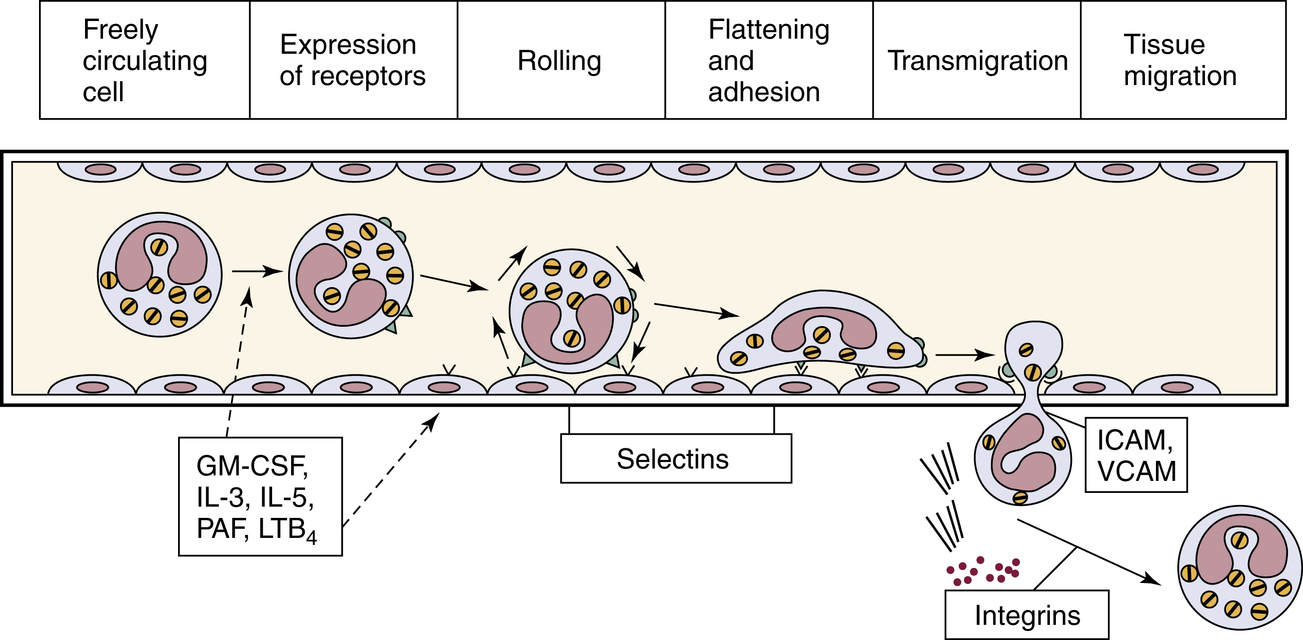
FIGURE 8.5 Cellular Adhesion and Recruitment.
GM-CSF, Granulocyte-macrophage colony-stimulating factor; ICAM, intercellular adhesion molecule; IL, interleukin; LTB4, leukotriene B4; PAF, platelet-activating factor; VCAM, vascular cellular adhesion molecule. (From Flint PW, Haughey BH, Lund VJ, et al. Cummings Otolaryngology—Head and Neck Surgery. 6th ed. Philadelphia, PA: Saunders; 2015, fig. 38-4; and Mygind N, Dahl R, Pedersen S, Thestrup-Pedersen K, eds. Essential Allergy. 2nd ed. Oxford: Blackwell Scientific Publications: 1996.)
• Symptoms: largely nasal symptoms, including rhinorrhea, sneezing, itching, and congestion, as well as other symptoms, including tearing, wheezing, and, potentially, laryngospasm and bronchospasm
• Late response: hours after exposure to antigen
• Mediators: leukotrienes and eosinophils
• Symptoms: congestion, increased rhinorrhea, and wheezing
Types of Hypersensitivities (Gell and Coombs Classification)
1. Type I: immediate/anaphylaxis (IgE)
2. Type II: cytotoxic (IgG, IgM)
3. Type III: immune complex (IgG, IgM, IgA)
4. Type IV: cell mediated (T cells)
Type I: Causes of Anaphylaxis
• Inhalants
• Foods
• Drugs
• Insect stings
Type I: Anaphylaxis Symptoms
• Upper respiratory: sneezing, itching, rhinorrhea, and congestion
• Lower respiratory: cough, bronchospasm, and wheezing
• Skin: urticarial, angioedema, itching, and whealing
• Systemic: hypotension, tachycardia, and feelings of impending doom
Type I: Anaphylaxis—Mechanism
• Cross linking of IgE on mast cells
• Degranulation of mast cells
• Release of histamine
Mast Cell Degranulation
• Vasodilation
• Increase capillary permeability
• Bronchoconstriction
• Tissue edema
Type II: Cytotoxic Reaction—Mechanism
• IgG or IgM mediated
• Antibody reaction with antigens on the cell surface
• Activation of complement
Type II: Cytotoxic Reaction—Examples
• Hemolytic anemia
• Transfusion reaction
• Acute graft versus host disease
• Goodpasture syndrome
• Myasthenia gravis
Type III: Immune Complexes—Mechanism
• Immune complexes form (binding of antibody to a soluble antigen)
• Complexes deposit in tissues
Type III: Immune Complexes—Examples
• Serum sickness
• Poststreptococcal glomerulonephritis
• Angioedema
• Gastrointestinal intolerance
Type IV: Cell Mediated—Mechanism
• Direct T-cell activation
• Cell-mediated inflammation
Type IV: Cell Mediated—Examples
• Dermatitis
• Tuberculosis
• Sarcoidosis
• Candidiasis
Allergens
Perennial Allergens
• Mites
• Cockroach
• Cotton particles
• Human skin scales
• Animal dander
• Molds
Seasonal Allergies
• Trees: winter and spring
• Grasses: spring, summer, and fall
• Weeds: summer and fall
Pollen as Allergen
• Windborne
• Lightweight
• Large quantities
• Allergenic in sensitive individuals
History and Physical Exam of Patient with Allergies
History
• Particular emphasis on:
• List of medications
• Co-morbidities
• Previous operations or treatments for allergy
• Childhood history
• Family history
Physical Exam
• Eyes: allergic “shiners” and long eyelashes
• Ears: erythema, postauricular fissures, desquamation of external auditory canal, tympanosclerosis, tympanic membrane retraction and/or perforation, and serous effusion
• Nose: discharge, edema of turbinates, polyps, and “allergic salute”
• Neck: lymphadenopathy
• Chest: wheezing
• Pharyngeal: high, narrow arched palate; lymphoid hyperplasia; cobblestoning of the posterior pharyngeal wall; hypertrophy of the lateral nasopharyngeal bands; chronic cough, and edema of the uvula and glottis
Stay updated, free articles. Join our Telegram channel

Full access? Get Clinical Tree


