9 OTHER INFECTIOUS DISEASES
Acanthamoeba Keratitis
Nicky R. Holdeman
 THE DISEASE
Pathophysiology
Acanthamoeba keratitis (AK) is a rare, sight-threatening disease, which results from colonization of the cornea with the free-living, microscopic, ubiquitous protozoa Acanthamoeba. These organisms have been isolated from treated and untreated tap water, swimming pools, salt water, air-conditioning units, air, dust, soil, vegetables, and animal waste.
Acanthamoeba inflammation is intensified by the release of proteolytic enzymes and stimulation of the immune system within the cornea. The organism is capable of existing in two forms: a free-living trophozoite, which is mobile and proliferates and feeds on bacteria and other unicellular organisms, and a double-walled cyst form that develops in response to a hostile environment, such as drugs, chemicals (chlorine), temperature, or other adverse stimuli. The cyst form is very hardy and can survive freezing temperatures and desiccation.
Etiology
AK results from exposure and subsequent infection of the cornea with Acanthamoeba. An epithelial break or bacterial infection may be necessary for Acanthamoeba infection, since the Acanthamoeba organism will initially sustain itself by ingesting bacteria. Contact lenses (CLs) may increase the ability of Acanthamoeba to bind to epithelial cells by altering the expression of certain proteins on the ocular surface.
The Patient
Clinical Symptoms
Symptoms commonly include pain, redness, foreign body sensation, photophobia, tearing, and decreased vision. The pain is often severe and out of proportion for the size of the corneal lesion and the extent of the inflammation. The accentuated pain (and decreased corneal sensitivity) may be a result of corneal nerve inflammation. However, in early cases, especially in the absence of keratoneuritis, pain may not be a significant symptom.
Clinical Signs
The keratitis may begin as an epithelial haze accompanied by epithelial breakdown, punctate staining, pseudodendrites, and elevated epithelial lines. The early presentation may be confined to the epithelium and is often a dendritiform epithelial pattern. Later in the infection, the more classic findings of a central stromal infiltrate, ring infiltrate, or satellite infiltrates may develop. Deep stromal involvement and presence of a ring infiltrate at presentation are often associated with a worse visual outcome.
Radial keratoneuritis—linear, branching infiltrates in the mid-stroma along the corneal nerves—which is considered the most specific sign for Acanthamoeba, may occur in less than 50% of patients.
Other conditions such as iritis, limbitis, scleritis, hypopyon, increased IOP, preauricular lymphadenopathy, and conjunctival follicles may be present. There is typically an absence of neovascularization.
A cataract may occur and progress during the management of AK. If left untreated, Acanthamoeba can potentially spread to the retina and cause serious chorioretinitis.
Note: Making the diagnosis is challenging, as AK is often misdiagnosed as a CL-related corneal epitheliopathy or the more common fungal or viral form of keratitis. The presence of conjunctival follicles, epithelial dendritiform pattern, PA nodes, and corneal hypothesia frequently leads to a misdiagnosis of herpes simplex keratitis. The lack of terminal end bulbs, typical in an HSV infection, is a good indicator that a patient may have AK.
Demographics
AK was first reported in the United States in 1973—an estimated 5,000 cases of AK have since occurred as of 2006; however, AK is so commonly misdiagnosed that the number could be much higher.
Although AK can occur in any age group, it is most often found in young adults who are immunocompetent.
CL wear has repeatedly been identified as a major risk factor for the development of AK. However, the condition is still very rare, affecting about 1.65 to 2.0/million CL wearers per year in the United States. Soft CL wearers are at 9.5 times greater risk of AK than rigid lens wearers.
The use of homemade saline and poor disinfection habits have been strongly associated with the condition.
Significant History
A history of corneal trauma, CL wear, and use of multipurpose solutions is important and comprises the primary risk factors for AK. CL wearers account for approximately 85% to 90% of cases, but AK can and does occur in non-CL patients.
Acanthamoeba has been found in contaminated CL cases and solutions. Inappropriate use of solutions, including homemade saline or rinsing of lenses in tap water or bottled water, puts a patient at significant risk.
A number of cases of AK have been reported in association with overnight orthokeratology and in patients swimming in a hot tub.
Ancillary Tests
Acanthamoeba can sometimes be cultured from corneal scrapings when plated on nonnutrient agar that has been overlaid with Escherichia coli. Samples should be incubated at approximately 37°C. Trophozoites feed on the bacterial overlay and usually appear within 3 days but can take up to 3 weeks. Cultures typically offer the most definitive means of diagnosis but are only positive in about 50% of clinically suspected cases.
The organism can be stained directly with Gram, Giemsa, methenamine silver, or Wright stain; however, identification is difficult. The chemofluorescent dye, calcofluor white, is often used to visualize the cysts and trophozoites. Periodic acid Schiff or acridine orange may also aid in the diagnosis.
PCR can be used as a diagnostic tool when very few cells are available for visual determination. This test has a reported sensitivity of 84% and specificity approaching 100%. Unfortunately, PCR techniques are expensive and require skilled technicians, which limit their usage.
Real-time fast-duplex TaqMan PCR (f-d-real-t PCR) appears to have a higher sensitivity and specificity than other techniques and may simultaneously detect ten different genotypes of Acanthamoeba. Future studies are needed to confirm the usefulness of this technique for disease management of AK.
Corneal biopsy is sometimes required to make a definitive diagnosis. If the patient is a CL wearer, the lenses and solutions should be cultured.
On occasion, the diagnosis of AK is made in retrospect by the failure of traditional therapy for bacterial and viral keratitis. If a clinician is confronted with a smoldering inflammation and if there has been no therapeutic response to antibiotics or antiviral agents, AK should be considered.
The Treatment
Treatment of AK is difficult, often with disappointing results; however, early and effective management may keep a patient from going blind. One should maintain a high index of suspicion for this condition, especially in CL wearers.
Treatment is most successful during the initial stages of the disease, so prompt diagnosis is essential and cytocidal agents must be used. If effective therapy is delayed for 3 weeks or more, the prognosis worsens. Bacterial super infection can be a serious concurrent problem and should also be addressed.
Topical Therapy
Propamidine 0.1% (Brolene) (not available in the United States, available OTC in Europe) and Neosporin gtts alternated q30–60min × 3 days around the clock; followed by the same drops alternated q1h during the day and q2h at night × 4 days. Doses are gradually tapered to q.i.d. with dibromopropamidine 0.15% ointment hs × 1 year.
Polyhexamethylene biguanide (PHMB) (Baquacil) 0.02% has been used with good results. The medication is cytocidal and is applied topically q1h until an objective and subjective improvement is noted at which time the drug is tapered in a similar fashion as is Brolene and Neosporin. Baquacil may also be used in combination with Brolene, Neosporin, or chlorhexidine.
Chlorhexidine is another cytocidal agent. Chlorhexidine 0.02% gtt q1h while awake and q2h while sleeping is often combined with Neosporin gtt q1h when awake and q2h at night. This regimen can be tapered over weeks to months as clinically indicated.
Topical cycloplegics and pain medication should be used as needed.
The role of topical steroids in the treatment of AK remains controversial. Mild steroids are sometimes used to reduce inflammation and pain, but they can simultaneously cause the transformation of the cysts into trophozoites, which can worsen the disease.
In general, steroids should be avoided until the infection has shown significant improvement, which would be at least 2 weeks completion of appropriate treatment. Judicious use of topical steroids can have a beneficial role in the management of some cases.
Antifungal agents, such as miconazole or clotrimazole instilled topically and ketoconazole (200 to 400 mg/d) taken orally, have been employed with some success. Topical (1%) and intrastromal (25 μg/mL) voriconazole may also be a promising adjuvant in the treatment of AK.
Note: Future treatments may include a proprietary formulation of N-Chlorotaurine (NCT), which is under investigation. NCT has demonstrated an amoebicidal effect on different strains of Acanthamoeba, including the cystic form.
Penetrating keratoplasty in an eye with an active infection usually has a poor prognosis. Quiet eyes that have been successfully treated often have a good visual outcome.
Demodicosis
Marcus G. Piccolo and Victor Malinovsky
ICD-9: 373.00
 THE DISEASE
Pathophysiology
Demodicosis is an infestation and overpopulation of the lash follicles and accompanying sebaceous glands with the mites Demodex folliculorum or Demodex brevis. Adult mites migrate to gland orifices, where they copulate. The female mite migrates back into the gland where it deposits its egg. Immature mites eventually migrate to the gland orifice. Infestation results in abnormal function of the glands, including blockage of the orifice and accumulation of keratin. In addition, the mites may consume the glandular epithelium.
Etiology
Overpopulation of the Demodex organisms.
The Patient
Clinical Symptoms
While many patients remain asymptomatic, some will report burning, itching, foreign body sensation, redness, and scaling of the eyelid margins. These symptoms are typically worse in warmer weather.
Clinical Signs
Clear keratin sleeves (or cylindrical dandruff) are found around the base of the lashes, which is highly suggestive of Demodex in the follicle. Keratin blockage of the meibomian glands may occur. The organism may act as a vector for bacteria, resulting in a concurrent bacterial blepharitis. The patient may present with lid thickening, scaling of lids, madarosis, trichiasis conjunctival inflammation, meibomian gland dysfunction, and dry eyes. The cornea may develop superficial vascularization, marginal infiltrates, a phlytenule-like lesion, and peripheral scars.
Demographics
Demodicosis increases with age. While this condition is rarely seen in children, 90% to 95% of adults over the age of 45 years have some Demodex infestation. The human is a natural host for the organism. Involvement in other areas such as the nose, cheeks, external auditory meatus, and forehead may be present.
Significant History
No particular history exists for demodicosis, although some infestations have been related to poor hygiene, perioral dermatitis, and rosacea. In fact, there appears to be a clinically significant increase of Demodex in patients with rosacea, suggesting that this ectoparasite may be involved in the pathogenesis of rosacea.
Ancillary Tests
The mite is an eight legged, spindle-shaped organism, approximately 0.3- to 0.4-mm long, which can only be viewed with microscopic magnification. Epilation of several lashes, which are then floated on a viscous fluid or 0.25% fluorescein drop, can be observed with a light microscope. One to two mites found on 16 epilated lashes (4 lashes from each eyelid) is considered normal. Overpopulation is considered when 6 to 8 mites or more are found on 16 epilated lashes.
The Treatment
Treatment is often problematic but is aimed at reducing the number of mites in the glands, preventing mating, and avoiding reinfestation. The use of bland ophthalmic ointments or ophthalmic antibiotic ointments may prevent the adult mites from copulating during the night, thereby reducing the population. The use of warm soaks and lid scrubs may also be beneficial. Some topical agents may be irritating to the eye and may not affect organisms deep within the glands.
Daily lid scrubs with 50% tea tree oil (TTO) and lid massage with 5% TTO ointment is recommended for eradicating ocular Demodex infestation.
The in-office application of ether to the lid margins with a cotton-tipped applicator will cause organisms within the glands to migrate to the surface. Repeated scrubs 5 minutes later will kill emerging organisms.
Phthiriasis Palpebrarum
Nicky R. Holdeman
ICD-9: 132.9
 THE DISEASE
Pathophysiology
Phthiriasis palpebrarum is a rare cause of blepharoconjunctivitis produced via an infestation of the eyelids by the crab louse, Phthirus pubis. Transference to the eye may occur by hand contact from the inguinal area, by contaminated clothing, combs, bedding, or towels, or by sexual activities. Children and infants may acquire the disease by close contact with the chest or axillary hair of an infested parent or caretaker.
Lice feed solely on human blood by piercing the skin, injecting saliva, and then sucking blood. The fecal material and saliva excreted by the parasite result in an inflammatory response.
Etiology
The etiology is infestation of the eyelids by lice (Fig. 9-1). The crab louse is a six-legged ectoparasite that typically prefers the anogenital area but may also involve facial hair such as the beard, mustache, eyebrows, and/or eyelashes. Nits (or eggs) may survive 3 weeks when removed from the host.
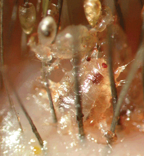
Figure 9-1. Adult louse with nits in a patient with phthiriasis palpebrarum.
Stay updated, free articles. Join our Telegram channel

Full access? Get Clinical Tree


