13 OCULAR EMERGENCIES AND URGENCIES
 Sudden Vision Loss
Anastas F. Pass
ICD-9: 362.34—TRANSIENT ARTERIAL OCCLUSION—AMAUROSIS FUGAX
ICD-9: 368.10—VISION DISTURBANCE; TRANSIENT VISUAL OBSCURATION
ICD-9: 368.11—SUDDEN VISUAL LOSS
ICD-9: 368.12—TRANSIENT VISUAL LOSS
 THE DISEASE
Pathophysiology
Sudden vision loss may indicate a serious vascular disorder and, as such, should be recognized as an ocular emergency. Reduced blood flow and, hence, reduced levels of oxygen at the cellular level result in metabolic compromise. The loss in function may be permanent or temporary. Longstanding vision loss may not be recoverable and may be a precursor to cerebrovascular accidents. The spectrum of sudden vision loss includes local and benign causes (e.g., retinal migraine or acephalgic migraine) (Table 13-1); however, these are considered diagnoses of exclusion. Vascular anomalies that give rise to vision loss are not only sight threatening but may be life threatening as well. When discussing vision loss, transient or otherwise, it should be remembered that there are various categories of vision loss with specific diagnostic coding.
- Transient visual obscuration—not a complete loss of vision but a “white-out” or “grey-out” of vision. Most common with presence of papilledema or increased intracranial hypertension and lasts seconds to minutes.
- Amaurosis fugax (AF)—a very brief loss of vision (typically unilateral) of seconds to minutes
- Transient vision loss (TVL)—a loss of vision (unilateral or bilateral) of minutes to longer
Etiology
Vision loss, when it is acute and transient (AF or TVL) should be considered secondary to a vascular disorder until proved otherwise. Retinovascular and/or cerebrovascular accidents may be caused by emboli that can occlude the retinal arterial supply (twig, branch, or central) or the cerebrovascular circulation (carotid or vertebral-basilar [VB]). These occlusions may be transient, causing little or no retinal damage, or may result in long-standing retinal nonperfusion and permanent sight loss.
Blood dyscrasias, hyperlipidemia, hyperviscosity syndromes, sickle cell disease, syphilis, systemic hypertension, idiopathic intracranial hypertension, diabetes, and inflammatory conditions (e.g., giant cell arteritis [GCA; arteritic ischemic optic neuropathy], nonarteritic ischemic optic neuropathy, and optic neuritis) can also lead to occlusion of the arterial system. The same diseases may also produce optic nerve head edema, resulting in vision loss. Connective tissue diseases and the vasculitidies (GCA, polyarteritis nodosa, Takayasu’s arteritis) have also been implicated in vision loss. Arteritic ischemic optic neuropathy typically presents as a painful vision loss and should also be considered a medical emergency (see “Temporal Arteritis” in Chapter 19).
TABLE 13-1 Clinical Differentiation of Common Etiologies Involving TVL
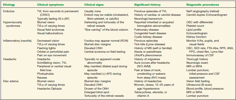
TVL, transient vision loss; AVM, arteriovenous malformations; ONH, optic nerve head; RON, retrobulbar optic neuritis; APD, afferent pupillary defect; CBC, complete blood count; ANA, antinuclear antibodies; PPD, purified protein derivative; MRI, magnetic resonance imaging; MRA, magnetic resonance angiography; CSF, cerebrospinal fluid.
Other etiological considerations include Uhtoff’s phenomenon (associated with MS or Leber’s optic neuropathy), branch retinal vein occlusion (BRVO)/central retinal vein occlusion (CRVO), venous stasis retinopathy, papillophlebitis, vasospastic AF, recurrent hyphema, orbital tumors, ocular ischemic syndrome, hypoperfusion, or dominant optic atrophy (now recognized as the most common heredodegenerative optic neuropathy).
Local causes, such as disc drusen, retinal detachment, and/or maculopathies, may result in sudden vision loss (or vision obscurations). Other considerations, as diagnoses of exclusion, include migrainous (scintillating) vision loss, drug toxicities, acute angle-closure glaucoma, or compressive lesions of the visual pathway.
The Patient
Symptoms and signs will vary depending on the circulatory system involved. Listed below are classic findings involving the retinal (R), carotid (C), and VB arteries, as well as inflammatory (I) causes.
Causes—Clinical Symptoms
- R/C/VB—Decreased vision, visual field defects or vision loss (complete or incomplete); typically unilateral
- R—Flashing lights
- C—Hemisensory loss
- C/VB—Diplopia
- C/VB—Dysphasia
- VB—Loss of equilibrium (ataxia)
- VB—Weakness of the extremities (hemiplegia)
- I—Pain (head, scalp tenderness, neck, periorbital)
- VB—Most likely bilateral involvement (visual, motor, cerebellar)
Clinical Signs
Prominent Signs
- Decreased acuity; typically unilateral (bilateral vision loss is an uncommon occurrence). With presentation of bilateral vision loss, one should assume a VB vascular anomaly (compression, embolus, occlusion), space occupying lesions, or a migrainous event; the latter on an exclusionary basis.
- Retinal emboli may be visible and can be broken down into three main categories:
1. Fibrin-platelet—which originate in the heart, large vessels, or from a thrombus. This form of embolus rarely results in retinal infarction.
2. Cholesterol “Hollenhorst” plaque— which typically originate from a plaque forming at the carotid bifurcation
3. Calcific—which originate from cardiac valves and associated with rheumatic heart disease. This form of embolus will result in the most severe retinal infarction.
- “Boxcarring” of blood flow (hyperviscosity syndromes)
- Retinal nonperfusion and edema (as in artery occlusion)
- Vein occlusion
- Tortuous vasculature
- Retinal arteriolar narrowing, focal constrictions, venous nicking
Subtle Signs
- Possible relative afferent pupillary defect
- Disc elevation/edema
- Nerve fiber layer infarct(s)
- Sensorium changes
- Horner’s pupil (possible in long standing atherosclerosis and carotid dissection)
Demographics
By radiographic studies, 56% to 100% of patients with central retinal artery occlusion (CRAO) demonstrate carotid occlusive disease. Individuals ≥40 years old demonstrate the highest incidence of CRAO. The mean presentation is in the sixth decade of life.
Men have a higher incidence of CRAO, retinal occlusive disease, and TVL (associated with atherosclerotic plaques) than do women; however, the incidence in postmenopausal women is equal to that of men. Life expectancy of patients with CRAO is 5.5 years, compared to 15.4 years for an age-matched population without CRAO.
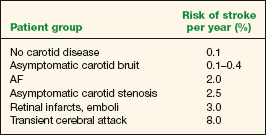
Individuals with retinal infarcts and emboli have a risk of stroke of 3% per year. Patients with TVL have a risk of stroke of 2% per year. (By comparison, patients without carotid artery disease [atherosclerosis] have a risk rate of 0.1% per year.) (See Table 13-2.)
Patients who report episodes of TVL have a high incidence of hyperlipidemia (60%) as well as ischemic heart disease (25%). The National Institute of Neurological Disorders and Stroke reports that approximately one third of individuals who experience a TVL will experience an acute stroke in the “near” future.
Incidence of retinal vascular occlusion is rare in children and young adults; however, the presence of TVLs and/or retinal occlusive disease in this group should be suspect for mitral valve prolapse, rheumatic heart disease, systemic lupus erythematosus, migrainous events, and recurrent hyphema. In patients younger than 40 years, an embolus from the heart is the most common cause of (central) retinal arterial occlusion. There is also suspicion that the use of oral contraceptives may cause TVLs in susceptible patients.
TABLE 13-2 Stroke Risk
Significant History
- Previous report of a transient ischemic attack or stroke
- Long-standing atherosclerosis
- Long-standing arteriolarsclerosis
- Cardiovascular anomalies (mitral valve prolapse, prosthetic valves, vasculitis, polycythemia, thrombocytosis)
- Systemic hypertension
- Diabetes mellitus
- Dyslipidemia
- Family history of transient ischemic attacks or vascular occlusive disease
- Oral contraceptives
- Smoking
Ancillary Tests
- Ocular assessment for TVL would include best visual acuity, pupil assessment, dilated fundus examination, and visual field assessment
- Laboratory studies: erythrocyte sedimentation rate (ESR), C-reactive protein (CRP) level, complete blood count (CBC) with differential, platelet count, lipid profile, antinuclear antibodies (ANA), anticardiolipin, antiphospholipid antibody, and lupus anticoagulant
- Noninvasive testing: blood pressure, carotid auscultation, cardiac evaluation (electrocardiogram, Holter monitor, echocardiogram), carotid duplex ultrasound, brain CT/MRI (consider magnetic resonance angiography [MRA] as a noninvasive carotid arterial evaluation), ophthalmodynamometry, or oculoplethysmography (for ocular perfusion pressures)
- Invasive testing: IV retinal flourescein angiography may show the presence of embolic particles in areas of vessel leakage. Carotid angiography should be limited to those patients who will undergo carotid surgery (with >90% stenosis).
The Treatment
Transient Vision Loss
Acute Angle-Closure Glaucoma
G. Richard Bennett and Fiaz Zaman
ICD-9: 365.22
 THE DISEASE
Pathophysiology
Acute angle-closure glaucoma is characterized by a rapid and large increase in the intraocular pressure (IOP), resulting from a sudden blockage of the anterior chamber angle.
Etiology
Angle closure can be precipitated by several different mechanisms. The most common cause is pupillary block. A resistance to flow of aqueous humor develops between the iris and lens that causes pressure to increase in the posterior chamber. This resistance causes the peripheral iris to bow forward, which obstructs the anterior chamber angle and causes the IOP to rise rapidly. Conditions and medications that place the pupil in a mid-dilated position (i.e., mydriatics to dilate the pupil, dim illumination, and/or anticholinergics such as antihistamines or antipsychotics) maximize lens/iris touch and can precipitate pupillary block in patients with a narrow anterior chamber angle. Acute angle closure is more common in patients who are hyperopic because of decreased axial length and a compressed lens-iris diaphragm.
Acute angle closure can also occur because of plateau iris syndrome, in which the peripheral iris obstructs outflow without preexisting pupillary block (see section on “Plateau Iris”).
Secondary causes of acute angle closure include posterior synechiae formation (chronic inflammation or neovascular membranes) or anterior displacement of the lens-iris diaphragm (CRVOs, scleral buckles, extensive panretinal photocoagulation, posterior segment tumors, choroidal detachments, or posterior misdirection syndrome).
The Patient
Clinical Symptoms
Symptoms on presentation include ocular pain, lacrimation, frontal headache, nausea, vomiting, photophobia, seeing colored halos around lights, and decreased vision. Patients can have a history of similar symptoms, suggesting prior episodes of intermittent angle closure.
Clinical Signs
- Decreased vision
- Conjunctival injection
- Corneal microcystic edema
- Shallow anterior chamber (deeper centrally than peripherally)
- Mid-dilated pupil (pupil may be miotic if acute angle closure is secondary to posterior synechiae formation)
- Significantly elevated IOP
Other signs that may be present include the following:
- Glaukomflecken (anterior subcapsular lens opacities) are indicative of previous attacks
- Iris atrophy (generalized or sectorial)
- Mild cells and flare in the anterior chamber
- Optic disc edema
Gonioscopy will demonstrate closure of the anterior chamber angle and usually a narrow angle in the contralateral eye.
Demographics
Groups at higher risk to develop acute angle-closure glaucoma include hyperopes, whites greater than blacks, women greater than men, people of Pacific Rim ancestry, Eskimos (from Canada, Greenland, and Alaska), and those who have had acute angle closure in the other eye. The most frequent age range is 55 to 65 years.
Acute angle-closure glaucoma is estimated to occur in 0.1% of whites.
Significant History
- Acute angle closure in the other eye, hyperopia, recent dilation, use of antihistamines, topiramate, or antipsychotic medications
- Recent retinal surgery or retinal photocoagulation
- Eskimo or Asian ancestry, previous episodes of intermittent blurred vision, and/or colored halos around lights
- Comprehensive medical history
Ancillary Tests
- Gonioscopy (if view of the angle is impaired, corneal edema can often be reduced by instilling a topical hyperosmolar agent, such as glycerin)
- A-Scan ultrasonography, ultrasound biomicroscopy, or anterior segment OCT
- Stereo disc photography and thorough retinal examination (once it is safe to dilate)
- Baseline visual fields
The Treatment
The initial management is aimed at lowering the IOP and breaking the pupillary block. The patient’s habitual medications, medicational allergies, and underlying medical conditions (cardiopulmonary status, electrolyte balance, etc.) should be considered prior to administering any topical or oral agents.
Initial Medical Therapy
- Place the patient in a supine position to allow the lens to locate posteriorly.
- Topical β-blocker (one drop of timolol 0.5%, levobunolol 0.25% to 0.5%, or carteolol 1.0%)
- Topical steroid (1% prednisolone acetate one drop q15min × 4)
- Topical apraclonidine, one drop 0.5% to 1.0%, or brimonidine 0.10% to 0.20%
- Oral or intravenous acetazolamide (250 to 500 mg orally or IV—not oral sequels)
- Oral or intravenous osmotic agent: One of the following may be indicated (may not be tolerated if the patient is experiencing nausea or vomiting):
1. Oral isosorbide 45% 1.0 to 1.5 g/kg (preferred in diabetics)
2. Oral glycerol 50% 1.0 to 1.5 g/kg
3. IV mannitol 20% 1 to 2 g/kg over 45 minutes
- Pilocarpine 1% to 2%, one to two drops after IOPs are lower than 40 mm Hg in cases of phakic pupillary block.
Note: Pilocarpine should be used judiciously, as it may potentially increase pupillary block by causing forward displacement of the lens/iris diaphragm. Pilocarpine should not be used in patients with mechanical closure of the angle.
Analgesics and antiemetics may be required to control pain and vomiting.
Gonioscopy should be repeated once the IOP is reduced to ensure that the angle is open.
Subsequent Management
- Angle-closure glaucoma is a surgical disease and appropriate surgical care is indicated. Cases of pupillary block require a peripheral iridotomy or iridectomy. If the IOP is successfully reduced with medical therapy, then postpone the iridotomy for a few days and continue the appropriate medications (topical β-blocker twice a day, pilocarpine 1% to 2% four times a day, apraclonidine 0.5% to 1.0% two to three times a day, or brimonidine 0.10% to 0.20% twice a day, prednisolone acetate 1% as indicated, and acetazolamide 250 mg orally four times a day or 500 mg sequels orally twice a day).
- Use pilocarpine 0.5% to 1.0% four times a day in the contralateral eye, if the angle is narrow, until a prophylactic peripheral iridotomy is performed.
- If the IOP cannot be controlled in cases of pupillary block with medical therapy, then attempt laser peripheral iridotomy (or surgical iridectomy) as soon as possible. In cases of angle closure secondary to anterior displacement of the lens/iris diaphragm, consider argon laser gonioplasty to help lower the IOP.
- If acute angle-closure glaucoma persists despite a patent peripheral iridotomy, plateau iris syndrome may be present (see section on “Plateau Iris”).
- Iridoplasty and immediate paracentesis may be appropriate in some cases.
- If the IOP remains elevated after a peripheral iridotomy is performed, medical treatment or filtration surgery may be necessary. Goniosynechialysis may also be helpful if chronic angle closure develops.
- Cataracts frequently develop after acute angle-closure glaucoma and may need extraction.
Chemical Injuries
Anastas F. Pass
ICD-9: 940.2—ALKALI BURN OF THE CORNEA AND CONJUNCTIVAL SAC
ICD-9: 940.3—ACID BURN OF THE CORNEA AND CONJUNCTIVAL SAC
 THE DISEASE
Alkali and Acid
Pathophysiology
Alkaline.The effect of lipophilic alkaline material (pH >10) on the cornea may be extremely invasive. As cellular destruction begins in the presence of high pH substances, secondary sequelae may occur that prolongs the alkaline injury. These sequelae often make the treatment more difficult and may result in sight-threatening complications.
Destruction to the cornea and contiguous tissues is dependent on the alkaline material involved. The ultimate effect of alkaline compounds is related to the number of hydroxyl (OH) groups. Additional OH ions increase the pH and the destructive capabilities of the compound. The alkaline substance reacts with the fatty acids of the cornea and converts these units to soaps (sopanification) as well as denaturing the collagen. This reaction results in cellular destruction and perpetuates corneal penetration.
Injuries to the adnexal, corneal, conjunctival, and intraocular tissues can create multiple problems in the management of an alkaline burn. Some alkaline material can penetrate into the anterior chamber within 15 minutes involving the iris, lens, and other intraocular structures, perpetuating further damage. The clinician must be aware of intraocular conditions (secondary glaucoma, cataracts, uveitis), as well as periocular damages (lid destruction and/or appositional anomalies, symblepharon and/or ankyloblepharon).
Acid. An acidic injury (pH <4) may present in similar fashion as an alkaline burn. With sufficient quantity and concentration, an acid burn can be as severe as an alkaline injury; however, an acid burn is usually limited to the superficial layers of the cornea. An exception would be hydrofluoric acid, which can cause liquefaction necrosis in a similar mechanism as an alkaline material.
As the proteins within the epithelial and superficial stromal layers coagulate in the presence of acidic compounds, a barrier is established, minimizing or halting destruction to the inner layers. Although superficial corneal opacification and tissue damage can be extensive, the resolution from an acid burn can be equally dramatic.
Injuries to the adnexal, corneal, conjunctival, and intraocular tissues must again be addressed. The clinician should monitor for intraocular damages as well as periocular trauma (see above).
Etiology
Alkaline. Alkaline chemicals are found in the home as well as in the worksite. Ammonia (a cleaning agent), ammonium hydroxide (a fertilizer), sodium hydroxide (lye), and calcium hydroxide (lime) are common causes of ocular alkaline injuries. Ammonia penetrates quickly and deeply as can ammonium hydroxide, the latter being considered one of the most severe alkaline injuries. Lye (which is found in cleaning agents and drain cleaners) and lime (used in construction and building materials) are less severe, but may still cause considerable damage to the cornea. Lime-containing material, though it does not penetrate the corneal stroma well, can persist in the conjunctival fornices prolonging contact to the eye.
Acid Acid injuries occur less frequently than alkaline burns, as stronger acids are not as common in households and are more limited to the work site. An exception would be automobile-battery accidents involving sulfuric acid. The most common acids include sulfuric acid, hydrochloric acid, nitrous acid, and acetic acid. Many of these acids are used in dilute solutions; however, given an adequate concentration or volume, severe ocular damage can occur. With blast injuries, such as a car battery explosion, it is essential to exclude extraocular and intraocular foreign bodies in addition to managing the burn.
The Patient
Clinical Symptoms
A cursory history and inspection will obviate the reason for the patient’s presentation. Treatment precludes an extensive history or detailed assessment, due to the nature of this emergent condition. The patient will often report the following:
- Severe pain
- Halos around lights
- Reduced vision
- Photophobia
Clinical Signs
Significant Signs
- Corneal opacification (the greater the degree of opacification, the more severe the burn) (Figs. 13-1 and 13-2)
- Limbal ischemia (the greater the degree of limbal ischemia, the more severe the burn) (Tables 13-3 and 13-4)
Associated Signs
- Conjunctival edema, injection, and hemorrhage
- Anterior chamber reaction
- IOP elevation
- Lid margin involvement/palpebral involvement
- Periocular skin and oral mucosal burns
- Breathing or swallowing difficulties (aspiration or ingestion of the chemical)
Demographics
- Incidence/prevalence in United States: estimated 300/100,000 per year
- Predominant age: 18 to 65—one study indicated that the average age of patients with ocular burns is 36 years
- Predominant sex: men > women
Significant History
- The patient will report a foreign substance being introduced to the eye.
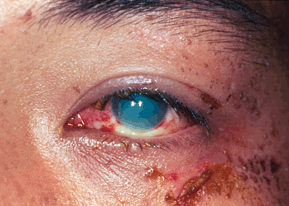
Figure 13-1. Alkaline injury of the face and eye.

Stay updated, free articles. Join our Telegram channel

Full access? Get Clinical Tree


