Ophthalmic Viscosurgical Devices (OVD): Physical Characteristics, Clinical Applications, and Complications
Stephen S. Lane
Dr. Lane has no commercial or proprietary interest in the products discussed and will not receive any remuneration resulting from their use.
The introduction of viscoelastic agents (now termed ophthalmic viscosurgical devices (OVD) by the International Standards Organization [ISO]) for uses in ophthalmic intraocular procedures has had a significant impact on the practice of ophthalmology. OVDs possess a unique set of properties, based on their chemical structure, that enable them to protect the corneal endothelium from mechanical trauma and to maintain an intraocular space, even in the face of an open incision. Viscosurgery,1 a term used to designate the procedures and manipulations performed with OVDs, has been used in a broad spectrum of ophthalmic procedures. The use of OVDs has become commonplace in anterior segment surgery, and it is likely that the widespread use and availability of these materials facilitated and helped ease the transition in the conversion first from intracapsular to planned extracapsular surgery and then to phacoemulsification.
The physical properties of OVDs are the result of chain length, intrachain, and interchain molecular interactions. It is important to realize that the diverse rheologic properties of any given OVD have a direct impact on the clinical characteristics of that particular material. A thorough understanding of these properties will allow ophthalmic surgeons the opportunity to choose an OVD that is task specific. For example, a specific substance may be selected because of its space maintenance qualities, its corneal endothelial protection qualities, or its coating qualities.
RHEOLOGIC AND PHYSICAL PROPERTIES
The rheologic characteristics of OVDs that are most relevant when considering their usefulness in ophthalmic surgery are viscoelasticity, viscosity, pseudoplasticity, and surface tension (Tables 9-1 and 9-2).
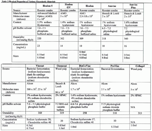
Table 9-1. Physical Properties of Viscoelastic Substances | ||||||||||||||||||||||||||||||||||||||||||||||||||||||||||||||||||||||||||||||||||||||||||||||||||||||||||||||
|---|---|---|---|---|---|---|---|---|---|---|---|---|---|---|---|---|---|---|---|---|---|---|---|---|---|---|---|---|---|---|---|---|---|---|---|---|---|---|---|---|---|---|---|---|---|---|---|---|---|---|---|---|---|---|---|---|---|---|---|---|---|---|---|---|---|---|---|---|---|---|---|---|---|---|---|---|---|---|---|---|---|---|---|---|---|---|---|---|---|---|---|---|---|---|---|---|---|---|---|---|---|---|---|---|---|---|---|---|---|---|
| ||||||||||||||||||||||||||||||||||||||||||||||||||||||||||||||||||||||||||||||||||||||||||||||||||||||||||||||
 |
Viscoelasticity
Elasticity refers to the ability of a solution to return to its original shape after being stressed. The rheologic property of viscoelasticity is the essence of the usefulness of these materials as surgical tools in ophthalmology. Elasticity allows the anterior chamber to reform after deformation by depression on the cornea when external forces are released. A nonelastic solution such as balanced salt solution (BSS) will show no such reformation after release of forces.
The terms viscosity and viscoelasticity are not synonymous. Viscosity, viscoelasticity, and pseudoplasticity are, however, interrelated. The amount of elasticity of an elastic compound increases with increasing molecular weight and greater chain length of the molecules. Unfortunately, comparison of the different OVDs with regard to elasticity is not easily made because of the different ways and nonuniform expression of values by the various manufacturers.
Viscosity
Viscosity (Table 9-1) reflects a solution’s resistance to flow, which is in part a function of the molecular weight of the substance. Viscosity of viscoelastics is measured in centipoise (cP) or centistokes (cSt), which are measures of the resistance to flow relative to a given shear force. Liquid solutions are generally considered to have viscosities of <10,000 cSt at rest, whereas solutions with resting viscosities >100,000 cSt are gel-like. The higher the solution’s molecular weight, the more it resists flow. Molecular weight, on the other hand, reflects the size of the solution’s molecules. Viscosity is dependent on the degree of movement of a solution, which is also known as the shear rate and varies inversely with temperature. The viscosity of a solution can be increased by increasing either the concentration or the molecular weight of the solution.
To facilitate optimal intraocular manipulation, an OVD should maintain space and protect tissues (possess a high viscosity at low shear rates), allow movement of instruments, aid in intraocular lens (IOL) implantation (possess a moderate viscosity at medium shear rates), and allow easy introduction into the eye through a small cannula (possess a low viscosity at high shear rates).2 At the present time, no OVD fulfills all of these requirements.
Pseudoplasticity
Pseudoplasticity refers to a solution’s ability to transform when under pressure, from a gel-like substance to a more liquid substance. The more pseudoplastic a material is, the more rapidly it changes from being highly viscous at rest to a thin, watery solution at high shear rates. A change in molecular structure accompanies this pseudoplastic behavior. In clinical terms, a high-molecular-weight, high-viscosity OVD at rest (zero shear force) acts as an excellent lubricant, and coats tissues and maintains space very well. When under the influence of stress (i.e., a high shear rate), however, the OVD will become an elastic molecular system behaving as an excellent shock-absorbing gel. The highest shear rates occur when a solution is passed through a cannula, and viscosity becomes independent of molecular weight. When the molecules align themselves in the direction of flow, the viscosity is determined solely by the concentration. Pseudoplastic solutions, therefore, have a low viscosity at high shear rates and can be extruded easily through a small-diameter cannular (27 or 30 gauge) (Fig. 9-1). It is important to emphasize that the viscosity of a viscoelastic substance at rest (0 shear rate) is a function of concentration, molecular weight, and the size of the flexible molecular coils of the material (Figs. 9-2A and B). At high shear rates, the viscosity is independent of molecular weight and is determined mainly by the concentration.3
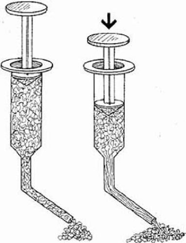 Figure 9-1. When shear is applied (flow through cannula), the large, randomly entangled coils begin to uncoil, allowing for flow. With increasing shear (more pressure on the syringe plunger) unfolding increases, entanglement drops, and the viscous solution flows easily through the cannula. |
Surface Tension
The coating ability of an OVD is determined not only by the surface tension of the material itself but also by the surface tension of the contact tissue, surgical instrument, or IOL. By measuring the angle formed by a drop of the OVD on a flat surface (the contact angle), the coating ability of a substance can be estimated. Lower surface tension and lower contact angle indicate a better ability to coat. In this respect a solution of sodium hyaluronate (HA) has a significantly higher surface tension and contact angle than does a solution of chondroitin sulfate, sodium hyaluronate/chondroitin sulfate in combination, or hydroxypropyl methylcellulose (HPMC), thus indicating these latter solutions provide superior coating.4
A comparison of the various physical properties of OVDs is summarized in Tables 9-1 and 9-2.
COHESION AND DISPERSION
In an attempt to help us better understand the interaction between these various rheologic properties and their clinical usefulness, Arshinoff and Jafari5 have divided OVDs into two categories:
Visco-cohesive OVDs are characterized by high-viscosity materials, which adhere to itself through intramolecular bonds, or intermolecular entanglement and resists breaking apart. In general, OVDs with long molecular chains will be more cohesive because the molecules become entangled. Cohesive OVDs possess a high molecular weight, a high degree of pseudoplasticity and high surface tension.
Visco-dispersive OVDs exhibit opposite characteristics. They possess lower viscosity and adhere well to external surfaces, e.g., tissues and instruments. These materials tend to break apart easily compared to cohesive materials and exhibit lower molecular weight, lower surface tension, and lower pseudoplasticity.
Although cohesiveness and dispersiveness are not measurable rheologic properties in themselves, they are useful constructs when considering the clinical behavior of OVDs as illustrated in the following table.
| OVD Characteristics | |
| Higher-Viscosity Cohesive OVDs | Lower-Viscosity Dispersive OVDs |
| Create and preserve spaces; displace and stabilize tissues | Selectively move and isolate tissues |
| Less protection of the corneal endothelium | Very protective of corneal endothelium |
| Clear | Less clear visualization |
| Easy to remove | More difficult to remove |
OPHTHALMIC VISCOSURGICAL DEVICE CHARACTERISTICS
With the introduction of Healon 5, a new descriptive term was introduced, “viscoadaptive”. This term refers to the ability of an OVD to adapt its behavior to the intended surgical task without the surgeon having to do anything except perform the task at hand. Unlike devices that fit one or the other of the above categories, the viscoadaptive agent ideally functions as both, adapting its behavior to a changing parameter in its environment. That changing parameter under most circumstances is the degree of turbulence present.
From a historical view, 1 million Daltons have been used as a convenient dividing line between a cohesive and a dispersive agent. With the introduction of DisCoVisc at 1.65 million Daltons, this older classification method would give the expectation of a cohesive product with little or no retention or protection.
However, although scientists understand that HA comes in various molecular lengths and weights, this information is not well known and is not intuitive to most clinicians. The combination of a medium-weight and medium-length HA molecule with chondroitin sulfate allows the product to have a triple negative charge, resulting in an agent that has the dispersive (retentive) characteristics of chondroitin sulfate while maintaining space like a traditional cohesive product. As a result, previous classifications of OVDs would not accommodate DisCoVisc because they all assumed a high correlation between viscosity and cohesion. In 1998 Poyer et al4 described how the rate of removal of different OVDs varied when they were exposed to increasing vacuum forces. They found that the rate of removal was typical for a given OVD and referred to this measurement as the cohesion-dispersion index (CDI) (Table 9-3). This index helped to establish an entirely new classification of OVDs as suggested by Arshinoff and Jafari,5 and as a consequence DisCoVis became the first viscous dispersive OVD because of its unique attributes of cohesive and dispersive characteristics. This classification was recognized by the FDA in DisCoVisc’s product insert and helps explain how one product can actually accomplish both dispersive and cohesive actions.
Table 9-3. | ||||||||||||||||||||||
|---|---|---|---|---|---|---|---|---|---|---|---|---|---|---|---|---|---|---|---|---|---|---|
|
DESIRED PROPERTIES OF OVDs
It is apparent from the previous discussion that the interplay between the various rheologic properties is responsible for the clinical characteristics we desire in performing ophthalmic surgery. As a corollary, the degree to which we can maximize these desirable clinical characteristics is, for the most part, based on our ability to optimize the unique rheologic and physical properties each OVD possesses. The desired properties of an ideal OVD are listed in Table 9-4.
Table 9-4. Clinical Uses of OVDs | |||||
|---|---|---|---|---|---|
|
VISCOELASTIC COMPOUNDS, THE BUILDING BLOCKS OF COMMERCIALLY AVAILABLE VISCOELASTIC MATERIALS
Sodium Hyaluronate
Sodium hyaluronate (NaHa) is a biopolymer occurring in many connective tissues throughout the body, including both the aqueous and vitreous humors. Its basic structural unit is a disaccharide, joined by a beta one-4 glucosidic bond, which is linked in a repeating fashion with beta glucosidic bonds to form a long unbranched chain. This mucopolysaccharide chain subsequently forms a random coil when placed in a solution such as physiologic saline. As the concentration of large sodium hyaluronate molecules is increased (>0.5 mg/mL), individual molecular coils start to overlap and are compressed (Fig. 9-2). This crowding of the chains increases the chances for various noncovalent chain–chain interactions. This, in turn, causes a considerable increase in the viscosity of the solution. For example, the kinematic viscosity of a 2-mg/mL concentration of sodium hyaluronate in physiologic buffer is only in the 100-cSt range: At 10 mg/mL, it is in the 100,000-cSt range. Therefore, a fivefold increase in the concentration causes a 1,000-fold increase in the viscosity of the solution. With this increase in viscosity, the elastic properties of the solution also increase. This forms the rationalization of Amvisc Plus and Healon GV. The elastic behavior of a concentrated (> 0.5 mg/mL) sodium hyaluronate solution is greatly dependent on the mechanical energy (shear force) applied to the solution. On a molecular level, this means that under the imposed strain, the polysaccharide coils slip by each other, and conformational and configurational rearrangements occur while the solution exhibits viscous flow (Fig. 9-2). The hyaluronate acid fraction (NIF-NaHa)6 used for ophthalmic procedures has a high molecular weight (2–5 million Da), a low protein content (<0.5%), and carries a single negative charge per disaccharide unit.
Stay updated, free articles. Join our Telegram channel

Full access? Get Clinical Tree


