Overall ophthalmic outcome
1. Improvement in one eye of at least one of the following measures, without worsening in any of these measures in either eye
(a) Eyelid aperture reduction by at least 2 mm
(b) Improvement by at least one grade in the NOSPECS classification for Class 2 signs [26]
(c) Exophthalmos reduction by at least 2 mm
(d) Ocular motility improvement by at least 8° in any duction
2. Worsening, if any of the following occurred
(a) Worsening by at least one grade in any of the NOSPECS classes [26]
(b) Appearance of a new NOSPECS class [26]
(c) Any sign of optic nerve involvement: decrease in visual acuity by at least one line on the Snellen chart, impaired color vision, presence of a relative afferent pupillary defect, or other concern for optic nerve compression
TED-specific Quality of Life (GO-QoL) questionnaire
1. Improvement, when an increase of six or more points on either one (or both) the GO-QoL scales (functioning and appearance) occurreda
2. Worsening, when a decrease of six points on any one of the two scales occurredb
The overall ophthalmologic outcome at the end of the treatment (6 months) was significantly better in the selenium group compared to the placebo group (P = 0.01) (Fig. 6.1). TED improved in 61 % of patients in the selenium group and in 36 % of patients in the placebo group and worsened in 7 % of the former and in 26 % of the latter group. The ophthalmologic outcome was similar when patients were evaluated 6 months after treatment withdrawal, indicating the persistence of the beneficial effect of selenium beyond the active treatment period (Fig. 6.1). Soft tissue involvement and eyelid aperture were the major determinants of the more favorable outcome in the selenium group (Fig. 6.2). Proptosis and ocular motility did not change; however very few patients had motility impairment at start of study (data not shown).
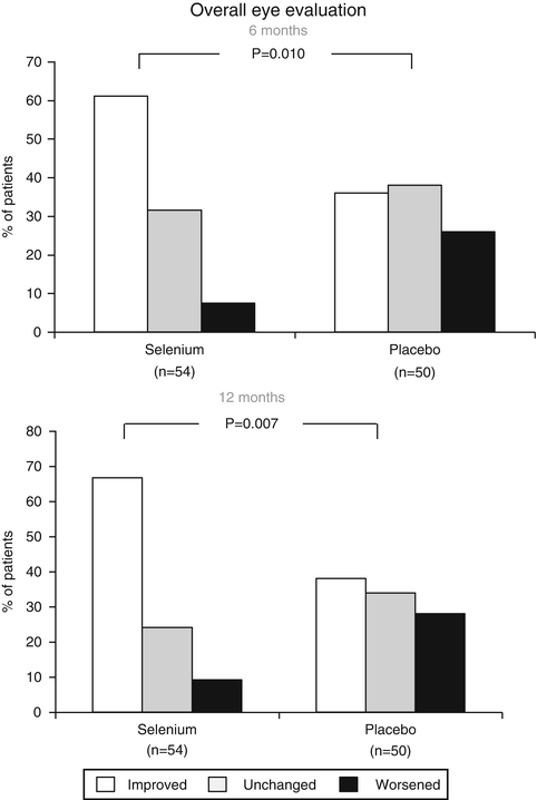
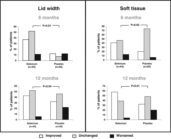
Fig. 6.1
Overall eye evaluation at 6 and 12 months in patients with mild TED treated with selenium (100 μg twice daily) or placebo (twice daily). Ophthalmologic outcomes were scored as improved, unchanged, or worsened according to the criteria reported in Table 6.1 (derived from data of Marcocci et al. [25]). P values were calculated using the Χ 2 test
Fig. 6.2
Changes in lid aperture and soft tissue at 6- and 12-month evaluation, in TED patients treated with selenium and placebo. Changes were scored according to criteria reported in Table 6.1, as improved, unchanged, or worsened. Χ 2 test was used to calculate P values (derived from data of Marcocci et al. [25])
The GO-QoL includes a “visual functioning” score and an “appearance” score. Baseline GO-QoL showed a mild to moderate impairment in quality of life with no differences between the two groups of patients. After 6 months of treatment, a greater proportion of patients receiving selenium showed an improvement of visual functioning (33/53, 62 %) and appearance (40/53, 75 %) scores compared with those treated with placebo. This better outcome in patients treated with selenium was confirmed by the overall GO-QoL evaluation, which includes changes in both subscales (Fig. 6.3).
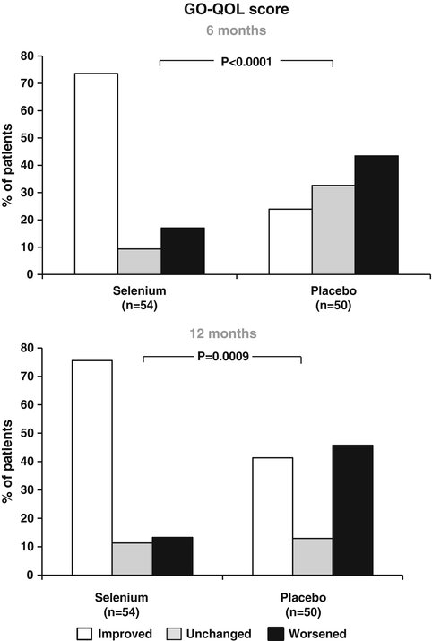
Fig. 6.3
Changes of GO-QoL at 6 and 12 months in patients with mild TED randomly assigned to treatment with selenium or placebo. Changes of QoL were scored as improved, unchanged, or worsened according to predefined criteria (Table 6.1). P values were calculated with the Χ 2 test (derived from data of Marcocci et al. [25])
The CAS improved after 6 and 12 months in all patients (Fig. 6.4). However, the reduction of CAS in selenium supplemented patients was significantly greater compared to the placebo group (p < 0.001). Diplopia score did not change significantly in any group during the study (data not shown).
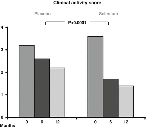
Fig. 6.4
Clinical Activity Score (CAS) evaluation at 6- and 12-month in patients supplemented with selenium or placebo. The Mann–Whitney test was used to calculate the P value (derived from data of Marcocci et al. [25])
No drug-related adverse effects were recorded.
In summary, this study reported a beneficial effect of selenium treatment in patients with mild TED both in terms of quality of life and ocular involvement. It supports the hypothesis that an intervention aimed at restoring the balance of the antioxidant/oxidant status could help patients with mild TED [25].
Two potential limitations of this study were the lack of information regarding the changes of serum selenium concentration after selenium selenite treatment and, secondly, the inclusion of patients coming from areas of marginal selenium deficiency. Therefore, further studies should be performed in selenium-sufficient areas in order to prove a beneficial effect of selenium supplementation in non-selenium-deplete patients.
Despite these limitations, selenium supplementation appears to reduce the severity of TED signs and symptoms in a selenium-deficient population. Whether this effect may also be seen with additional antioxidant supplements remains to be seen.
Some concerns on the safety of selenium supplementation have been raised particularly for patients living in selenium-sufficient areas. The Nutritional Prevention of Cancer trial, performed in the United States, reported a potential risk of developing type 2 diabetes in patients supplemented with 200 μg selenium daily for up to 12 years [8]. Assessment of diabetes was the secondary end point of the study and diabetes occurrence was self-reported. The study showed an increased incidence of diabetes in patients supplemented with selenium compared with those given placebo; the development of diabetes correlated with higher serum selenium concentration [8].
Population studies on serum selenium levels and diabetes have provided conflicting results. A National Health and Nutrition Examination Survey, designed to evaluate the association of serum selenium concentration and type 2 diabetes in US population, found a higher prevalence of diabetes in patients with high serum selenium levels [31]. On the other hand, a study performed in the elderly French population found a sex-specific (male) protective effect of higher selenium levels on the occurrence of dysglycemia [32]. An association of selenium supplementation with glaucoma has also been reported [33].
Stay updated, free articles. Join our Telegram channel

Full access? Get Clinical Tree


