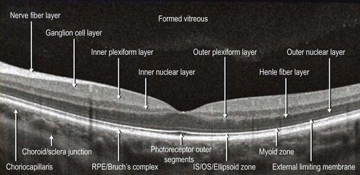
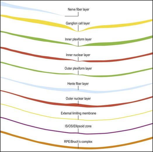
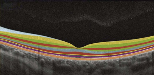
4.1 Commercially available SD-OCT scanners have an axial resolution of between 4 µm and 7 µm and a transverse resolution of approximately 15 µm. This high resolution allows for exquisite viewing of the retinal detail. Due to the limited penetration of light beyond the pigmented RPE and the drop-off of the OCT signal with depth, the image at the level of the choroid has lower resolution. The layers of the normal retina are labeled in Figure 4.1.1. Discrete hyporeflective spaces are noticed primarily in the outer retina, but usually span multiple layers (Fig. 4.1.3). The differential diagnosis includes:
Normal Retinal Anatomy and Basic Pathologic Appearances
Normal Retinal Anatomy
General Appearance of Retinal Pathology on SD-OCT
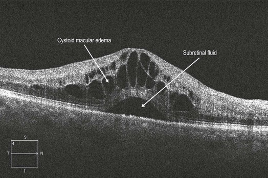
Cystic Changes in Outer Retina
Normal Retinal Anatomy and Basic Pathologic Appearances