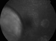
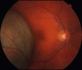
21.2 The most common appearance is a pigmented, elevated choroidal lesion that will enlarge without treatment (Fig. 21.2.1 and Fig. 21.2.2). Without documented growth, features such as overlying lipofuscin (orange pigment), associated subretinal fluid, larger size, and proximity to the optic nerve help to differentiate from benign lesions such as choroidal nevus. On a clinical basis, the diagnosis can be made with greater than 99% accuracy. Biopsy is rarely necessary, but can confirm the diagnosis. Radiation retinopathy can often develop after treatment with external radiation (Fig 21.2.3). Figure 21.2.1 Color fundus photograph of a large, elevated pigmented choroidal melanoma. The lesion is so elevated that the neighboring macula and optic nerve are out of focus.
Choroidal Melanoma
Clinical Features:
Choroidal Melanoma