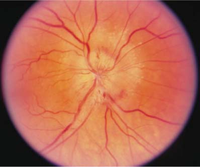
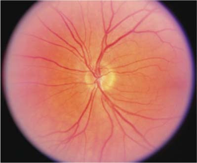
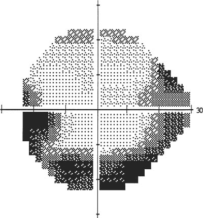
Chapter 19 Nonarteritic anterior ischemic optic neuropathy (NAION) is the most common cause of acute optic neuropathy in persons over 50 years of age. Despite the longstanding recognition of this entity, NAION remains a disease without treatment or prophylaxis. Only recently has new information been gained about the natural history of the disease from the Ischemic Optic Neuropathy Decompression Trial and the Follow-Up Study.1–4 The patient with suspected NAION should be evaluated by an ophthalmologist within 24 hours. NAION strikes both men and women, usually over the age of 50 years. The incidence is estimated as 2.3 to 10.2 per 100,000 persons over the age of 50 years per year in the United States, or if extrapolated, 1500 to 5700 new events per year.5–7 Visual loss is abrupt and occurs without warning symptoms. The loss is characterized as a scotoma in 45% of patients and “blurred vision” in 36%. Forty-five percent of patients subjectively complain of progression between the onset of symptoms and the ophthalmic examination. Twenty-nine percent of patients with visual acuities better than 20/64 will be documented to fall below 20/64 within 30 days.2 Although pain is not a common feature, as it is in optic neuritis, periocular discomfort, dull or aching in nature (not exacerbated with eye movements), has been reported in 10% of patients.2 Visual acuity is usually affected, although nearly 50% of patients have visual acuity better than 20/64 initially. Unfortunately, 29% of these patients progressively lose vision within the first 30 days, and ultimately only 35% have acuities better than 20/64. Thirty-five percent have visual acuity between 20/64 and 20/200 and 35% of patients have acuities worse than 20/200.2 Color vision is typically reduced but may be normal. Subjective comparison may reflect color desaturation. The most common visual field defect is the so-called altitudinal defect, one that respects the horizontal meridian. It is most commonly inferior (Fig. 19–1). Arcuate or other nerve fiber bundle defects, central scotomas, and a variety of other patterns may also be seen. An afferent pupillary defect is present in the affected eye unless the contralateral eye was previously affected. Optic nerve edema is necessary for the diagnosis for NAION. Diffuse disc edema, usually with flame hemorrhages, is seen in the majority of patients (Fig. 19–2), although sectoral edema also occurs. Confirmation of the diagnosis is aided by examination of the unaffected contralateral eye, which should have a small cup-to-disc ratio (“disc at risk”) (Fig. 19–3). FIGURE 19–1 Inferior altitudinal visual field defect in a patient with nonarteritic anterior ischemic optic neuropathy on automated visual field testing. The presence of any other neuroophthalmic findings suggest an alternate diagnosis. Occlusion or hypoperfusion of small branches of the short posterior ciliary arteries is thought to be the precipitating event. Theoretically, vaso-occlusive diseases such as hypertension and diabetes mellitus may narrow blood vessel lumina below a critical threshold in an already compromised vasculature in small cup-to-disc optic nerves. FIGURE 19–2 Diffuse disc edema with flame hemorrhages in a patient with nonarteritic anterior ischemic optic neuropathy. FIGURE 19–3 Small cup-to-disc ratio (“disc at risk”) in the unaffected fellow eye of a patient with nonarteritic anterior ischemic optic neuropathy. Sixty percent of patients with NAION are identified as having one or more risk factors associated with atherosclerotic disease such as hypertension and diabetes mellitus.2
NONARTERITIC ANTERIOR ISCHEMIC OPTIC NEUROPATHY
URGENCY OF EVALUATION
DIAGNOSIS
SYMPTOMS
Demographics
Visual Loss
Pain
SIGNS
Visual Acuity
Color
Visual Field
Pupils
Optic Disc
OTHER ABNORMALITIES
ETIOLOGY
RISK FACTORS
![]()
Stay updated, free articles. Join our Telegram channel

Full access? Get Clinical Tree


