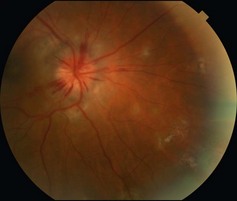
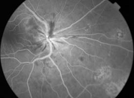
17.1 Presentation of MCP is variable. Most patients complain of decreased visual acuity, but photopsia, floaters, blurring of central vision, and enlargement of the blind spot can occur. Anterior segment inflammation may be present but is typically mild. Vitritis of variable severity, and optic disc edema may be present. Multiple small, round to ovoid, pale lesions occur at the level of the outer retina, retinal pigment epithelium (RPE) and choroid, usually 50–350 µm in size, variable in number, and involve mainly the posterior pole (Fig. 17.1.1). Older lesions become pigmented and ‘punched-out’, resembling histoplasmosis lesions. RPE metaplasia and choroidal neovascularization (peripapillary and macular) are common sequelae. CME, epiretinal membrane and subretinal fibrosis may be seen later.
Multifocal Choroiditis
Clinical Features:
Multifocal Choroiditis