Chapter 61 Minimally Invasive Video-Assisted Parathyroidectomy
 This chapter contains additional online-only content, available on expertconsult.com.
Introduction
Bilateral neck exploration (BNE) with the identification of at least four parathyroid glands and the removal of pathologic parathyroid tissue has represented for several decades the standard of treatment of primary hyperparathyroidism (primary HPT) (see Chapter 59, Standard Bilateral Parathyroid Exploration).1,2 In experienced hands, this approach has a cure rate of more than 95% with minimal morbidity, usually less than 3%.1
In spite of the excellent results obtained with BNE, since the early 1980s less invasive procedures (unilateral neck exploration [UNE]) have been introduced, with the aim to reduce the surgical trauma and the already low complication rate of parathyroidectomy.3,4
The application of minimally invasive parathyroidectomy was initially limited. Only since the early 1990s have these procedures been widely developed because of the evolution of the techniques of preoperative localization (ultrasound, sestamibi scan)5 and the introduction of quick intraoperative PTH (IOPTH) assay.6 Preoperative localization studies allow for a more targeted approach, and the IOPTH assay is able to intraoperatively confirm the success of surgery.7,8
Preoperative Localization Studies
In the case of concordant ultrasonography and scintigraphy, the overall accuracy in parathyroid localization is higher than 95%, whereas in the case of negative localization studies the likelihood of multiglandular disease (MGD) is higher than 30%.16,17
Obviously, the availability of accurate preoperative localization studies allows the planning of minimally invasive surgical procedures targeting the identified affected gland(s). Imaging studies are concordant in up to 65% of patients with primary HPT.7,11 It has been well demonstrated that if more than 51% of the patients are eligible for a unilateral exploration or a focused approach, the utilization of preoperative localization studies is cost effective.10 A “targeted” approach can also be proposed in cases of positivity of only one localization study.17 In such a circumstance, the risk of a multiglandular disease is about 17%.17
 Please see the Expert Consult website for more discussion of this topic.
Before the advent of the new localization imaging techniques, the sensitivity of preoperative localization studies was 60% to 70%.7 For this reason and because of the high success rate of BNE in experienced hands, the guidelines of the National Institutes of Health (NIH) established that preoperative localization studies for patients with hyperparathyroidism were not necessary before initial surgery and should be performed only in the reoperative cases.9 The introduction of sestamibi scintigraphy has substantially modified this paradigm. Actually, sestamibi scan is able to localize a parathyroid adenoma in 80% to 95% of cases, with a specificity of about 90% in studies at high-volume centers,5,7,8,10,11 although it may have a sensitivity lower than 50% when interpreted by nondedicated operators.7 Sestamibi scan is less accurate in cases of multiglandular disease (double adenoma, hyperplasia), which is usually the cause of false-negative results7,8,11 False-positive results are usually related to the presence of nodular thyroid disease.11Single-photon emission computed tomography (SPECT) sestamibi allows a tridimensional reconstruction that can be useful for a spatial localization of the affected gland(s), giving additional information to the surgeon7,8 (see Chapter 57, Guide to Preoperative Parathyroid Localization Testing).
Ultrasound with a high-resolution probe is an accurate, inexpensive, noninvasive, and reproducible procedure, which allows for the precise localization of a parathyroid adenoma in about 80% of the cases, giving additional information about position, size, relationship with adjacent structures, and coexisting thyroid diseases5 (see Chapters 13, Ultrasound of the Thyroid and Parathyroid Glands, and 57, Guide to Preoperative Parathyroid Localization Testing). Even better results are obtained if the ultrasound examination is performed by experienced endocrine surgeons, even as a single localization procedure.12–14 It has been demonstrated that surgeon-performed ultrasound had a sensitivity of 82% with a specificity of 90%, compared with radiologist-performed ultrasound, which had a sensitivity of 42% and a specificity of 92%.15
Intraoperative PTH Assay
Similar to the progress in the field of preoperative imaging techniques that allowed targeted approaches, the development and the availability of the IOPTH assay gave surgeons the opportunity to intraoperatively verify the completeness of the surgical resection as an alternative to the complete visualization of all four glands6,8,18–21 (see Chapter 63, Intraoperative PTH Monitoring during Parathyroid Surgery). The half-life of intact PTH is 3 to 5 minutes. As a consequence, after the resection of a single adenoma, the removal of all hyperfunctioning parathyroid tissue should be confirmed by a significant reduction of PTH levels. Because rapid techniques have been developed, the IOPTH assay appeared an attractive method to intraoperatively verify the success of the surgical resection: a “biochemical” frozen section. The turnaround time has now become very short (less than 10 minutes) and can be performed by laboratory personnel in the operating room using portable machines. Several studies demonstrated a positive correlation between an adequate IOPTH decrease and postoperative eucalcemia.7
As a consequence, IOPTH assay emerged as an important adjunct to localizing studies when a focused approach has been planned. Indeed, by delivering immediate feedback during surgery, IOPTH monitoring allows the surgeon to decide whether unilateral exploration has been successful or if four-gland exploration is necessary.6–8,18–20 It has been demonstrated that IOPTH increases the cure rate of minimally invasive procedures from 95% to 98%, although it has also been associated with an increase of about 13% of unnecessary BNE.16,22
Most authors agree that IOPTH monitoring is an important, even essential, complementary tool in cases of minimally invasive procedures.18–20,23,24 However, some authors have questioned its usefulness in intraoperative decision making, especially in cases with concordant localization studies, suggesting one gland disease.25–27 The positional statement of the European Society of Endocrine Surgeons suggests it may be best reserved for patients undergoing targeted parathyroidectomy on the basis of a single preoperative localization study (i.e., sestamibi scan28 or ultrasonography29) or in cases of discordant preoperative localization studies, when the risk of multiglandular disease is higher.17,30
In a retrospective nonrandomized comparative study, Barczynski et al.31 found that the routine use of IOPTH significantly improves cure rates of minimally invasive, either open or video-assisted parathyroidectomy, in comparison to open image-guided unilateral neck exploration without IOPTH. This study also suggested that IOPTH offers value by adding to surgical decision making regarding further neck exploration, especially in cases of only one positive imaging study.31 Moreover, one paper demonstrated that, at least in an endemic goiter region, IOPTH monitoring seems necessary even in patients with “localized” single-gland disease, as abandoning it would significantly increase persistent disease (from 0.9% to 5%).32
 Please see the Expert Consult website for more discussion of this topic.
There are no standardized guidelines about the site, the number, and the timing of blood samples (see Chapter 63, Intraoperative PTH Monitoring during Parathyroid Surgery).30 Most authors agree that is preferable to obtain samples from a peripheral vein because samples obtained from internal jugular veins seem less accurate as they are more prone to be affected by variations in PTH levels related to manipulation.33 Most of the authors agree that obtaining samples at baseline (when anesthesia is induced—preincision sample), at the identification of the adenoma (preexcision sample), and 5 and 10 minutes after the removal of the adenoma (postexcision samples).20 Some authors prefer to reduce the number of samples in order to reduce cost, obtaining samples preincision and 15 to 20 minutes postexcision.27 A later sample (20 to 30 minutes postexcision) may be necessary in cases of inadequate or slow PTH decrease.6,20,34 Moreover, we35,36 and others33,34 found that in order to obtain a higher specificity, it is necessary to extend the sampling period in all the cases.
Obtaining two “basal” samples (preincision and preexcision samples) is considered necessary because gland manipulation is usually responsible for an increase in PTH levels. In absence of the preexcision sample, postexcision results might be difficult to interpret. To avoid the need for a preexcision sample, some authors propose avoiding any pressure on parathyroid glands before ligating or clipping the vascular pedicle of the adenoma.37
The most intriguing and debating point concerns the interpretation criteria. Indeed, no complete and univocal “curative criteria” of IOPTH decline has yet been established38 (see Chapter 63, Intraoperative PTH Monitoring during Parathyroid Surgery). Most groups use a 50% reduction from the highest basal level (either the preincision or preexcision sample) 5 to 10 minutes following parathyroidectomy as the criteria for successful surgery.6,7,18–20,23,38,39 Others recommend a greater than 50% reduction from the preincision baseline value alone or even an absolute normalization regardless of the preexcision levels.21,32,34,40,41 Still others prefer kinetic analysis of PTH, as the PTH half-life can vary from patient to patient.42,43 Nonetheless, Carneiro et al.39 reported that the Miami criterion (an IOPTH drop > 50% from the highest basal level at 10 minutes after gland excision) has the highest accuracy (97%) in predicting postoperative calcium levels.
It seems that IOPTH works least well when it is needed the most. Indeed, the role of IOPTH monitoring in identifying patients with multiglandular disease (MGD) is still not clear. Some surgeons report a high incidence of false positives with intraoperative PTH measurements. It has been reported that with an appropriate fall in PTH levels, there is still a 14% to 30% incidence of additional enlarged glands on bilateral neck exploration44,45 In one retrospective study, IOPTH failed to predict the presence of additional abnormal glands in 75% of patients with MGD (6 of 8 patients) when the “50% rule” was applied.46 Sokoll et al.47 also reported false-positive results of IOPTH in patients with multiglandular disease (primary, secondary, and tertiary HPT) with a > 50% fall, despite the presence of other enlarged glands. Miller et al.48 reported that adherence with the 50% rule may lead to several missed cases of MGD, a determination that depends in part on the initial baseline PTH level.48 One particular source of false positivity seems to be double adenomas. In a retrospective study on 287 patients, PTH levels decreased more than 50% in 12 out of 15 patients with double adenomas, with a false-positive rate of 57%.45 In combination with sestamibi scan and ultrasonography, IOPTH monitoring still only correctly predicted the presence of 80% of the double adenomas.45 In another retrospective study of 20 patients with double adenomas, IOPTH was found to have a false-positive rate of 55% when the 50% rule was applied to predict cure.40
Because obtaining a definitive and durable cure is the primary goal for the treatment of patients with primary HPT, stricter cure criteria have been variably evaluated and proposed. In this context the authors of this chapter have evaluated sampling and interpretation criteria that could minimize the rate of false-positive results.35 We found that to optimize the accuracy of the IOPTH, postexcision samples should be collected 10 and 20 minutes after gland removal. We found that the best three predictors of operative failure and MGD are (1) PTH drop < 50% at 20 minutes, (2) a residual (at 20 minutes) PTH level above the normal range, or (3) a significant increase of the PTH levels between the sample obtained 20 minutes after gland excision with respect to the 10 minute samples.35 We evaluated the effectiveness of the new interpretation criteria compared with the Miami criterion in a prospective large series analysis.36 We found that the application of the new interpretation criteria results in a lower false-positive rate, markedly reducing the risk of missing a MGD. Obviously, this result was obtained at the cost of a higher rate of false-negative results and consequent unnecessary bilateral exploration (15.9% versus 4.8%). A retrospective study comparing different interpretation criteria confirmed that our criteria best predicts MGD even if at the cost of a significantly higher rate of false-negative results.34
It is interesting to note that the reported proportion of patients with MGD is considerably lower in recent series using focused approaches combined with IOPTH measurement compared with series where bilateral exploration was performed using a gross morphologic definition of a hyperfunctioning gland.18–20 Irvin et al.20 also found that using intraoperative PTH measurements as a functional study of disease led to a much lower incidence of MGD, but with an equal cure and recurrence rate. These data suggest that size may not be the most reliable predictor of diseased glands and that some enlarged glands may not be hyperfunctioning. To support this view, a study by Yao et al.49 demonstrated that the weight of normal parathyroid glands removed at parathyroidectomy may be greater than previously reported in autopsy studies.
Besides interpretation criteria, there are other controversial aspects of the IOPTH assay. In particular, some authors have questioned the cost effectiveness of IOPTH.50 In a cost analysis study based on a meta-analysis of the literature, IOPTH was responsible for a 4% increase in costs, in spite of a marginal increase in cure rate.22 However, institution-specific factors seemed to influence the value of IOPTH.22
Anesthetic propofol agent was thought to possibly interfere with the PTH assay, but a randomized trial has shown that the PTH assay can be employed during propofol sedation.51
In spite of these controversial aspects, IOPTH has emerged has a very useful intraoperative adjunct for parathyroidectomy, especially in cases of targeted parathyroidectomy relying on one single preoperative localization study or discordant localization studies and in the reoperative parathyroidectomy.30,52
IOPTH has been also proposed as an adjunct to intraoperative lateralization of hyperfunctioning parathyroid tissue by samples obtained from internal jugular vein on both sides.18–20 It offers also the possibility to measure PTH concentration in fine-needle aspiration materials obtained from lesions suspected of being pathologically enlarged parathyroids. A high PTH concentration in the washing fluid of the aspirate has a 100% specificity for a parathyroid lesion and may replace frozen section examination.21
Minimally Invasive Parathyroidectomy
The application of endoscopic techniques to neck surgery during the late 1990s resulted in the development of minimally invasive techniques for parathyroidectomy.53 The general trend toward less invasive procedures for parathyroidectomy is well demonstrated by the results of an international survey driven in 2000 among the members of the International Association of Endocrine Surgeons (IAES). Fifty-nine percent of the participants used a minimally invasive approach.54 We feel this percentage may have further increased since that time.
Even if a minority of the authors still consider a standard BNE performed by an experienced endocrine surgeon to be the best treatment for patients with primary HPT,55 others maintain that BNE should be relegated to the past. Clearly, minimally invasive procedures for parathyroidectomy are assuming a more and more important role and may in time represent the new gold standard for the treatment of primary hyperparathyroidism, at least in its sporadic form.
The consensus statement of the European Society of Endocrine Surgeons (ESES) assumed that even if BNE has excellent results and is always an option for the surgical treatment of primary HPT, minimally invasive parathyroidectomy is a safe and cost-effective procedure to treat selected patients with sporadic primary HPT, especially in cases of positive preoperative localization tests.30 Similarly, the proceedings of the third international workshop on primary hyperparathyroidism (Orlando, Florida, 2008) reported that “unlike previous dogma that mandated surgical identification of both pathologically enlarged and normal parathyroid glands, the current paradigm in many centers is to identify and excise the incident enlarged gland and to confirm operative cure employing a rapid intraoperative PTH assay.”52 Despite these statements, an audit from the Scandinavian quality register for parathyroid surgery showed that BNE is still performed in two thirds of parathyroid procedures.57 Indeed, it is true that not all patients with hyperparathyroidism can be treated by a selective minimally invasive approach. Thus, we feel BNE does still maintain a relevant role in the treatment of patients with primary HPT.
Minimally invasive (or focused, targeted, or selective) parathyroidectomy encompasses a number of different techniques, including open approaches (open minimally invasive parathyroidectomy [OMIP]58,59), minimally invasive radio-guided parathyroidectomy (MI-RP),60,61 video-assisted parathyroidectomy (VAP),62–64 and purely endoscopic parathyroidectomy (EP).53,65–69 As a consequence, there is no strict or unequivocal definition of what a minimally invasive parathyroidectomy (MIP) is. The term minimally invasive should be reserved for procedures that allow performance of a traditional operation through access that minimizes trauma of the surgical exposure and dissection. BNE is associated with a very low morbidity (<3%) and high success (>95%) rates by experienced surgeons. A minimally invasive procedure should obtain at least the same results, with the main advantage of reducing the skin incision and, consequently, allowing better cosmetic results.71 MIP was thus used to indicate parathyroid procedures performed through a mini-incision, usually less than 2.5 to 3 cm.71 In other words, they would be mini-incision and associated with minimal dissection targeted parathyroid procedures.
Many studies comparing MIP/focused parathyroidectomy with standard BNE suggest that the focused techniques are safe and at least as good as BNE, with some advantages, especially in terms of less postoperative hypocalcemia, shorter operative time, earlier discharge, better cosmetic result, and reduced postoperative pain. These results were strongly confirmed by five randomized trials with short-term results71–75 and one with long-term results76 demonstrating some distinct benefits for MIP over standard BNE (see the Evidence-Based Recommendations section).
Techniques for MIP
Several variants of minimally invasive procedures have been described since the late 1990s, including minimally invasive procedures without (OMIP, MI-RP)8,60,61,77–81 and with the utilization of the endoscope (EP and VAP).53,62–69
Minimally Invasive Radio-Guided Parathyroidectomy
In MI-RP, a handheld gamma probe is used to facilitate intraoperative localization, identification, and dissection of the pathologic gland(s) and to confirm the removal of all hyperfunctioning parathyroid tissue60,61,81 (see Chapter 64, Radio-Guided Parathyroid Exploration). This approach implies that technetium 99 m-sestamibi is injected intravenously 2 to 4 hours prior to surgery. Obviously, a prerequisite for this approach is the precise coordination between the operating room, the nuclear medicine department, the surgeon, and the nuclear medicine radiologist.27 The anterior portion of the patient neck is scanned on the operating table, and the site with highest count is explored using the handheld probe as a guide toward the pathologic gland(s). An excised parathyroid adenoma should contain more than 20% of the postexcision background radioactivity.82 This approach may result in reduced operative time83 and may eliminate the need for IOPTH.61 Although this technique has been refined and validated, it has been adopted only by a minority of endocrine surgeons worldwide, mainly because of the logistic requirements. Moreover, it is widely believed that MI-RP adds little information to preoperative sestamibi scan and IOPTH measurement,27,52,77 although there is no prospective study on this topic. Some data suggest the gamma probe may be potentially misleading in certain circumstances.79,84 At present, MI-RP is considered an alternative minimally invasive technique, with potential advantages in reoperative cases.30
Open Minimally Invasive Parathyroidectomy
OMIP represents the most widespread minimally invasive technique.52,54,85 In the IAES survey, 92% of the surgeons dealing with MIP adopted an OMIP, whereas only a minority of them (35%) relied on endoscopic (13%) or video-assisted procedures (22%).54 A focused parathyroidectomy, performed through a small (2.5 to 5 cm) central52 or a lateral (over the site of the adenoma and overlying the anterior border of the sternocleidomastoid muscle)59 incision, guided by preoperative localization studies and IOPTH, seems therefore to be the most attractive and widespread technique for the surgical treatment of primary HPT.* Indeed, experienced endocrine surgeons seem to find the procedure easy to learn and reproduce in different surgical settings, and it can be performed under locoregional anesthesia, with reduced operative time and as a short stay procedure.7,24 On the other hand, the main limitation of OMIP resides in the potential poor visualization of the neck structures, because of the small size of the skin incision or, conversely, the need for a larger skin incision when compared with video-assisted or endoscopic techniques. Because coexistent thyroid nodular disease is relatively common, associated thyroid resection can be performed as well, especially by this approach, typically after conversion from regional block to general anesthesia.24
Video-Assisted and Endoscopic Techniques
Techniques that imply the utilization of the endoscope can be classified in endoscopic65–69 and video-assisted procedures (see Chapter 36, Robotic and Extracervical Approaches to the Thyroid and Parathyroid Glands: A Modern Classification Scheme).62–64,86,88
Endoscopic Parathyroidectomy
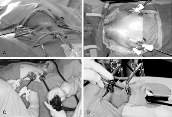
Gagner first described the totally EP in 1996,53,89 and other authors subsequently utilized and modified it.65,66 It is carried out entirely under steady gas flow, using a 5-mm endoscope introduced through a central trocar, and two or three additional trocars for the instruments (Figure 61-1). The dissection is first performed beneath the platysma to obtain a good working space. The midline is then opened and the strap muscles retracted to expose the thyroid lobe and explore the parathyroid glands after dissecting the thyroid from the fascia.
< div class='tao-gold-member'>
Stay updated, free articles. Join our Telegram channel

Full access? Get Clinical Tree


