Purpose
To compare meibomian gland morphology between children and adults.
Design
Cross-sectional, observational case series.
Methods
In an institutional setting, healthy pediatric (n = 78, 30 boys, 48 girls; mean age ± standard deviation, 4.1 ± 3.4 years; range 1 month – 12 years) and adult (n = 25, 11 men, 14 women; 31.3 ± 4.8 years; range 24-39 years) volunteers participated in the study. A noninvasive mobile pen-shaped infrared meibography device was used to observe the meibomian glands. Lost meibomian gland area (meiboscore) was scored from grade 0 (no meibomian gland loss) through grade 3 (>two-thirds of the total area lost). The number of meibomian glands in each eyelid was counted by reviewing the video. Eyelid width was measured using a ruler. Meibomian gland density was calculated as the number of meibomian glands divided by the eyelid width.
Results
The numbers of meibomian glands in the upper/lower eyelids were 26.9 ± 3.9 / 22.0 ± 2.2 in the pediatric group and 28.1 ± 2.7 / 24.4 ± 2.6 in the adult group ( P = .22, P < .0001). Eyelid width was 24.4 ± 2.4 mm in the pediatric group and 27.9 ± 1.57 mm in the adult group ( P < .0001). Meibomian gland densities in the upper/lower eyelids were 1.09 ± 0.17 / 0.91 ± 0.11 in the pediatric group and 1.01 ± 0.12 / 0.88 ± 0.10 in the adult group ( P = .03, P = .45).
Conclusions
The mobile pen-shaped infrared meibography device is useful for obtaining information on the meibomian gland structure, not only in adults but also in children, including infants. The morphology of the meibomian glands in children was the same as that in adults, distributing across the whole tarsal plates in both the upper and lower eyelids.
Meibomian glands are sebaceous glands embedded in the tarsal plates of the eyelids that supply the main components of the outer layer of the tear film. They are the largest sebaceous glands in the human body and are essential for maintaining the structural and refractive integrity of the ocular surface. Their growth after birth, however, has been unclear.
Infants occasionally stare without blinking for almost a minute, which requires great tear film stability. Isenberg and associates investigated the lipid layer and stability of the tear film in infants using interference fringe biomicroscopy and found that in the first 6 postnatal months, the lipid layer of the tear film is much thicker than that in adults, contributing to the greater tear film stability. Some clinical and in vitro studies revealed that infants have a longer tear breakup time and greater tear film stability than adults. Although there are a couple of reports on in vivo observations of normal pediatric meibomian glands using the noncontact meibography system developed by our group, there are no reports on the meibomian gland morphology of subjects under 2 years old. The reasons for that may be that these subjects are too young to sit still for slit-lamp microscopy and there have been no devices available for observing infant meibomian glands.
The development of a noninvasive mobile pen-shaped infrared meibography device enabled us to noninvasively observe infant meibomian glands. Moreover, this system can capture the images of the whole eyelid at the same time. Therefore, we can easily count the number of meibomian glands. The purpose of the present study was to examine meibomian gland morphology in children and to compare the morphology with that in adults.
Methods
In this cross-sectional study, all participants were healthy Japanese volunteers. The pediatric group comprised 78 eyes of 78 children (30 boys and 48 girls; 4.1 ± 3.4 years; range 1 month – 12 years) and the adult group comprised 25 eyes of 25 people (11 men and14 women; 31.3 ± 4.8 years; range 24-39 years). The numbers of pediatric subjects in each age group are as follows: 1-11 months, 15 (6 boys and 9 girls); 1-3 years, 22 (6 boys and 16 girls); 4-6 years, 22 (11 boys and 11 girls); 7-9 years, 12 (5 boys and 7 girls); and 10-12 years, 7 (2 boys and 5 girls). Because age-related morphologic changes in the meibomian glands tend to occur after the age of 40, we considered adults under 40 years old to be appropriate controls to compare with children. Before the examination, written informed consent was obtained from all participants or from the parents of children under 20 years of age. Subjects with any eye symptoms, allergies, or history of contact lens use or eye surgery were excluded from the study. Approval for all procedures mentioned below was obtained prospectively from the institutional review board of the University of Tokyo School of Medicine (No. 3760). This study adhered to the tenets of the Declaration of Helsinki.
The noninvasive mobile pen-shaped infrared meibography device (MeibomPen; Japan Focus Inc, Tokyo, Japan) contains an infrared light–emitting diode as the light source and a highly sensitive complementary metal oxide semiconductor image sensor camera. This device has been validated to provide meibomian gland images of the same quality and quantity as the noncontact meibography system equipped with a slit lamp.
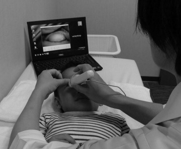
Examinations were performed sequentially as follows: We observed and recorded the meibomian glands using the MeibomPen ( Figure 1 ). The upper and lower eyelids were turned over and the meibomian glands were observed with the subject in a seated or supine position in a semi-dark room. The width of the eyelid (ie, the horizontal distance from the medial canthus to the lateral canthus) was measured using a ruler. The number of meibomian glands was determined by counting the main ducts connecting to the lid margin on the videos obtained with the MeibomPen. Partial or complete loss of meibomian glands was scored according to the following grades (meiboscore) for each eyelid, as previously reported: grade 0, no meibomian gland loss; grade 1, area loss less than one-third the total area of the meibomian glands; grade 2, area loss between one-third and two-thirds of the total area; grade 3, area loss greater than two-thirds of the total. Meiboscores for the upper and lower eyelids were summed to obtain a total meiboscore from 0 through 6 for each eye. Meibomian gland density was calculated as the number of meibomian glands divided by the eyelid width. Data from the right eyes were used unless these data were not available. All examinations were performed by a skilled examiner (R.S.).
Correlations among age and various factors were evaluated using Spearman correlation analysis. Sex differences in each age group and differences in each measurement between the adult group and pediatric group were evaluated using the Mann-Whitney U test. A P value of less than .05 was considered significant. Data are shown as mean ± standard deviation unless otherwise specified.
Results
Images of the meibomian glands could be obtained for all lower eyelids. Images of the meibomian glands in the upper eyelids could not be obtained in 13 infants, aged 3 months to 3 years, because of subject refusal or inability to turn over the upper eyelid. Representative images of infant meibography are shown in Figure 2 . Less than 1 minute was required to capture the video of both the upper and lower eyelids. No complications were reported after the examination.
Eyelid width, total meiboscore, and number and density of the meibomian glands in the pediatric and adult groups are shown in the Table . The eyelid width was significantly larger in the adults than in the children. The total meiboscore in adults was significantly larger than that in children. Meibomian gland number in the upper eyelid was not significantly different between the adult and pediatric groups, whereas that in the lower eyelid was significantly larger in the adults than in the children. Meibomian gland density in the upper eyelid was significantly greater in children than in adults, whereas that in the lower eyelid was not significantly different between the adult and pediatric groups.
| Group | Width of Eyelid (mm) | Total Meiboscore | No. of Upper MG | No. of Lower MG | Density of Upper MG (No. of MG/mm) | Density of Lower MG (No. of MG/ mm) |
|---|---|---|---|---|---|---|
| Pediatric | 24.4 ± 2.4 | 0.10 ± 0.31 | 26.9 ± 3.9 | 22.0 ± 2.2 | 1.09 ± 0.17 | 0.91 ± 0.11 |
| Young adult | 27.9 ± 1.6 | 0.48 ± 0.59 | 28.1 ± 2.7 | 24.4 ± 2.6 | 1.01 ± 0.12 | 0.88 ± 0.10 |
| P value | <.0001 | .0001 | .22 | <.0001 | .03 | .45 |
Correlations between age and other parameters (eyelid width, meibomian gland number, and meibomian gland density) in the pediatric group are shown in Figure 3 . Eyelid width increased with age from 0 through 12 years ( Figure 3 , Top left). The number of meibomian glands in the upper/lower eyelids was weakly positively correlated with age ( Figure 3 , Top center and Top right). Meibomian gland density in the upper eyelid was not significantly correlated with age, whereas that in the lower eyelid was negatively correlated with age ( Figure 3 , Bottom left and right).


