FIGURE 10.1 A. The pectoralis major muscle is supplied by the pectoral branch of the thoracoacromial artery. The vascular anatomy of the pectoralis major muscle is demonstrated here. B. Demonstrates the design of the skin paddle for the pectoralis flap (blue) and the preservation of the DP flap (black dotted line).
Usually, the reconstruction of a defect in the lateral pharyngeal wall is performed immediately following the resection. Secondary reconstructions may be necessary to repair a fistula, radiation necrosis, or stricture or due to the medical stability of each patient. Prereconstruction clearance of surgical margins, meticulous hemostasis, and identification/preservation of important recipient bed neurovascular structures are important before embarking on the reconstruction.
The chest is usually widely prepped and draped concurrent with the head and neck region. The use of barrier drapes can help separate any gastrostomy tube from the donor site. Until the harvest occurs, the area is usually covered with sterile towels and drapes to prevent contamination from the aerodigestive dissection superiorly as much as possible. Typically, during harvest, a separate OR table and instruments are used after the surgeon regowns to minimize contamination into the donor site or any tumor seeding potential. If the patient can be safely pharmacologically paralyzed during this aspect of the procedure, it is desirable to minimize muscular contraction during harvest.
For this example, a pectoralis flap is the best tissue to use to reconstruct an inferior lateral pharyngeal defect resulting from local recurrence and fistula formation after total laryngectomy (Figs. 10.2 and 10.3).
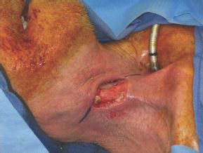
FIGURE 10.2 This photograph demonstrates a pharyngocutaneous fistula. Reconstruction of this defect is best accomplished with a pedicle flap of vascularized muscle and skin.
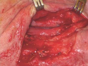
FIGURE 10.3 To reconstruct the defect, the edges of the fistula are prepared for reconstruction. An elliptical incision is made around the fistula to prepare for reconstruction.
The representative case in the accompanying figures is a 68-year-old male with fistula formation of the right lateral pharynx after total laryngectomy with neck dissection and postoperative radiation therapy. After neck dissection and excision of nonviable tissue, the right lateral pharyngeal defect is measured.
Accurate assessment of the defect is the first important step in the reconstruction. Once the dimensions of the defect and its location and character are known, the size of the PMMF skin paddle needed can be determined. Proper placement of the skin paddle incisions is crucial. Finding the most inferior skin paddle margin is done first, in measuring the arc of rotation needed to reach the most superior edge of the defect without tension. This is accomplished by using either an unrolled 4 × 4 gauze or the umbilical tape that would have been used with the tracheotomy. The pivot point of the arc of rotation is located at the emergence of the vascular pedicle to the PMMF, at the middle third of the inferior border of the clavicle. This measurement is important to assure that the edge of the skin paddle will reach the superior aspect of the defect without tension, when the flap is rotated into position. It also alerts the surgeon as to the amount of skin paddle that may be needed beyond the pectoralis major muscle and thus may require inclusion of the rectus sheath for vascular perfusion.
The skin lying most medial and inferior on the pectoralis major has the fullest arc of rotation but often needs to be extended more inferiorly below the border of the pectoralis major muscle. The blood supply to this portion of most inferior skin is usually random but may have some supply from the lateral thoracic artery previously mentioned. It is important, however, to include the rectus fascia to capture as much random blood supply as possible. Knowing then the placement of the inferior edge of the skin paddle and the dimensions of the defect to be closed, a skin paddle is designed for the pedicled PMMF (Fig. 10.4).
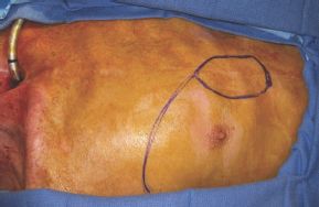
FIGURE 10.4 The appropriate skin paddle is designed on the chest wall to prepare for harvest of the flap.
Similar evaluation of the superior aspect of the skin paddle is tested using the gauze measurement technique to make sure that the superior aspect of the flap approaches the location of the inferior aspect of the defect. The center of the paddle is usually placed midline between the nipple and sternum but can extend to include both structures laterally and medially as needed. Too small a cutaneous paddle should not be drawn, as this will limit the number of cutaneous perforators to the paddle and compromise the entire cutaneous portion of the PMMF. Most skin paddles are drawn in a boat shape with the “pointy” bow of the boat being the superior aspect of the skin paddle and the “boxy” stern of the boat being the inferior aspect of the skin paddle. This paddle shape will help facilitate the closure of the chest wall defect. Elliptical-shaped paddles are also useful to help have an easier and more cosmetic donor site closure. As a general rule, the paddle should remain above the costal margin to ensure the best vascular supply to the distal portion of the PMMF.
The skin paddle is then drawn to size based on the accurate measurements of the defect. An oblique incision line from the superior lateral corner of the skin paddle to the anterior axilla is drawn. This incision line is important for two reasons. It helps to locate the lateral border of the pectoralis major muscle during flap harvest, thereby aiding in identifying the neurovascular boundaries rather than cutting across them with the skin incision. This incision is also placed, to avoid crossing into the region of the DP flap blood supply if this should be needed in a future surgery (Fig. 10.5).
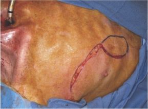
FIGURE 10.5 The incisions to harvest the pectoralis muscle are performed in such a way that the skin of the DP flap is left uninterrupted in the event that it is required for later reconstruction.
The first incision made is from the anterior axilla to the superior apex of the skin paddle. This is then continued down the lateral portion of the skin paddle. These lateral incisions are made first to enable visualization of the lateral border of the pectoralis muscle, especially its most inferior extent. The inferior extent determines the extent of the cutaneous paddle that has the best axial blood supply. The remaining portion of the skin paddle beyond the inferior aspect of the pectoralis muscle is generally random in nature but usually reliable within limits. Determination of the inferior border of the pectoralis muscle may also allow further inferior placement of the skin paddle (redraw the paddle) as no other paddle incision have been made. This may result in less tension once the PMMF is rotated into a higher defect. It may also require more superior paddle harvest if the muscle is very short to ensure adequate blood supply to the inferior skin paddle.
Care is taken not to incise the pectoralis major fascia, while identifying the lateral border of the muscle. This also helps to avoid separating the fibers of the pectoralis major muscle and inadvertently injuring the lateral thoracic blood supply to the muscle. This also helps to distinguish the pectoralis muscles from the external oblique and serratus anterior muscles. The lateral incisions are subsequently undermined laterally enough in a plane superficial to the plane of the pectoralis fascia.
Stay updated, free articles. Join our Telegram channel

Full access? Get Clinical Tree


