Purpose
To describe the outcomes and medical management necessary to achieve successful lid surgery in patients with biopsy-confirmed and presumed mucous membrane pemphigoid.
Design
Retrospective, interventional case series.
Methods
We included patients with positive biopsy results and cases with a typical clinical active bilateral presentation with negative biopsy results but classic features. We identified 11 operated eyes of 7 patients with lid malposition resulting from mucous membrane pemphigoid, particularly cicatricial entropion, that required surgical correction. Complete ophthalmologic history and examination were performed. The main outcome measures were control of ocular inflammation, progression of disease, and surgical success.
Results
A bandage lens was used in 8 (72.7%) eyes to protect the cornea while immunosuppression and control of disease activity were achieved. Control of ocular inflammation before lid surgery was achieved in all cases. Immunosuppressive treatment before lid surgery was used in all cases for a mean of 15.1 months (range, 8.2 to 33.1 months) and after surgery for a mean of 6.6 months (range, 3.0 to 11.2 months). The oral immunosuppressive drugs used were mycophenolate and cyclophosphamide. Prednisone was used concomitantly in 4 (57%) patients. Full surgical success was achieved in all patients, with 1 patient requiring a second intervention because of residual disease. The mean postoperative follow-up period was 20.8 months (range, 6.0 to 30.5 months).
Conclusions
Successful entropion repair in patients with mucous membrane pemphigoid can be achieved if control of inflammation is attained before the procedure. Ocular surface protection while achieving disease control is essential in the management of these patients.
Chronic cicatricial conjunctivitis describes a group of diseases that have a common end point, which is progressive scarring of the ocular surface. Autoimmune diseases such as mucous membrane pemphigoid (MMP) are a very important cause. MMP is characterized by the presence of recurrent blistering of mucous membranes and skin, followed by scar tissue formation. When clinical manifestations are found only in the eye, the term ocular cicatricial pemphigoid sometimes is used. MMP remains a challenging condition to diagnose and manage. Appropriate treatment, if initiated early, can prevent devastating damage to the ocular surface and blindness. The overall treatment approach needs to be tailored to each specific patient and to address both the systemic immune process and the local ocular sequelae.
Trichiasis, distichiasis, entropion, lagophthalmos, and symblepharon are the most common eyelid abnormalities that can develop during the course of the disease. These may need to be managed surgically, because they exacerbate local inflammation and potentially can cause further scarring. However, this should be attempted only after ocular inflammation is under control, because manipulation of the conjunctival surface during the active course of the disease can promote progression of cicatrization. This can lead not only to surgical failure, but also to the worsening of the patient’s initial condition. Contemporizing measures to control local causes of inflammation must be taken while the systemic disease is brought under control. This allows us to distinguish inflammatory activity resulting from the underlying immune disease from that secondary to lid abnormalities. Even after addressing local inflammation caused by lid disease, exposure, dry eyes, infection, and medication toxicity, almost 75% of patients require systemic immunosuppression. Also, 46% of patients require long-term systemic treatment to avoid disease reactivation or progression. The objective of this study was to describe our current strategy for patients with confirmed or presumed MMP who require eyelid surgery involving cicatricial entropion repair and to demonstrate that a good surgical outcome is possible when local and systemic treatment to control disease activity are implemented and the cornea is protected.
Methods
The study protocol was approved by the institutional review board (Medical Science Institutional Review Board A, University of Miami, no. 20130743; December 10, 2013). Patient data were collected and maintained in accordance with Health Insurance Portability and Accountability Act guidelines. The institutional review board reviewed and approved the management of data. All patients or their legal representatives signed an informed consent form for their corresponding surgeries. The described research adheres to the tenets of the Declaration of Helsinki. This was a retrospective, interventional case series that included 11 consecutive eyes of 7 patients with confirmed or presumed MMP who underwent surgical lid repair at the Bascom Palmer Eye Institute, Department of Ophthalmology, University of Miami Miller School of Medicine, from January 2010 through December 2013.
Data from each patient were recorded on the basis of a detailed ocular history and examination. Previous ophthalmologic records were reviewed, and data collected included demographic data, medical history, ocular history, best-corrected visual acuity, history of systemic drug toxicity, and anterior segment examination findings at the slit lamp.
Disease Diagnosis
Proven MMP cases were defined as those having positive immunofluorescence results on bulbar conjunctival biopsy (deposition of immunoglobulin G, immunoglobulin A, complement C3, or a combination thereof along the basement membrane zone) and by its characteristic clinical phenotype for ocular disease (a typical conjunctival fibrosis pattern). We included only patients who sought treatment for a bilaterally active phase of the disease.
Possible or presumed MMP cases were defined using the same criteria as for proven MMP, but with negative direct or indirect immunofluorescence results. In addition, these cases had documented absence of other identifiable disorders causing bilateral progressive cicatrizing conjunctivitis, including, but not limited to, drug-induced conjunctival cicatrization, atopic keratoconjunctivitis, Sjögren syndrome, Stevens-Johnson syndrome, sarcoidosis, vasculitis, or lichen planus.
Staging of MMP was determined by the stages proposed by Foster: stage I, chronic conjunctivitis with subepithelial conjunctival fibrosis; stage II, fornix foreshortening; stage III, symblepharon; and stage IV, ankylophlepharon and a frozen globe.
Immunosuppressive Treatment
A step-up approach was used to define the appropriate immunosuppressive treatment for each patient. Unless patients already were receiving an effective systemic treatment, the standard initial regimen consisted of oral steroids (1 mg/kg daily) plus mycophenolate (1 g twice daily), escalating the dose until disease activity was under control or the maximum dosing of 3 g daily was achieved. In cases where disease activity persisted after increasing the mycophenolate dose, it was discontinued and cyclophosphamide was initiated (begun orally at a dose of 100 mg daily). The initial dose of it could be increased by 25 mg every 2 weeks up to a maximum of 150 mg daily. In some cases with severe inflammation or a history of a previous failed response to mycophenolate, cyclophosphamide was initiated immediately. Cyclophosphamide was offered in the oral form or as an infusion to all patients, discussing the benefits and risks of each approach. All patients in this series chose oral therapy. Oral steroids, when used, were used for at least 3 months and were tapered slowly after disease activity was under control. After what was considered successful surgery, systemic immunosuppressive treatment was maintained for at least 3 months. Chemomonitoring of patients was carried out according to standard of care usually consisting of complete blood count, renal function testing, liver function testing, and urinalysis.
Local and Topical Therapy
Before surgical intervention, a bandage contact lens was used in some cases in which exposure or trichiasis was presumed to be causing inflammation of the ocular surface. A 16- to 18-mm Kontur lens (Kontur Kontact Lens Co, Hercules, California, USA) was used to protect the cornea from lashes, lid trauma, or exposure until surgical management was implemented. Topical therapy in the form of antibiotics, topical steroids, and lubricants was used as medically necessary in each patient.
Surgical Technique
All surgical procedures were performed using local anesthesia under monitored anesthesia care. When a bilateral procedure was required, it was performed the same day. For the upper eyelid, an upper eyelid tarsotomy procedure usually was performed. Three 4-0 silk sutures were used to evert the upper eyelid over a large chalazion clamp, thus exposing the entire tarsal conjunctiva. Then, an incision was made through the conjunctiva and tarsus 3 to 4 mm from the lid margin with a 15 Bard-Parker blade. Dissection was carried out over the anterior surface of the tarsus superiorly and also between the tarsus and muscle toward the lid margin for several millimeters on each side of the incision. After this, 3 double-armed sutures of 5-0 Vicryl (Ethicon US, LLC. Somerville, New Jersey) were placed in a lamellar fashion first through the superior edge of the tarsal incision. Each of these double-armed sutures then was brought through the groove at the lid margin and out through the skin anteriorly a few millimeters above the lid margin. These sutures then were tied over a foam or cotton bolster, which was left in place for 2 weeks.
For the lower eyelid, a Wies procedure was preferred. After a full-thickness blepharotomy incision 4 to 5 mm from the lid margin was made, 3 double-armed 5-0 chromic sutures were placed through the lower lid retractors. Then, these were passed anterior to the tarsal plate so as to exit the skin just below the lid margin. The blepharotomy incision was closed; then, the chromic sutures were tied without using bolsters and were left in place until they dissolved.
The symblepharon repair and fornix reconstruction was carried out according to the clinical presentation of the patient. Several drops of 1:1000 epinephrine were applied to the ocular surface to achieve vasoconstriction and prevent excessive bleeding. A 4-0 nylon suture was placed in the mid portion of the tarsal plate of the lower (or upper) lid to apply traction. The subconjunctival scar tissue was dissected from the perilimbal area, and the remaining epithelium-lined conjunctival flap was freed from the underlying bulbar sclera and the adjacent conjunctiva. This pedicle flap was recessed to the deep fornix so that it covered the palpebral surface of the lid. Mitomycin C, tissue glue, suture, and amniotic membrane were used at the surgeon’s discretion.
Postoperative Period and Evaluation of the Results
After surgery, all patients received topical antibiotic and steroid eye drops. The former was discontinued when epithelialization was completed, and the latter was tapered off according to the duration of conjunctival inflammation.
Main Outcome Measures
The primary outcomes were surgical success, disease progression, and the control of ocular inflammation before and after lid surgery. Inflammation was measured at the slit lamp and was classified into 2 categories: quiet (no inflammation) or active (any degree of inflammation). Secondary outcomes consisted of the time required to achieve a complete regression of ocular inflammation, systemic medication use, relapse in inflammation, and best-corrected visual acuity.
Lid surgical outcome was classified into 2 categories: success (complete repair of both anatomic features and function of the eyelid without recurrence) or failure (defined as the persistence or recurrence of entropion or symblepharon). In case of failure, a second surgery was attempted.
Results
Demographic and clinical characteristics of patients with mucous membrane pemphigoid before treatment are presented in Table 1 . The mean age at presentation was 64 ± 9 years. Four patients were women and 3 were men. All patients had bilateral active disease at presentation. The most common eyelid pathologic findings at the slit-lamp examination were cicatricial entropion, trichiasis, forniceal shortening, and symblepharon. Bulbar conjunctival biopsy results for MMP were 42.8% positive (3/7 patients). In all positive cases, the direct and indirect immunofluorescence results were compatible for MMP. Immediately before lid surgery was performed, best-corrected visual acuity in these patients ranged from 20/15 to 20/400 (mean, 20/50). Five patients had stage 3 disease on the Foster scale in both eyes; 1 patient had stage 2 disease in both eyes, and another had stage 2 disease in the right eye and stage 3 disease in the left eye.
| Patient No. | Age (y) | Sex | Surgical Eye | BCVA (Right to Left) | Lid Abnormalities | IF Results | Disease Staging (Right Eye/Left Eye) | Surgery Performed |
|---|---|---|---|---|---|---|---|---|
| 1 | 54 | M | Both | 20/20 to 20/25 | Entropion, trichiasis, fornix shortening, symblepharon | Positive | 3/3 | BLL tarsorrhaphy |
| 2 | 65 | M | Both | 20/150 to 20/400 | Entropion, trichiasis, fornix shortening, symblepharon | Positive | 3/3 | BUL tarsotomy, BLL Wies |
| 3 | 64 | F | Right | 20/60 to 20/50 | Entropion, fornix shortening | Negative | 2/2 | Wies inferior right lid |
| 4 | 81 | M | Both | 20/25 to 20/25 | Entropion, trichiasis, punctal stenosis, symblepharon | Negative | 2/3 | Fornix reconstruction, Frost suture, fornix reconstruction with MMG |
| 5 | 54 | F | Both | 20/25 to 20/50 | Entropion, trichiasis, fornix shortening, symblepharon | Negative | 3/3 | BLL Wies |
| 6 | 61 | F | Left | 20/50 to 20/40 | Entropion, trichiasis, fornix shortening, symblepharon | Negative | 3/3 | Fornix reconstruction, Wies inferior left lid |
| 7 | 74 | F | Left | 20/30 to 20/60 | Entropion, fornix shortening, symblepharon | Positive | 3/3 | Frost, Quickert, and symblepharon repair with AMG |
Three of the patients in this group were using IOP-lowering drugs before their arrival at our institution. Two of them had biopsy-confirmed MMP and the other patient had started his medication many years after his cicatrizing conjunctivitis was first diagnosed. Three of the 7 patients in this series had undergone previous lid surgery. Two patients had 2 previous unsuccessful surgeries each for entropion repair before receiving any systemic immunosuppression. One patient had a history of successful bilateral cosmetic lower lid surgery, without conjunctival manipulation, some years before the manifestation of ocular disease.
The immunosuppressive agents used to control inflammation effectively were mycophenolate mofetil in 4 cases and oral cyclophosphamide in the other 3 cases. One patient initially assigned to mycophenolate had to cross over to cyclophosphamide treatment because of persistent disease activity after 6 months of treatment.
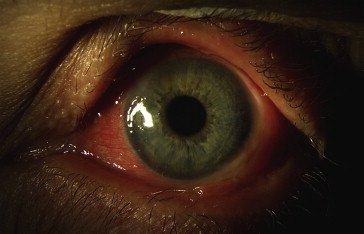
Control of ocular inflammation before surgery was achieved in all cases ( Figures 1, 2 , and 3 ). All 7 patients received systemic immunosuppressive treatment before the lid surgical procedure for a mean of 15.1 months (range, 8.2 to 33.1 months). On average, it took 5.0 months (range, 1.0 to 11.3 months) to achieve disease inactivity after starting systemic treatment ( Figure 3 ). Control of disease was maintained for a mean of 10.1 months (range, 1.8 to 29.9 months) previous to the corrective surgical intervention.