Otorrhea or otorhinorrhea arises from disorders of the temporal bone that may be divided into (i) trauma, (ii) congenital defects, (iii) infection of the middle ear and mastoid, and (iv) tumors of the skull base (most commonly as a complication of their surgical removal).
Trauma
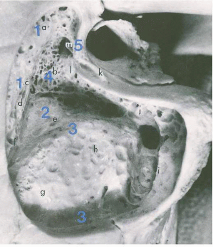
Trauma of the temporal bone may result in a longitudinal or transverse fracture or a combination of both.
Longitudinal fractures follow a blow to the front or side of the head. They extend along the longitudinal axis of the petrous bone, passing through the external auditory canal, tegmen tympani, and anterior surface of the petrous pyramid, usually sparing the inner ear structures of the otic capsule. Longitudinal fractures account for 80% of temporal bone fractures. If hearing is affected, it is usually a conductive loss due to middle ear fluid (CSF or blood). Occasionally, the ossicular chain may be disrupted. Injury to the facial nerve occurs in about 15% of cases and is usually at the level of the geniculate ganglion. Cerebrospinal fluid leakage, when it occurs, is by way of a laceration of the dura at the tegmen tympani. Most often this type of leakage is self-limiting and does not
require surgical repair. Cerebrospinal fluid otorrhea occurs when the tympanic membrane or external ear canal is breached; otherwise the CSF flows down the eustachian tube and is evident as rhinorrhea. Vertigo is not common.
Transverse fractures of the temporal bone follow a blow to the occiput, which produces a line of fracture perpendicular to the long axis of the petrous pyramid; they account for 15% of temporal bone fractures. Often this fracture crosses the vestibule and internal auditory canal and results in a complete sensorineural hearing loss and loss of vestibular function. The site of injury to the facial nerve is medial to the geniculate ganglion and occurs in approximately 45% of transverse fractures. Ecchymosis may develop within hours over the mastoid (Battle’s sign). Examination of the tympanic membrane may reveal blood or CSF in the middle ear. Injury to the ossicles and tympanic membrane are rare. Spinal fluid leakage and vertigo are quite common. A combined longitudinal and transverse fracture of the temporal bone follows severe trauma and represents 5% of temporal bone fractures.
Penetrating wounds, either directly through the skin into the temporal bone or indirectly through the external auditory canal and tympanic membrane, can result in CSF otorrhea or otorhinorrhea. Barotrauma, such as descending rapidly in an airplane or a rapid change in pressure while scuba diving, can occasionally result in CSF leakage. A congenital defect is most likely a predisposing factor in these cases.
Congenital Defects
Congenital defects resulting from malformation and incomplete closure of fissures can result in communications between the middle ear space and the middle and posterior cranial fossae. These defects can occur both in the mastoid portion of the temporal bone and the bony labyrinth. Congenital absence of bone and dural barriers between the subarachnoid space and the mastoid air cells are common causes of CSF leak in infants and children who have experienced recurrent bacterial meningitis. Congenital malformations resulting in CSF leakage occur most commonly in the cochlear aqueduct and internal auditory canal and permit direct communication between the subarachnoid space and the perilymph. These defects usually remain asymptomatic until a defect in either the round or oval window occurs as a result of an external trauma or surgery.
Spontaneous CSF fistulas in adults most commonly occur through defects in the floor of the middle cranial fossa (
4). It is theorized that constant, physiologic intracranial pressure and CSF pulsations cause focal ischemia, which weakens the dura to the point of rupture (
5). More recent theories propose the formation of arachnoid granulations, macroscopic enlargements, or distensions of minute projections of arachnoid villi. These project into the intradural venous sinuses, allowing for resorption of CSF into the bloodstream (
6,
7,
8).
The necessity for dural injury in the pathogenesis of CSF leakage by way of the temporal bone appears to be evident in the work by Åhrén and Thulin (
9). They reviewed the autopsy findings in 94 skulls in patients more than age 40 years. Defects in the tegmen were found in 21% of these skulls. There were multiple defects in 6% of the skulls. Another 16% had thin transparent bone in the tegmen tympani. There were no meningeal or cerebral herniations in these cases. A small to large area of exposed dura during mastoid surgery, whether primary or secondary, does not necessarily preclude a cerebral herniation. One must be particularly careful to avoid full-thickness cauterization of dura using monopolar cautery and avoid tearing the dura with a cutting bur or curette. Herniations usually occur through the central portion of normal exposed dura. Encephaloceles can herniate through a defect as small as 2 mm. Herniation of dura may occur in the area where brain abscesses were drained through the tegmen in the past.
Infection
Cerebrospinal fluid leakage as a complication of both acute and chronic infection occurs because the mastoid air cells are in close contact with the posterior fossa and the roof of the middle ear (tegmen tympani), which is the floor of the middle cranial fossa. Consequently, infection can destroy this thin, bony communication and erode dura to establish a communication between the subarachnoid space and the tympanomastoid compartment. Chronic middle ear and mastoid infection can result in spontaneous CSF leakage by formation of an erosive granulation tissue or cholesteatoma. Cerebrospinal fluid leakage occurs much more commonly following the surgical treatment of this disease.
The bacterial flora of chronic otitis media and mastoiditis are distinct from the usual respiratory pathogens. Gramnegative organisms, both aerobes (Proteus, Klebsiella, and Pseudomonas) and anaerobes (Bacteroides species) predominate. Mixed infections are common. Chronic infections rarely produce meningitis, presumably because the chronicity permits adhesions of the meninges to the infected bone. Ipsilateral brain abscesses of the temporal lobe or cerebellar hemisphere with mixed organisms are the more common intracranial complications of chronic otitis media and mastoiditis. Other complications include subdural abscesses, sigmoid sinus thrombophlebitis, and otitic hydrocephalus.
Skull Base Tumors and Surgery
Tumors of the temporal bone are a rare cause of spontaneous CSF leakage. However, CSF leak remains a fairly common postoperative complication following skull base surgery. Subsequent meningitis adds a significant source of morbidity and mortality to these patients, many of whom are already handicapped by neurologic dysfunction, including cranial nerve deficits. Factors that contribute to the formation of CSF fistulas include suboptimal wound closure, formation of granulation tissue, and elevated intracranial pressure. The dura of the posterior fossa is tightly adherent to the temporal and occipital bones. A partial or total excision of the temporal bone almost invariably results in a dural defect.
Early surgeries for large acoustic neuromas were reported to be complicated by CSF leak in up to one third of cases. Subsequent advances in neurotologic technique, including waxing of air cells at the internal auditory canal petrosotomy site and fat obliteration of the mastoid defect, have reduced this incidence to less than 20% in more current series, with some reporting rates lower than 10% (
10,
11,
12,
13). Hoffman reviewed several previous series and calculated an overall incidence of 12% for both translabyrinthine and suboccipital approaches (
14). In a multicenter review of the Acoustic Neuroma Registry, 175 of 1579 patients (11.1%) developed CSF leak. Meningitis was seen in 39 patients (2.5%) (
15). Some studies have shown even higher rates of meningitis (
1,
16).
Cerebrospinal fluid leaks are more likely to occur following the removal of larger tumors (
1,
16,
17). The onset of CSF leaks following skull base surgery is often delayed, with more than one third diagnosed more than a week postoperatively (
1,
18,
19).
We reviewed our own experience in 504 skull base cases managed between 1988 and 1996 (
20). There were 63 cases of CSF leakage in this series, for an overall incidence of 12.5%, as shown in
Table 35.1. Rates for meningitis are depicted as well. The mean postoperative interval of diagnosis of CSF leak was 5.7 days, with a range of presentation from immediately postoperatively to 33 days. An additional three cases of late postoperative presentation of CSF leak were seen. Seven of the 63 patients with CSF leak were noted postoperatively to have hydrocephalus, two of whom required placement of ventriculoperitoneal shunts. Noninvasive expectant management was associated with cessation of CSF leakage in only two of eight cases (25%), suggesting that observation alone is not a reasonable option. Lumbar drainage was successful in 30 of 41 patients (73%). Standard mastoid obliteration, with or without obliteration of the eustachian tube orifice via a posterior tympanotomy, was successful in 7 of 10 cases (70%). Extended tympanomastoid obliteration was used in 24 cases, including those patients who failed the aforementioned techniques. All 24 of these patients (100%) had no recurrent or persistent leak.