Lens Design, Fitting, and Evaluation


Lens Design, Fitting, and Evaluation


Edward S. Bennett

Luigina Sorbara


The ability to successfully fit rigid gas-permeable (GP) contact lenses is often what separates the good contact lens practitioner from the average one. Certainly there are numerous patients who benefit from the quality of vision and ocular health provided by GP lenses. This chapter will discuss the importance of fitting GP lenses and the lens design, fitting, evaluation, and ordering procedures.


▪ HOW TO OPTIMIZE INITIAL COMFORT

The first time a patient experiences contact lens wear can be quite traumatic. This is especially true with GP lenses because of the smaller diameter and greater lens movement with the blink as compared to soft lenses. This experience, in itself, can affect the fitting habits of practitioners who may decide to fit soft lenses—even when they are not the best option for patients—in an effort to provide a more initially comfortable and less time-consuming experience.

It is important, if not imperative, for the initial patient experience to be positive for the patient to be successful. The common perception by patients is that the initial comfort with GP lenses is poor, and this represents the primary reason why patients discontinue GP lens wear.1 It is evident that if the patient has a poor initial experience with GP lenses, he or she will influence others away from considering this option. If the clinician—via his or her educational background (or lack thereof) or employment environment—is not motivated to fit GP lenses, it is likely that, despite the many benefits of GP lenses, patients will not be fitted into the mode of correction that would be indicated because of quality of vision, eye health, or some other reason.

The authors have recommended a fourfold approach to optimizing initial comfort.2 These factors include (a) presentation, (b) topical anesthetic, (c) initial vision, and (d) lens design.


Initial Presentation

Most contact lens patients rely on their practitioner to recommend an appropriate lens material and design. It is important for practitioners to recognize that when presenting patients with a choice, they may, without realizing it, bias that choice. The doctor’s language creates and colors patient perceptions. Such terms as discomfort, pain, or always seems to feel like there is something on the eye set up strong negative expectations. Prescribing practitioners are powerful authority figures. What they say and how they say it can easily influence patients.

Simple words like soft, hard, or rigid can influence a patient’s contact lens preferences. Even nonverbal cues such as facial expression and eye contact can communicate an attitude to the patient. It is always important to begin with the assumption that a new patient has somewhere been given the impression that rigid lenses are uncomfortable. GP lenses can then be described in the following way: “GP lenses typically provide excellent vision and eye health. They are also quite wettable and durable. However, they do not feel the same way a soft lens does at
first. Because they are smaller, they move more on the eye. The lids sense this movement initially and gradually adapt, typically resulting in comfortable lens wear.” The use of the term gas permeable as compared to the previous rigid gas permeable terminology was recently proposed by the Contact Lens Manufacturers Association (CLMA) to minimize the focus on “rigid” lenses.

The impact of presentation methods was confirmed by a study in which subjects who were not previous contact lens wearers were divided into three groups before initiating GP lens wear.3 Group one subjects, as part of the diagnostic fitting process, observed a videotape of a doctor discussing GP lenses with a new wearer using terms such as discomfort, possible pain, intolerance, and failure when describing GP lenses. Group two subjects observed a videotape of a doctor discussing GP lenses using neutral terms such as lens awareness and initial lid sensation. However, this doctor did not appear to be particularly positive about the GP lens option. In group three the doctor discussing GP lenses used the same terminology as in the videotape observed by group two but exhibited a positive attitude toward GP lenses. Eight subjects in this 1-month study discontinued lens wear; six of these subjects were in group one, and the other two were in group two. No subject provided with a neutral content and enthusiastic presentation discontinued lens wear. Likewise, the group three subjects were significantly more compliant in returning daily questionnaires than the other two groups.

It is important for patients to know the benefits of GP lenses, that they will adapt over time, and that good comfort is a very realistic goal.4 Certainly experienced GP fitters do not have a problem with the comfort issue, and as one acquires more experience in fitting GP lenses, greater confidence in presenting this option in a positive but realistic manner results.


Topical Anesthetic Use

A second important technique for obtaining initial GP patient comfort and satisfaction is the use of a topical anesthetic during contact lens fitting. This has been considered somewhat controversial because of concerns pertaining to its potential for softening the epithelium, resulting in a greater incidence of corneal staining.5,6 In addition, there is always the potential for misleading the patients who will ultimately experience the typical lens awareness with GP lenses. Fortunately, although these are legitimate concerns, they have not been confirmed by clinical research with GP lenses.7,8

With these issues in mind, an 80-subject multicenter study was performed to evaluate the effect of topical anesthetic use on initial comfort and patient satisfaction among first-time GP lens wearers.7 Forty subjects were administered a topical anesthetic at the fitting visits, while a second group of 40 received a placebo. One month later, 70 of the 80 subjects were still wearing lenses. Eight of the 10 who discontinued lens wear were in the placebo group. In addition, at the completion of the study, subjects who had been given anesthetic rated their experience during adaptation, their comfort, and their overall satisfaction significantly higher than those subjects who had received placebo.

The benefits of topical anesthetic are significant. All patients are, at minimum, mildly apprehensive about the initial application of contact lenses. If the first few minutes of lens wear are acceptable, it makes sense that patient satisfaction, and the potential for successful longterm wear, will be greatly enhanced. The topical anesthetic should be allowed to wear off, and the patient will gradually experience lens awareness. In addition, in busy clinical practice environments, where time is a precious resource, the ability to evaluate the fluorescein pattern soon after diagnostic lens application is invaluable.8 This is also important because of the fact that soft lenses often require little chair time during the fitting process and GPs benefit from being competitive with soft lenses whenever possible.

Nonsteroidal anti-inflammatory drugs (NSAIDs) have also been used to reduce awareness during adaptation.9,10 Because NSAIDs reduce production of prostaglandins, which are mediators of pain, reducing their production reduces pain. The most effective drug within this class
at inhibiting prostaglandin synthesis appears to be Voltaren. A recommended NSAID dosage for GP lens adaptation is as follows10:


  • Instill one drop of Voltaren in each eye 30 minutes and then 15 minutes before lens insertion.


  • Instill a third drop just before lens insertion.


  • A fourth drop can be instilled 1 hour after insertion.


  • This regimen can be maintained for 3 to 5 days or until adaptation is completed.

Topical anesthetic use is certainly not a requirement for GP fitting. However, for apprehensive practitioners as well as patients—particularly young people, people with keratoconus, and soft lens refits—anesthetic use can mean the difference between success and failure.


Initial Vision

Providing patients with an important benefit from GP lenses, good quality of vision, can be very important for initial patient satisfaction. When a patient is fitted with GP lenses in their correct power—either via empirical fitting or from an inventory—the resulting “wow” factor may reduce apprehension about lens awareness.


Lens Design

This will be discussed in more detail later in this chapter; however, a well-designed, alignment fitting lens with an optimum edge design and shape will contribute to a more positive initial patient experience.


▪ FITTING AND EVALUATION

There are numerous important components to fitting lenses successfully. These include such factors as diagnostic fitting, fluorescein application and evaluation, and an accurate and compatible lens design.11


Methods of Fitting


Empirical Fitting

Empirical fitting refers specifically to designing lenses empirically or without using diagnostic lenses. Practitioners who utilize empirical fitting methodologies claim that manufacturers’ recommendations (supplying the laboratory with such minimal prefitting information as keratometry values and refraction) and fitting guides provide effective means to obtain maximum lens performance and fit. Empirical fitting also means that a new, unworn lens will be fitted to one patient only. Empirical fitting has the attraction of eliminating a fitting visit and, consequently, enabling a simplistic fitting approach.12 As indicated previously, however, empirical fitting often provides a very important benefit, good initial vision. The newer automated manufacturing equipment makes the success of empirical designs more likely than 10 to 20 years ago because of higher-quality lenses, aspheric and pseudoaspheric peripheries, thinner center thicknesses, and more consistent edge designs. Likewise, topography software programs allow the practitioner to better match corneal shape with the recommended computer-assisted design.13,14,15,16


Diagnostic Fitting

Diagnostic fitting is still a popular method of fitting GP lenses. It allows the practitioner to feel confident in the final lenses to be ordered as multiple diagnostic lenses can be applied (if necessary) and those that result in the best lens-to-cornea fitting relationship will be ordered. Greater patient compliance with the follow-up schedule in addition to significantly fewer reordered lenses have been found with diagnostic versus empirical fitting.17 Although a fitting visit and a sufficient number of diagnostic lenses are required, diagnostic fitting allows
practitioners the opportunity to evaluate the lens-to-cornea fitting relationship and to make the changes necessary to obtain a good fit and provide acceptable vision to the patient. It is also apparent that the fitting visit provides patients with an opportunity to become familiar with their particular lenses. Finally, such factors as lens centration and residual astigmatism can be evaluated.18 The primary limitations pertain to (likely) not leaving the office with lenses, as compared with most soft lens patients, and the fact that, in most cases, satisfactory vision will not be obtained with the first pair of GP lenses applied. The 6 D myopic patient who is fitted with 3 D power diagnostic GP lenses will not only experience initial awareness, but also blurred vision. Nevertheless, with most special design GP lenses, including bifocal, keratoconic, and postsurgical designs, it is important to use diagnostic fitting sets because of the greater challenges involved in the fitting process and the more custom nature of the designs. When diagnostic fitting sets are to be used, it is important that the lenses are in the same design and material as the lenses to be ordered. A comparison of the factors involved in deciding between empirical and diagnostic fitting is provided in Table 5.1.


Diagnostic Fitting Sets/Inventories


Specific Fitting Sets

Having available several different diagnostic fitting sets is important, if not essential. For example, 20 lens diagnostic fitting sets in a −3.00 D power would be beneficial in materials of both low (<50) and high (≥50) oxygen permeability (Dk). An example of such a fitting set is provided in Table 5.2. In addition, similar diagnostic sets in a +3.00 D power in a high-Dk material with a minus lenticular edge design and in a −8.00 D power in a low-Dk material and a plus lenticular edge design would be recommended. Keratoconic, bitoric, aphakic, and bifocal diagnostic sets are also recommended and are discussed in other chapters of this text (Chapters 14, 15, 17, and 18).

For the diagnostic fitting sets, a good average overall diameter is 9.4 mm with an 8.0-mm optical zone diameter. However, for steeper than 44.50 D, you may find a 9.0/7.6-mm design to provide an optimum fitting relationship. Base curve radii can range from 40.75 D (8.28 mm) to 45.50 D (7.42 mm) in 0.25 D steps. A relatively constant edge lift of 0.09 to 0.11 mm is recommended for the diagnostic lenses; therefore, the flatter base curve radii will have a greater flattening of the peripheral curve radii, and steeper base curve radii will have a greater steepening. Finally, the appropriate center thickness should be ordered. For example, a low-Dk material may have a center thickness of approximately 0.14 mm in a −3.00 D power, although this can vary from material to material. All of the diagnostic lenses of the same power should have equal and
appropriate center thicknesses; these parameters—and especially edge shape—should be verified upon receiving them from the laboratory. With the increasing popularity of ultrathin designs, a diagnostic set of this design in a minus power (i.e., −3.00 D) would be recommended as well.


TABLE 5.1 DIAGNOSTIC FITTING VERSUS EMPIRICAL FITTING


Diagnostic Fitting Advantages


Fewer reorders


Practitioner confidence in fitting relationship


Greater patient satisfaction


Better patient compliance


Empirical Fitting Advantages


Good initial vision experience


Easier method


Minimizes transfer of diagnostic lens contaminants


Less initial chair time


Allows topography software to assist in lens design


TABLE 5.2 RECOMMENDED PARAMETERS FOR A 20-LENS DIAGNOSTIC SET, LOW-AND HIGH-DK GAS-PERMEABLE MATERIALS


Overall diameter


9.2 mm


Optical zone diameter


7.8 mm


Center thickness


0.14 mm


Power


−3.00 D


LENS


BCR (mm)


SCR/W


ICR/W


PCR/W


1.


7.42


8.00/.3


8.80/.2


10.00/.2


2.


7.46


8.10/.3


8.90/.2


10.10/.2


3.


7.50


8.20/.3


9.00/.2


10.20/.2


4.


7.54


8.20/.3


9.00/.2


10.20/.2


5.


7.58


8.30/.3


9.10/.2


10.30/.2


6.


7.63


8.30/.3


9.20/.2


10.40/.2


7.


7.67


8.40/.3


9.30/.2


10.50/.2


8.


7.71


8.50/.3


9.40/.2


10.60/.2


9.


7.76


8.50/.3


9.50/.2


10.60/.2


10.


7.81


8.60/.3


9.60/.2


10.70/.2


11.


7.85


8.60/.3


9.60/.2


10.80/.2


12.


7.89


8.70/.3


9.70/.2


10.80/.2


13.


7.94


8.70/.3


9.70/.2


10.90/.2


14.


7.99


8.80/.3


9.80/.2


11.00/.2


15.


8.04


8.80/.3


9.90/.2


11.10/.2


16.


8.08


8.90/.3


10.00/.2


11.20/.2


17.


8.13


8.90/.3


10.10/.2


11.30/.2


18.


8.18


9.00/.3


10.20/.2


11.40/.2


19.


8.23


9.10/.3


10.30/.2


11.50/.2


20.


8.28


9.20/.3


10.40/.2


11.60/.2


BCR, base curve radius; ICR/W, intermediate curve radius/width; PCR/W, peripheral curve radius/width; SCR/W, secondary curve radius/width.


Inventories

The use of a large (100-200 lenses) inventory system, which has been very popular with hydrogel lenses, is also an alternative available to practitioners. The advantages of using a large inventory to fit rigid lenses are many and include the following: (a) some patients can be fitted out of stock; (b) lens replacements can be provided to patients without delay, so patient satisfaction is enhanced; and (c) lens parameter changes can be made in the office without delay. Unlike hydrogel lenses, because of the custom or multiparameter design—especially base curve radius (BCR)-inherent with successful fitting of GP lenses, a minimum of 200 lenses is necessary to directly fit the majority of patients without having to order the lenses from a laboratory. Such a 200-lens inventory is given in Table 5.3.


TABLE 5.3 PARAMETERS FOR GAS-PERMEABLE INVENTORY LENS SET
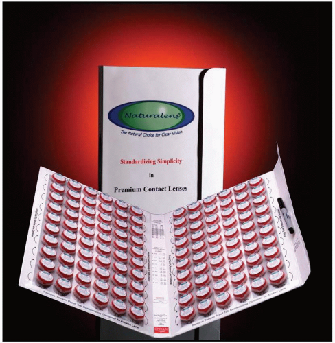


BASE CURVE RADIUS (mm)


RX


7.42


7.50


7.54


7.58


7.63


7.67


7.71


7.76


7.80


7.85


7.89


7.94


7.99


8.04


8.13


−1.25


94


110


123


136


149


162


175


188


−1.50


79


95


111


124


137


150


163


176


189


−1.75


80


96


112


125


138


151


164


177


190


−2.00


1


14


27


40


53


66


81


97


113


126


139


152


165


178


191


−2.25


2


15


28


41


54


67


82


98


114


127


140


153


166


179


192


−2.50


3


16


29


42


55


68


83


99


115


128


141


154


167


180


193


−2.75


4


17


30


43


56


69


84


100


116


129


142


155


168


181


194


−3.00


5


18


31


44


57


70


85


101


117


130


143


156


169


182


195


−3.25


6


19


32


45


58


71


86


102


118


131


144


157


170


183


196


−3.50


7


20


33


46


59


72


87


103


119


132


145


158


171


184


197


−3.75


8


21


34


47


60


73


88


104


120


133


146


159


172


185


198


−4.00


9


22


35


48


61


74


89


105


121


134


147


160


173


186


199


−4.25


10


23


36


49


62


75


90


106


122


135


148


161


174


187


200


−4.50


11


24


37


50


63


76


91


107


−4.75


12


25


38


51


64


77


92


108


−5.00


13


26


39


52


65


78


93


109


Overall diameter = 9.4


Optical zone diameter = 8.2


Secondary curve radius (SCR) = base curve radius (BCR) + 1.0 mm/0.3 mm wide; peripheral curve radius (PCR) = BCR + 3.0 mm/0.3 mm wide


FIGURE 5.1 The Naturalens inventory (Advanced Vision Technologies).

Some of the manufacturers, including those who manufacture the Boston Envision lens design (Bausch & Lomb) and the Naturalens (Advanced Vision Technologies, Fig. 5.1), can provide smaller inventories because of the philosophy that their respective designs can be successfully fitted to the great majority of patients with fewer base curve radii.2

Some GP lens manufacturers are unwilling to manufacture such large inventories because of the labor and expense necessary to do so. However, they are often available to practitioners who fit a high volume of GP lenses. As with soft lenses, the initial expense to the practitioner is minimal; however, the laboratory usually requires the practitioner to meet the following agreement provisions19: (a) maintain an inventory of lenses equal to the original consignment, (b) fit a certain number of lenses within a specified period, and (c) use manufacturer’s lens design parameters. Nevertheless, it is a valuable alternative that can increase your success with GP lenses while also providing a valuable service to many GP patients.


Storage of Diagnostic Lenses

Storing diagnostic lenses in the hydrated state has the advantage of providing good initial wettability while maintaining the lenses in a somewhat sterile state. However, depending on the frequency with which the solution is changed, there are many advantages to keeping the lenses in the dry state.20 It is both efficient and convenient to store the lenses dry because they can be kept in flat-pack cases that occupy very little space in the office. If the lenses are stored in the hydrated state, it is possible for the solution to either dry up in the case or leak out, both of these problems resulting in a lens that may adhere to the case or even change in base curve radius because of variation between the hydrated and unhydrated states. In addition, the dried solution may be difficult to remove from the lens surface. Whenever a diagnostic lens has been applied, however, it is recommended to carefully clean the lens and blot it dry with a soft tissue prior to disinfection and placement in the case and into the appropriate diagnostic lens set. The Centers for Disease Control and Prevention (CDC) recommends ophthalmic-grade hydrogen peroxide for GP lenses; therefore, AOSept or Clear Care (Ciba Vision) for a 5- to 10-minute soak has been recommended.21 GP lenses that are going to be dispensed to a patient should be hydrated for a minimum of 24 hours prior to application to enhance surface wettability and maintain the base curve radius in the hydrated state (similar to the “on-eye” condition as the back surface rests against the tear film). Obviously, patients should likewise be advised to maintain the lenses in the appropriate soaking/disinfecting solution upon removal (see Chapter 6).


Fluorescein Application and Evaluation


Description

Sodium fluorescein is an organic compound that is inert and harmless to tissue.22 The application of fluorescein enables the practitioner to evaluate the lens-to-cornea fitting relationship. In fact, it would be appropriate to indicate that fluorescein has an invaluable, if not essential, role in rigid lens fit assessment.

To perform the procedure, the fluorescein strip is wetted with an ophthalmic irrigating solution. The strip is then gently applied against the superior bulbar conjunctiva with the patient viewing inferiorly. It is important to reassure the patient that this procedure is painless. In addition, the thumb should carefully pin back the upper lid to prevent the possibility of the lid pushing the strip toward the superior cornea, which could result in superior corneal staining and accompanying subjective discomfort.

The use of an ophthalmic irrigating solution has numerous advantages for wetting the fluorescein strip, including the following:


  • Sterility.


  • Slightly alkaline in pH, which assists in fluorescence.


  • Reduced risk of burning and stinging caused by pH.


  • Less viscous than use of a wetting solution (which may result in an abnormally thick tear layer).

However, for optimum fluorescence, the use of liquid fluorescein has been recommended.23


Methods of Observation

The fluorescein pattern can be evaluated with both a Burton lamp and a biomicroscope.


Burton Lamp:

The traditional method of evaluating the fluorescein pattern is by use of an ultraviolet fluorescent lamp that utilizes a +5.00 D magnification lens to assist in viewing (Fig. 5.2). This method has the following advantages:


  • Inexpensive.


  • Easy to use.


  • Overall field of view and ability to directly compare fluorescein pattern of both eyes simultaneously.
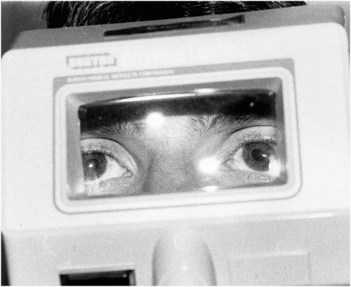


    FIGURE 5.2 The Burton lamp for fluorescein pattern evaluation.


However, the Burton lamp is very limited in its abilities. It does not allow for variable magnification or illumination. In addition, it is an ineffective method of observing the fluorescein pattern of rigid lens materials with ultraviolet-absorbing capabilities. Therefore, it would not be advantageous or appropriate to use this as the only method to evaluate a fluorescein pattern. However, it is a useful adjunct to the biomicroscope because of the overall field of view. This is especially beneficial in observing some of the more distinctive patterns, such as those pertaining to high corneal toricity and keratoconus.


Biomicroscope:

The most popular method of evaluating the fluorescein pattern of a rigid lens is with the biomicroscope. The primary advantage of this over other observational methods is flexibility. It allows the practitioner the opportunity to vary the magnification, illumination, and slit-beam width while observing the fluorescein pattern. Proper use of a biomicroscope for GP fitting and evaluation is essential for patient success.

As biomicroscopes vary considerably from manufacturer to manufacturer, it is important for a good illumination source and variable magnification to be present to effectively evaluate the fluorescein pattern. In fact, it has been determined that with many biomicroscopes it is not possible to use <10× magnification and still retain an adequate field of view.24

Once fluorescein has been properly instilled, the patient should be instructed to blink several times for adequate distribution on the eye. The fluorescein pattern should be initially observed under low magnification with a wide (diffuse) slit-beam and high-intensity illumination. The central and peripheral fluorescein pattern should be relatively easy to determine after several seconds. An optic section with the angle of illumination equal to 45 to 60 degrees can also be used to observe the pooling of tears in relation to the contact lens. It will appear as a green layer representing the outer layer of tears on the lens; then a wider dark layer, which is the contact lens; next another green layer, which represents the tear layer between the lens and cornea; and finally a bright grayish layer, the cornea.22 The lens-to-cornea fitting relationship can be evaluated by viewing the thickness of the tear layer along the optic section.

Typically, the fluorescein pattern is viewed with the assistance of a cobalt blue filter, which, in effect, transmits blue light that will activate the fluorescein dye. It is important to use a Wratten no. 12 yellow filter (or equivalent) that can be attached to the observation system to serve as a barrier filter, screening out all but the wavelengths of interest.25 The importance of the yellow filter cannot be underestimated since it makes an easily observable improvement in fluorescein pattern evaluation. The use of a yellow filter, in combination with a good illumination source, is especially important in the evaluation of GP materials that contain ultraviolet inhibitors because, as the material absorbs wavelengths that correspond to the illumination source, there is an apparent reduction or even absence of fluorescence behind the lens unless the appropriate filters and illumination source are used. It is hoped that biomicroscopic manufacturers will begin to incorporate the yellow filter into their respective instruments.


Pattern Evaluation:

The fluorescein pattern assumes a variety of forms. Areas of fluorescein pooling appear green; areas in which fluorescein is absent or where the tear layer is too thin to detect, having the contact lens in direct contact with the cornea, appear as dark or black. In between these extremes, the varying thickness of the tear layer is observed as varying shades of green.

An alignment fit is observed when the lens evenly contours the cornea with a light, even tear pooling (Fig. 5.3). Apical clearance exists when a steep central fit with excessive fluorescence or central tear pooling is present (Fig. 5.4). This can result in midperipheral bearing and sealoff with a reduced ability to remove cellular debris and mucus that may be an important precursor to rigid lens adherence to the cornea. Apical clearance has also been found to induce corneal steepening, even after short-term wear.26 Apical bearing exists when there is direct contact of the lens against the central cornea or the amount of tear pooling is too shallow to detect
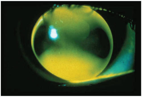
with the instillation of fluorescein (Fig. 5.5). Excessive apical bearing can potentially result in corneal molding with resultant distortion or warpage. In addition, the gradual formation of a central corneal abrasion is also possible.
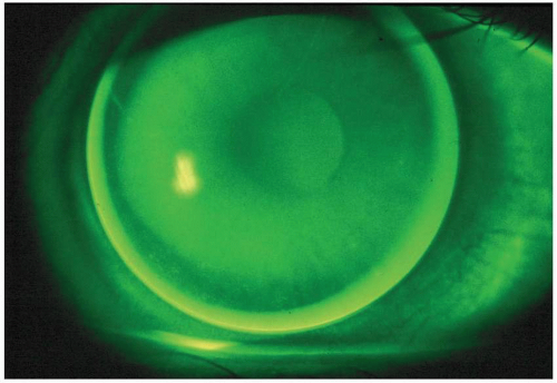


FIGURE 5.3 An alignment fluorescein pattern.
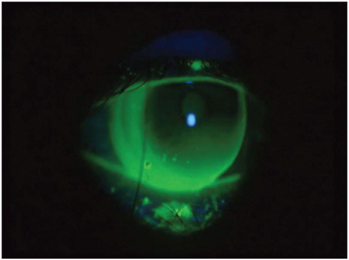
With corneal astigmatism greater than one diopter, a dumbbell-shaped fluorescein pattern will be observed (Fig. 5.6). Typically, along the steeper meridian of the cornea, the tear layer thickness gradually increases toward the edge and the lens does not touch the cornea.

Along the flatter meridian, however, the tear layer thickness decreases toward the periphery and the lens comes in contact with the cornea at the edge of the optical zone. As corneal astigmatism increases, the difference in tear layer thickness between the two primary meridians becomes greater, the area of alignment becomes smaller, and the astigmatic, or dumbbell-shaped, fluorescein pattern becomes exaggerated.27 If the cornea exhibits with-the-rule corneal astigmatism, the pooling is in the vertical meridian with alignment or bearing in the horizontal meridian. If the cornea exhibits against-the-rule astigmatism, the opposite is true: the pooling is in the horizontal meridian with alignment or bearing in the vertical meridian. In high corneal astigmatism—typically greater than two diopters—the use of a high-Dk material with a steeper than K base curve radius will result in excessive flexure and reduced visual acuity. In addition, the “rocking” of the lens during the blink process may result in discomfort, mechanical corneal staining, and possible lens adherence. The selection of a lower oxygen-permeable material, and perhaps a flatter base curve radius, is recommended. Another option would be a bitoric design, especially if the high amount of corneal toricity results in inferior decentration of the lens (see Chapter 14).


FIGURE 5.4 An apical clearance fluorescein pattern.


FIGURE 5.5 An apical bearing fluorescein pattern.
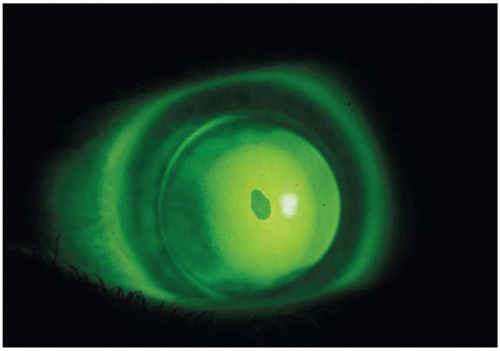
It is important to evaluate the fluorescein pattern after the blink since the amount of pooling and bearing will vary during the blink process. If the lens is decentered, the position of the lens relative to the cornea must be considered prior to evaluating the fluorescein pattern. For example, an inferior decentering lens will typically exhibit excessive superior pooling since the flatter peripheral bevel is adjacent to the steeper central cornea.

The evaluation of the fluorescein pattern at the lens periphery is also beneficial. There should be sufficient clearance peripherally—typically greater than apically—to allow sufficient tear exchange and debris removal while avoiding mechanical irritation as the lens moves across the cornea. If fluorescein pooling is minimal or absent peripherally and seal-off exists, the peripheral curve(s) should be flattened.

Fluorescein pattern evaluation of the rigid lens-to-cornea fitting relationship should be performed both at the fitting visit and at all subsequent follow-up visits. A practitioner’s ability to properly assess fluorescein patterns occurs with experience and frequent evaluation. There are several educational resources available from the GP Lens Institute (www.gpli.info) including an educational CD-ROM entitled “GP Fitting, Evaluation, and Problem-Solving,” a GP Lens Management Guide, and a Fluorescein Pattern Identification laminated card. It would be erroneous to believe that fluorescein pattern evaluation is not as important with GP materials as with polymethylmethacrylate (PMMA) as a result of the reduction in edema-related complications. The lens material is only as good as the practitioner’s ability to properly evaluate it; a poor lens-to-cornea fitting relationship can result in numerous problems, including desiccation, adhesion, and abrasion. In particular, the fluorescein pattern evaluation is invaluable in the difficult-to-fit cases such as high corneal toricity, irregular/distorted corneas, and keratoconus.28


FIGURE 5.6 The dumbbell-shaped fluorescein pattern present with a highly astigmatic patient.


False Fluorescein Patterns:

Occasionally, the fluorescein pattern is contrary to the expected appearance. This phenomenon can occur as a result of a variety of causes:


  • Corneal topography. This varies between patients; for example, a patient with a small corneal cap (defined as the region within 0.50 D of the corneal apex) will exhibit a somewhat steeper fluorescein pattern than a patient with a larger-than-average cap.29,30


  • Selection of a steep BCR may result in poor tear exchange and a misleading small amount of fluorescein centrally.


  • If the peripheral curve is too steep, peripheral seal-off can occur and the fluorescein pattern can exhibit apical clearance.


  • In certain individuals—particularly dry-eye patients—the fluorescein will dissipate quickly and may create a “pseudoapical flat” relationship; therefore, the pattern should be evaluated immediately after fluorescein instillation.


  • A “pseudosteep” pattern has been reported in high minus fluoro-silicone/acrylate (F-S/A) lenses.31 Apparently the edge thickness blocks the fluorescence, giving an appearance of central pooling. Likewise, one would expect that a high plus lens may demonstrate a flatter than actual base curve fitting relationship because the thick center would attenuate the light more.


Designs/Fitting Philosophies

There are numerous available methods for determining the rigid lens design parameters for diagnostic fitting. In this section of the chapter two fitting philosophies will be presented.

There are two primary fitting philosophies for designing and fitting GP lenses, both based on lens position on the eye. The first approach is to design a lens so that it positions consistently under the upper eyelid (i.e., a lid-attached fit) and the second is to design a lens that achieves an interpalpebral fit on the eye.

Studies have been done to compare these two lens designs with respect to comfort, vision, and physiologic response.32,33,34 Results are mixed about which design performs the best because of individual variation in corneal topography, lid/cornea interaction, and lid tension.

Only gold members can continue reading. Log In or Register to continue

Stay updated, free articles. Join our Telegram channel

Jul 5, 2016 | Posted by in OPHTHALMOLOGY | Comments Off on Lens Design, Fitting, and Evaluation

Full access? Get Clinical Tree

Get Clinical Tree app for offline access