Lasik Complications: Etiology, Prevention, and Management
Neda Shamie
Kourosh Eghbali
Peter J. Mcdonnell
Laser in-situ keratomileusis (LASIK) has gained international popularity among ophthalmic surgeons and patients as the procedure of choice for refractive surgical correction of myopia and low to moderate degrees of hyperopia (1,2). Its popularity has surpassed that of photorefractive keratectomy (PRK) with its benefits including more rapid recovery of vision, less postoperative discomfort, and lower risk of postoperative stromal haze in high refractive corrections (3, 4, 5). This benefit lies in the creation of a corneal flap and maintaining the surface epithelial and Bowman’s layers prior to laser ablation of the stromal bed in LASIK.
As with other refractive surgical procedures, LASIK may be associated with both intraoperative and postoperative complications. These include ablation-related complications, suboptimal refractive corrections, postoperative visual aberrations, and loss of best corrected visual acuity. With the creation of the corneal flap in LASIK, however, a new category of flap-related complications unique to corneal lamellar surgery may occur, including intraoperative flap complications such as free caps or buttonholes and postoperative complications such as flap striae and diffuse lamellar keratitis.
Complications occur infrequently, with skilled surgeons reporting a rate of 1% to 2% incidence of minor complications and 0.2% to 0.3% incidence of major sight-threatening complications (6, 7, 8, 9). With experience we are recognizing preoperative patient-related factors that may increase the rate of known complications. By modifying the surgical technique, surgical parameters, and patient selection, a surgeon can decrease, but not eliminate, the incidence of complications and patient dissatisfaction. More importantly, a careful preoperative assessment and recognition of a high-risk patient can help counsel the patient appropriately to allow for a more informed consent and more realistic expectations. Although some complications can be avoided with careful planning and attention to detail, others are unavoidable. When complications do occur, early recognition of the problem with prompt management is key in restoring an optimal outcome.
INTRAOPERATIVE COMPLICATIONS
Poor Exposure
Inadequate globe exposure can prevent proper placement of the suction ring leading to inadequate suction and related flap complications. In addition, with poor exposure, a smooth passage of the microkeratome may not occur, leading to further intraoperative complications. This problem is one that can often be anticipated and prevented with proper preoperative evaluation. Inadequate exposure is related to orbital and facial anatomy in patients with a history of facial/orbital fractures, sunken or small eyes, prominent brows, or narrow palpebral fissures (10). A lid speculum with a posteriorly angulated hinge and a locking mechanism can improve the exposure in some patients. Also, careful draping and proper patient head positioning may help alleviate this problem (10). When necessary, the assistant may exert downward pressure on the lid speculum to proptose the eye during the microkeratome pass. Incomplete passes of the microkeratome due to interference from the drape, lids, or redundant conjunctiva can be prevented. Occasionally, the microkeratome pass can be performed without using a lid speculum, with tape used to isolate the lashes. In rare cases, some suggest doing a lateral canthotomy in patients with narrow palpebral fissures or a retrobulbar injection in patients with sunken eyes or prominent brows (6). Lastly, PRK should be considered in this challenging subset of patients.
Inadequate Suction
Inadequate suction or loss of suction can lead to serious problems during the microkeratome pass such as thin flaps, perforated flaps, or free caps (6,10). The proper placement of the suction ring requires adequate exposure and the surgeon’s attention to detail. The surgeon and the technician must be familiar with the workings of the microkeratome and the suction mechanism. They may be falsely reassured by the readings on the suction console when in fact adequate
suction has not been obtained. It is therefore advisable to look for the following signs indicating an intraocular pressure of at least 65 mm Hg: pupillary dilation, transient loss of the patient’s vision, and sufficient intraocular pressure (IOP) measurement using a Barraquer tonometer or pneumotonometer (6,10).
suction has not been obtained. It is therefore advisable to look for the following signs indicating an intraocular pressure of at least 65 mm Hg: pupillary dilation, transient loss of the patient’s vision, and sufficient intraocular pressure (IOP) measurement using a Barraquer tonometer or pneumotonometer (6,10).
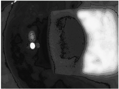 FIGURE 82-1. A central area of buttonhole is shown in this flap, which occurred as a result of a partial loss of suction during the microkeratome pass. (Courtesy of Farid Eghbali, O.D., with permission.) |
Redundant conjunctiva or chemotic conjuctiva (as a result of repeated failed attempts of obtaining adequate suction) can result in pseudosuction as the conjunctiva obstructs the orifice of the suction ring. If redundant conjunctiva is noted, the redundancy can be decreased by expanding the lid speculum and stretching the conjunctiva. Rarely, a conjunctival peritomy needs to be done to place the suction ring directly onto bare sclera for adequate suction. When chemotic conjunctiva occurs iatrogenically, patience is often the best remedy. After 30 to 60 minutes, minor chemosis may resolve (6,10). A small incision in the conjunctiva may also allow drainage of the fluid, as can “milking” the fluid away from the limbus using a blunt instrument such as a Merocel sponge (10). The safest option remains delaying the surgery for several days.
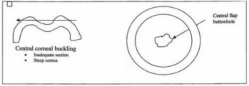 FIGURE 82-2. Central corneal buckling can occur in very steep corneas or when inadequate suction results in decreased intraocular pressure during the microkeratome pass. This can result in a thin or buttonhole flap. |
Microkeratome-Related and Flap Complications
Thin Flaps and Buttonholes
Buttonhole flaps (Fig. 82-1) and especially thin flaps are among the more common LASIK flap-related intraoperative complications encountered, with incidence ranging from 0.1% to 0.2% depending on surgeon experience (11, 12, 13, 14, 15, 16). In addition to surgical expertise, other factors that may affect the incidence of this complication include inadequate suction, corneal anatomy, and microkeratome malfunction or defect (17,18).
With loss of suction or with insufficiently elevated IOPs during the microkeratome pass, buckling of the central cornea may result in thin or perforated buttonhole flaps (Fig. 82-2). This problem can be minimized by assuring an IOP of at least 65 mm Hg using a Barraquer tonometer or pneumotonometer, verifying from the patient that the vision has dimmed or observing dilatation of the pupil intraoperatively after obtaining suction (18). Patients with excessive vitreous syneresis or history of vitrectomy may be at increased risk due
to the difficulty of maintaining high enough IOPs using the suction ring and should be counseled appropriately (19).
to the difficulty of maintaining high enough IOPs using the suction ring and should be counseled appropriately (19).
Abnormally steep corneal curvature can also predispose a patient to intraoperative flap complications. Corneas steeper than 47 D or with irregular surface (i.e., history of previous penetrating keratoplasty or scleral buckle) may be at increased risk of developing buttonhole flaps or flaps with central thinning. As in the case of inadequate suction, this is thought to occur as a result of central corneal buckling during the microkeratome pass (Fig. 82-2) (19). The surgeon can opt to use a thicker plate and a smaller ring on these steeper corneas. Another option is to avoid the lamellar cut and proceed with PRK instead.
Probably the most avoidable factor that may predispose to this complication is microkeratome malfunction and poor blade quality (20). Meticulous attention to cleaning, assembly, and preoperative assessment of the blade and the microkeratome system can help minimize intraoperative complications. Additionally, avoidance of significant manual torque of the cornea during the microkeratome pass can also help avoid irregular and possibly perforated flaps (18).
In the case when a buttonhole flap is created, a consensus exists that the surgeon should not proceed with laser ablation of the stromal bed, and should instead replace the flap and abort further surgery until at least 3 months later, when a new, thicker flap can be recut (18,19). In the case when a thin flap without a buttonhole is created, the decision to abort further surgery is not as clear. Ablation can be safety done if the central flap appears to be of adequate thickness. Some have proposed converting to immediate transepithelial photorefractive keratectomy after repositioning the defective flap (21). The relative merits of this approach and the possibility of increased risk of haze with PRK in this setting are not yet completely understood. Of concern in the case of both buttonhole flaps and thin flaps is the increased risk of epithelial ingrowth (Fig. 82-3), irregular astigmatism, stromal scarring, and flap striae (17,18).
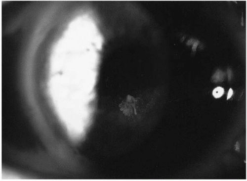 FIGURE 82-3. An area of epithelial ingrowth (inferiorly) starting from the edge of a buttonhole flap. (Courtesy of Farid Eghbali, O.D., with permission.) |
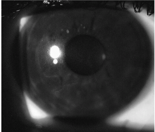 FIGURE 82-4. An incomplete flap due to inadequate suction causing an incomplete pass of the microkeratome. The edge of the flap is easily seen when one follows the outer edges of the fibrosed flap. (Courtesy of Farid Eghbali, O.D., with permission.) |
Incomplete Flap
Incomplete primary cuts (Fig. 82-4) can occur as a result of a variety of causes interfering with a smooth and complete pass of the microkeratome. One of the most common causes is interference of the forward movement of the microkeratome by the speculum, eyelids, eyelashes, conjunctiva, or drapes. Additionally, electrical power outage, loss of suction, a problem with the gear mechanism, or premature release of the foot pedal can lead to shortened cuts. Other mechanical causes include improper assembly of the microkeratome system and salt crystal accumulation causing excess friction in the forward movement of the microkeratome through the suction ring slots (18,19).
Ensuring good exposure and proper maintenance, cleaning, and preoperative assembly of the microkeratomesuction ring system can reduce the incidence of incomplete flaps. In the case when an incomplete flap does occur, the burden is on the surgeon to decide whether or not to proceed with laser ablation. If the hinge is near but outside the outer diameter of the planned treatment zone, laser ablation can be performed. If the hinge is more central, most authors believe the flap should be repositioned and further surgery delayed until at least 3 months later when a LASIK flap recut can be safely performed (22,23). More importantly, the surgeon should avoid the temptation to manually complete the lamellar cut as this can lead to irregular astigmatism and scarring. If the etiology of the incomplete pass was due to anatomic reasons such as a narrow palpebral fissure, every effort should be made to prevent a repeat event, including possibly creating a lateral canthotomy (18,19). Photorefractive keratectomy should also be considered as an alternative in the case where the risk of flap
complications is not thought to be preventable even at the later date.
complications is not thought to be preventable even at the later date.
Free Cap
Free caps or flaps with a 360-degree cut and no hinge are uncommon intraoperative complications and usually do not prevent an excellent surgical outcome (17,22). Possible causes are either mechanical or anatomic in nature. Careful attention needs to be paid to the assembly of the microkeratome/suction ring system to ensure sufficient advancement of the stopper or proper assembly of the blade and the microkeratome onto the track. Flat corneas less than 41 D of mean keratometry are at increased risk of developing free caps. With the larger flat corneas, an insufficient amount of cornea is presented to the microkeratome through the suction ring, which may result in a smaller diameter, free cap (18,19).
Prevention is the key to avoiding this as well as other intraoperative flap-related complications. Understanding the risk factors leading to this complication can help the surgeon in counseling the patient appropriately. If on preoperative evaluation the mean corneal curvature is found to be less than 41 D and the diameter is larger than 14.5 mm, the physician should counsel the patient about the increased risk of flap complications, use a larger suction ring, and consider PRK instead of LASIK (18).
If a free cap does occur, every effort should be made to maintain the orientation of the cap and to keep the cap protected. The cap could be kept covered in the microkeratome or placed in an antidesiccation chamber, epithelial side down, while the ablation is being performed. Upon completion of the ablation, the cap should be replaced onto the stromal bed in the correct orientation, taking advantage of corneal markings made prior to the microkeratome cut or repeat marks made upon realization that a free cap has been cut. Interface irrigation can be done with careful attention so as not to overly hydrate the cap. To ensure good adherence of the cap to the stromal bed, some recommend a prolonged drying time of at least 5 minutes prior to removing the lid speculum and allowing blinking. A striae test can help ensure good adherence as can making note of a symmetrically small gutter. Rarely is it necessary to suture the cap in place. Although the use of the bandage contact lens is controversial as some propose that it may result in dislodgment of the free cap, it may be helpful if disruption of the epithelium has occurred during the procedure (18,19).
Epithelial Defects
Epithelial defects can occur both preoperatively as well as intraoperatively. Patients with a history of dry eyes, basement membrane disorder, or recurrent erosions are at greatest risk for development of this complication. Some surgeons opt to test epithelial adherence in the preoperative evaluation at the slit lamp using a dry cotton tip applied to the surface epithelium after instillation of topical anesthetic. If the patient is deemed to be at high risk due to epithelial surface disease, PRK may be offered as an option.
In preparation for the surgery, avoidance of excessive topical anesthetic eyedrops is necessary to reduce the risk of possible epithelial toxicity predisposing to sloughing. Intraoperatively, ensuring proper assembly of the microkeratome system, lubricating the surface of the cornea and the microkeratome tracks, and keeping the patient calm to avoid excessive movement of the eye can all help protect the epithelium. The use of toothed forceps in reflecting the flap, aggressive marking of the flap, or improper use of dry sponges on the corneal surface can also result in disruption of the surface epithelium.
If epithelium is disrupted during the surgery, every attempt should be made to smooth the epithelium back into its original position. This can be done using a cellulose sponge. If repositioning of the epithelium cannot be done in a way to ensure a smooth surface, the epithelial tags should be removed. Care must be taken to avoid introducing epithelial remnants under the flap. If severe epithelial disruption occurs, a loose fitting bandage contact lens can be used until the surface is fully epithelialized. Frequent topical nonsteroidal or steroidal eyedrop use should be avoided as these may delay epithelialization of the surface.
Intraoperative Bleeding
Many patients who are considering laser refractive surgery have been long-term contact lens users and may have related peripheral corneal neovascularization. This predisposes the patient to having intraoperative bleeding during the microkeratome pass (Fig. 82-5). Although not of great
concern, intraoperative bleeding can be a nuisance during the surgery and can result in an increased risk of diffuse lamellar keratitis (DLK) or epithelial ingrowth, impede uniform stromal laser ablation, and possibly cause blood staining of the flap (18,24).
concern, intraoperative bleeding can be a nuisance during the surgery and can result in an increased risk of diffuse lamellar keratitis (DLK) or epithelial ingrowth, impede uniform stromal laser ablation, and possibly cause blood staining of the flap (18,24).
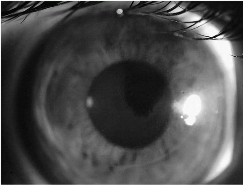 FIGURE 82-5. Interface blood under the flap minutes after the procedure. The source of the blood is the superior pannus as a result of extended soft contact lens wear. (Courtesy of Farid Eghbali, O.D., with permission.) |
A thorough preoperative evaluation and attention to the presence of and location of a peripheral corneal pannus can help guide the surgeon in modifying the size of the flap or the location of the hinge to try to avoid this complication. Preoperative argon laser ablation of large peripheral vessels has been reported to close the vessels (18). Additionally, the use of topical brimonidine has been suggested to decrease the risk of subconjunctival hemorrhage, bleeding from the micropannus, and postoperative hyperemia (25).
Intraoperatively, if bleeding occurs, every effort should be made to first avoid extension of the blood into the interface. Prior to lifting the flap, a sponge soaked with 2.5% or 10% phenylephrine drops can be placed over the area of bleeding to vasoconstrict the vessels (2). Manual pressure can also be applied onto the suction ring for a short period of time to encourage coagulation of the vessel. A Gimbel-Chayet sponge may be used to prevent tracking of the blood into the interface or onto the stromal bed (19). If during the laser stromal bed ablation bleeding continues, laser treatment should be interrupted while the blood is wiped off of the bed. Intermittent irrigation may help minimize blood in the interface (19). Often flap replacement and interface irrigation tamponades any oozing vessels at the conclusion of the procedure (18,19). Given the need for more aggressive irrigation of the interface, readherence of the flap may be delayed and drying time should be increased. In the authors’ experience, bleeding rarely prevents an excellent outcome.
Decentered Flap
Decentered flaps can occur as a result of surgeon decentration of the suction ring, globe torque, and loss of suction, lack of patient cooperation, and error in centering the optical axis. Often there is only a slight decentration of the flap, and laser ablation can safely be done if the ablation zone centered on the optical axis falls within the margins of the flap. When the decentration is to a degree that the surgeon cannot safely proceed with the laser ablation, the flap should be repositioned and the surgery postponed until 3 to 4 months later when a new flap can be recut (18).
Corneal Perforation
Corneal perforation is a devastating complication of LASIK that rarely occurs with the newer generation of microkeratome systems. It has been described in the context of improper microkeratome assembly with failure to properly place the depth plate into the microkeratome assembly. With meticulous assembly of the microkeratome system, as well as the current use of newer generation microkeratomes with fewer components, this devastating complication is highly preventable (26,27).
If corneal perforation occurs during LASIK, sudden expulsion of the intraocular contents can take place as a result of markedly elevated IOP. Rapid response time in this setting is critical. Aqueous leakage, if recognized early, can prompt the surgeon to immediately stop the power and suction and, it is hoped, limit the ocular damage. In the event of perforation, the eye should be protected with a shield and the patient immediately transferred to the operating room to undergo anterior segment reconstruction with possible anterior vitrectomy, lensectomy with or without intraocular lens implantation, iridoplasty, corneal laceration repair, and possibly penetrating keratoplasty. A retinal specialist may be required, as these eyes often suffer vitreoretinal complications (18).
Laser Ablation-Related Complications
Central Islands
With newer software and with scanning beam and flying spot lasers, central islands occur infrequently. The occurrence of central islands in association with the earlier generation broad-beam lasers resulted in loss of best corrected visual acuity and poor visual quality (28). According to Kang et al. (29), the diagnosis of central islands is made based on topographic examination with a central area 2.5 mm or more in diameter of a higher refractive power of more than 1.5 D as compared to the midperiphery (Fig. 82-6). Patients present clinically with halos, glare, ghosting, and residual myopia. Most cases present within the first week after LASIK and more than 75% of cases persist more than 6 months (28). Excimer laser phototherapeutic keratectomy has been performed on the stromal bed in the persistent cases with some positive results but with unpredictability of the refractive outcome (28,30). A small-diameter shallow PRK can also be effective in managing these islands.
Decentered Ablation
Decentered ablation (Fig. 82-7) can result in postoperative irregular astigmatism, loss of best spectacle-corrected and uncorrected visual acuity, and visual aberrations (i.e., glare, halos, ghost images) (18). It can occur as a result of poor fixation by the patient or poor centration of the laser beam by the surgeon. Prior to initiating the laser ablation, the surgeon and the assistant should confirm proper patient head positioning. With reassurance and calm instruction of the patient throughout the laser ablation, the surgeon can guide good patient fixation. If the patient loses fixation, the surgeon must stop the ablation and continue only after adequate fixation is regained. Rarely a fixation ring is warranted. Mild to moderate amounts of decentration (up to 1 mm) are typically well tolerated. It is possible that eye-tracking systems will reduce
the incidence of decentration, but most studies to date have not documented a lower risk of decentration with eye-tracking delivery systems (31,32).
the incidence of decentration, but most studies to date have not documented a lower risk of decentration with eye-tracking delivery systems (31,32).
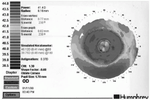 FIGURE 82-6. An axial map showing a central area of steepening adjacent to an area of flattening post myopic LASIK. This is known as a central island. (Courtesy of Farid Eghbali, O.D., with permission.) |
Irregular Astigmatism
Irregular astigmatism is defined as one with an irregular topographic map (18). It can result from a decentered ablation, incorrect flap repositioning, epithelial ingrowth or interface debris, irregular or incomplete lamellar keratectomy, and preexisting irregular astigmatism. Usually minor postoperative irregular astigmatism resolves within several months and only 1% to 2% of patients develop enough irregular astigmatism to cause loss of vision (18). With irreversible irregular astigmatism, rigid gas-permeable contact lenses can be prescribed to obtain the best corrected visual acuity. The utility of wavefront-guided excimer lasers in treating postLASIK irregular astigmatism has not yet been
evaluated in the initial clinical trials, but may be an enticing option for this difficult to treat subset of patients.
evaluated in the initial clinical trials, but may be an enticing option for this difficult to treat subset of patients.
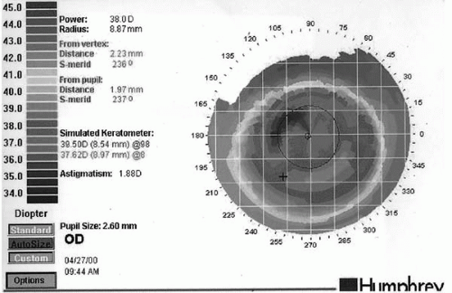 FIGURE 82-7. This topography map shows an ablation decentered inferiorly by approximately 1 mm. Each of the boxes on the map corresponds to 1 mm on the cornea. (Courtesy of Farid Eghbali, O.D., with permission.) |
Over- and Undercorrection
Residual refractive error may result from undercorrection, overcorrection, or regression with an overall re-treatment rate of 5.5% to 28% (33,34). Undercorrection or regression is seen more frequently especially after high myopic corrections. It may result from difficult preoperative refraction, unstable ametropia, or simply patient-specific factors affecting the healing process, and the expected results are based on population nomograms. Assuring a stable preoperative refraction can help decrease the chance of postoperative refractive surprises. This is especially true in patients who have a long history of contact lens use. The recommendation is for patients using soft contact lenses to discontinue use for at least 2 weeks and those using rigid gas permeable lenses to discontinue use for at least 3 to 4 weeks prior to a refractive surgery evaluation. This helps avoid instability of refractive error as a result of corneal warping. If there is any question, some recommend reevaluating in 1 month to prove a stable refractive error (within 0.50 D of sphere or cylinder and minimal change in axis) prior to proceeding.
Overcorrection occurs less frequently partly because surgeons often err on the side of undercorrection to avoid the risk of postoperative hyperopia in their myopic patients. At high altitudes with lower humidity levels in the laser room, the corneal stroma dehydrates, resulting in more tissue ablation for each laser spot delivered; this in turn results in overcorrection. Regardless of the cause, postoperative hyperopia as a result of overcorrection leads to more patient dissatisfaction and is more difficult to treat than residual postoperative myopia.
Regression results in unstable postoperative refractive outcome with continued loss of effect over several months. It is thought to occur as a result of epithelial hyperplasia and remodeling of the stroma in response to surgery (10). More regression is often noted with greater depth of ablation and smaller treatment zone. Usually stability is noted within 3 months after surgery for moderate refractive errors and within 6 months for high refractive errors (10). It is important not to confuse progressive keratectasia and related myopic shift with regression.
In the instance that postoperative refractive outcome is not ideal and is proven to have stabilized, the patient may opt to undergo an enhancement. The approach for the retreatment can follow either lifting the original lamellar flap or recutting a new flap. Most relift the original flap especially if done within 1 year of the original surgery, but recutting a flap, especially if a larger ablation zone is necessary, can offer a safe and effective alternative. Although studies have shown no statistically significant difference in complication rates between these two approaches, each incurs its own set of risks, which is outside the scope of this chapter.
POSTOPERATIVE COMPLICATIONS
Interface Debris
Nonorganic and organic materials can be introduced into the interface during LASIK. Talc from surgical gloves, lint from the drapes, metal shavings from microkeratome blades, and sponge fibers have all been described (35). Organic materials such as mucus or oil from the tear film are also commonly seen deposited in the interface (18,35). If the debris is noted during or immediately after the procedure, it probably should be immediately removed by irrigation of the interface. If noted on follow-up visits, interface debris such as mucus or tear film and even metal filings or sponge particles rarely cause inflammation, irregular astigmatism, or loss of best corrected or uncorrected visual acuity and need not be removed. Inorganic materials such as talc might result in inflammation and loss of best corrected visual acuity, and their removal should be contemplated (36). Regardless of the source of the debris, if there is loss of visual acuity or quality due to inflammation, irregular astigmatism, and flap irregularity deemed secondary to the debris, every attempt should be made to irrigate or if needed lift, irrigate, and reposition the flap. Of importance is the ability to differentiate debris from diffuse lamellar keratitis (DLK), infectious keratitis, or epithelial ingrowth, all of which require immediate attention and intervention as discussed further in this chapter.
Flap Displacement
Flap displacement is an immediate postoperative complication that normally occurs within 24 to 48 hours. The incidence ranges from 0.85% to 2.0% (8,15,37). The direction of the displacement is usually a function of the location of the hinge. For example, a nasal hinged flap often displaces inferiorly, whereas a superior hinged flap displaces laterally. The etiology of the displacement is mainly mechanical in nature. Eye rubbing, dry eyes, and hitting the flap with the tip of postoperative medication bottles are thought to account for mechanical shifting of the flap in some patients. Other causes include a poorly adherent flap due to poor endothelial pump function or excessive intraoperative stromal hydration. Delayed traumatic flap displacement has also been reported as late as 38 months after surgery (38). A review of the literature has demonstrated some unexpected causes such as accidental self-induced amputation of the flap or accidental trauma during sports (39, 40, 41).
Many surgeons evaluate the flap biomicroscopically immediately after surgery to ensure that the flap has not been displaced from blinking. Some surgeons recommend at least a 2-minute drying time at the conclusion of the surgery prior to removing the lid speculum to allow for flap adherence to the stromal bed, whereas others believe it is best to maintain a well-lubricated surface (18). Also, the use of an eye shield for three nights after the procedure results in a very low risk of inadvertent nocturnal flap displacement.
Flap displacement exposes an area of stroma that as a result can increase the chance of DLK, infectious keratitis, or epithelial ingrowth. It is therefore important to surgically reposition the flap promptly. Shifted or dislodged flaps can be managed by lifting the affected area, cleaning the epithelium or debris from the stromal bed, and refloating the flap into its correct position. If the repositioning is excessively delayed, the flap can become markedly edematous with a shrunken diameter, increasing the difficulty of repositioning and resulting in delayed adherence to the stromal bed and a large asymmetric gutter. Sutures may be needed to secure the flap in the correct alignment.
When a small flap displacement occurs, the rapid epithelial growth over the exposed stroma can sometimes confuse the diagnosis, mimicking flap striae. Careful slit-beam inspection of the gutter can help differentiate flap displacement from flap striae and guide the surgeon toward the correct management.
Corneal Neurotrophic Epitheliopathy and Dry-Eye Syndrome
Corneal sensation is mediated by axon terminals of the long ciliary nerves derived from the ophthalmic division of the trigeminal nerve (42). Large radial branches (stromal nerve roots) of the ciliary nerves penetrate the corneal domain from the limbus in the anterior third of the stromal depth, with most entering from the 3 and 9 o’clock meridia. As these nerves course to the center of the cornea, they branch horizontally and vertically, giving rise to the dense subepithelial plexus above Bowman’s layer (42, 43, 44, 45). Fibers from this plexus pass into the epithelium and form a network called the basal epithelial nerve plexus, from which intraepithelial nerve terminals emerge between the epithelial cells (46). When these stromal nerve roots are transected proximally, as occurs in the formation of the lamellar LASIK flap, the distal nerve endings degenerate and corneal sensation is lost (47, 48, 49, 50, 51). This has led to the term postLASIK neurotrophic epitheliopathy, describing a focal phenomenon in which central keratopathy occurs within the margins of the LASIK flap (52). This condition is thought to be due to alterations of the normal epithelial physiology as a result of a decreased blink reflex, tear flow, and local neuromodulatory factors (47,48,52, 53, 54).
Stay updated, free articles. Join our Telegram channel

Full access? Get Clinical Tree


