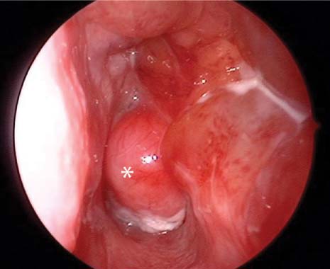
72 An 11-year-old boy was referred to a tertiary care center with a history of recurrent self-limiting epistaxis. The epistaxis was spontaneous and usually left sided. He also complained of 6 months of left-sided nasal obstruction. There was no history of trauma. He has had no change in vision or facial sensation. He denied headache. His medical history was unremarkable. He has no allergies and takes no medication. His surgical history was significant for a previous endoscopic procedure to remove a similar mass 1 year ago. On physical examination, obstruction of the left nasal passage was demonstrated by retained secretions. His extraocular motion was intact. He had no diplopia. His visual acuity was grossly intact. The other cranial nerves were intact. His oral cavity and oropharynx were unremarkable. Nasopharyngoscopy demonstrated a fleshy red mass in the left posterior choanae, and it filled the nasal cavity. The nasal septum was pushed to the right, and the medial maxillary wall was partially taken down (Fig. 72.1). The differential diagnosis of a child with a nasal mass includes nasal polyps, antrochoanal poly, nasopharyngeal angiofibroma, hemangiopericytoma, squamous papilloma, inverted papilloma, sinus mucocele, lymphoma, encephalocele, nasal glioma, teratoma, esthesioneuroblastoma, lymphoid hyperplasia, rhabdomyosarcoma, or carcinoma. Fig. 72.1 Nasal endoscopic image of posterior nasal choanal obstruction with recurrent vascular mass. JNA may be secondary to a desmoplastic response of the embryonic chondrocartilage during the development of the cranial bones. The tumor is found at the superior margin of the developing sphenopalatine foramen formed by the bifurcation of the palatine bone, the horizontal ala of the vomer, and the root of the pterygoid process. From this location the tumor has easy access to the pterygopalatine fossa through the pterygomaxillary fissure. The tumor may pass into the nasopharynx, the infratemporal fossa, or through the skull base intracranially. As the tumor grows, it expands the pterygomaxillary fissure, causing anterior bowing of the posterior wall of the maxillary antrum. A hormonal theory for development of a JNA has been suggested because of the lesion’s occurrence in adolescent males. Etiology from nonchromaffin paraganglionic cells of the terminal branches of the maxillary artery has also been suggested. Deletions of chromosome 17, including regions for tumor suppressors, are present. Patients typically present with nasal obstruction and epistaxis. As the tumor progresses, hyponasal speech and cheek and palate deformities may occur. The mass presents as a red, smooth, mucosa-covered compressible mass in the nasal cavity and nasopharynx. JNA is an aggressive benign neoplasm. Recurrence rates after treatment vary from 0 to over 50%. The plain radiographic signs thought to be classic for juvenile nasopharyngeal angiofibroma are found in Table 72.1. However, in most centers computed tomography (CT) will be the primary diagnostic modality.
Juvenile Nasopharyngeal Angiofibroma
History
Differential Diagnosis—Key Points
Test Interpretation
Nasopharyngeal mass Anterior bowing of the posterior wall of the maxillary antrum Erosion of the sphenoid bone Erosion of the hard palate Erosion of the medial maxillary sinus Displacement of the nasal septum |
The anterior bowing of the posterior wall of the antrum seen on lateral skull film is known as the Holman-Miller sign and is pathognomonic for juvenile nasopharyngeal angiofibroma.
CT with contrast or magnetic resonance imaging (MRI) is helpful for diagnosis and staging. With these imaging studies, the extent of involvement and intracranial extension may be determined. The location and radiographic appearance of the tumor are generally diagnostic.
Stay updated, free articles. Join our Telegram channel

Full access? Get Clinical Tree


