and Yi Ning J. Strube2
(1)
Wright Foundation for Pediatric Ophthalmology and Adult Strabismus Medical Center, Los Angeles, CA, USA
(2)
Queen’s University, Kingston, Ontario, Canada
Keywords
Infantile esotropiaCongenital esotropiaCiancia’s syndromeAccommodative infantile esotropiaCongenital esotropia observational studyOptokinetic nystagmus asymmetrySmooth pursuit asymmetryResidual esotropiaMobius syndromeInferior oblique overactionDVDDHDAn esotropia (ET) presenting during the first 6 months of life is termed infantile esotropia. Of the various presentations of infantile esotropia, these are the most common:
Small-angle neonatal esotropia
Congenital esotropia
Ciancia’s syndrome
Accommodative infantile esotropia
More than 70 % of normal neonates typically have a small exotropia, which usually resolves by 4–6 months of age. Infantile esotropia, on the other hand, is rare and usually does not resolve spontaneously.
3.1 Small-Angle Neonatal Esotropia
3.1.1 Clinical Features
Esotropia 15–35 PD; variable angle
Onset birth to 2 months of age
Approximately 30 % will resolve spontaneously by 6 months of age
3.1.2 Etiology
Unknown etiology.
3.1.3 Clinical Evaluation
3.1.3.1 Amblyopia
Unusual, as the deviation is often intermittent and there is some binocular fusion. Unless strong fixation preference is present, do not treat with patching, which could break down weak fusion. Treat amblyopia by patching the dominant eye 2–4 h a day until the patient holds fixation well with the nondominant eye. Follow up every 1–2 weeks to test for change in fixation preference. A week or two of patching can reverse fixation preference in these young infants.
3.1.3.2 Cycloplegic Refraction
Use cyclopentolate 1 % (one or two doses, 5 min apart). Refract 30 min after the last dose.
3.1.3.3 Complete Ocular Examination
A complete ocular examination, including a dilated retinal exam, is important to rule out a sensory esotropia. Sensory esotropia can be caused by an infantile cataract, retinoblastoma, optic nerve hypoplasia, or any other cause of infantile visual loss.
3.1.4 Management
The Congenital Esotropia Observational Study (CEOS) sponsored by the National Institutes of Health has shown that infants with a small-angle (≤35 PD), variable, or intermittent esotropia have a high rate of spontaneous resolution, as approximately one third will resolve by 6 months of age [1]. These infants should have a cycloplegic refraction, and if hypermetropic of +3.00 sphere or more, give spectacles with the full correction. (See Sect. 3.4 later in this chapter.) If the infant is not significantly hypermetropic, observe for spontaneous resolution until the infant is 6–9 months old. Some patients will show an increasing esotropia and they should be considered for early surgery if the deviation becomes constant and is ≥40 PD on at least two consecutive exams. (See Sect. 3.2 below.)
Premature infants will frequently have a small, variable esotropia. There is not much in the literature to guide us in these cases. It is probably best to watch these patients for several months for spontaneous resolution. Consider surgery at 9–12 months of corrected age if a constant esotropia >15–20 PD persists, and the child is not a significant anesthesia risk.
3.1.4.1 Surgical Procedure
Surgery is based on the near deviation, as this is the most reliable measurement in these young children. Bilateral medial rectus (MR) recessions are preferred because the deviation is usually comitant. If there is unilateral vision loss, then a monocular recession-tightening procedure is performed on the poor-seeing eye.
3.2 Congenital Esotropia
3.2.1 Clinical Features
Large-angle constant esotropia (>40 PD)
Onset from birth to 6 months of age
Spontaneous resolution is rare
Amblyopia is common (50 %)
Associated motor phenomena (usually present after 2 years of age):
Inferior oblique overaction (IOOA) (60 %)
Dissociated vertical deviation (DVD) (40 %)
Latent nystagmus (40 %)
3.2.1.1 Smooth Pursuit Asymmetry/Optokinetic Nystagmus Asymmetry
Normal children and adults have precise and symmetrical smooth pursuit for following an object moving slowly from side to side. Infants, however, have smooth pursuit asymmetry, with a deficiency in nasal-to-temporal smooth pursuit, compared with pursuit following an object moving in a temporal-to-nasal direction. The nasal-to-temporal–directed smooth pursuit will lag behind the target, and saccadic movements intrude to catch up. This asymmetry is demonstrated only during monocular viewing. Smooth pursuit asymmetry is a manifestation of visual motor immaturity and naturally resolves as motor fusion develops by 4–6 months of age. Patients with disorders that disrupt binocular visual development, such as congenital esotropia or unilateral congenital cataract, retain smooth pursuit asymmetry throughout life, despite surgery [2]. Thus, the presence of smooth pursuit asymmetry in older children and adults with esotropia is a sign of neonatal onset with early disruption of binocular visual development.
3.2.2 Etiology
Unknown etiology.
3.2.3 Preoperative Evaluation
3.2.3.1 Ductions
Mild (−1) limitation of abduction is common and does not indicate a lateral rectus paresis. Try the doll’s head maneuver or spinning the child (vestibular stimulation) to elicit full abduction. Intact abduction saccadic eye movements in the face of mild limitation of abduction indicate good lateral rectus function and a tight medial rectus muscle.
3.2.3.2 Differential Diagnosis of Infantile ET With Limited Abduction (In Order of Decreasing Incidence)
Ciancia’s syndrome (tight medial rectus muscles) (See below)
Duane’s syndrome
Congenital fibrosis syndrome
Congenital sixth nerve palsy (very rare—usually transient, resolving by 4 months of age)
Infantile myasthenia gravis
3.2.3.3 Versions
Check for inferior oblique overaction and V pattern.
3.2.3.4 Amblyopia
Check fixation preference. Strong preference for one eye indicates amblyopia.
3.2.3.5 Measure Deviation
Prism alternate cover testing is best, but use Krimsky testing for verification or if prism cover testing is unobtainable. Surgery is usually based on the near deviation, which is the most reliable in infants. When possible, measure the distance and near deviation.
3.2.3.6 Cycloplegic Refraction
Use cyclopentolate 1 % (one or two doses, 5 min apart). Refract 30 min after the last dose.
3.2.4 Management
In general, congenital esotropia is a surgical disease and requires strabismus surgery. If the cycloplegic refraction shows ≥+3.00 sphere, then prescribe the full hypermetropic correction. If an esotropia of more than 10–15 PD persists after full hypermetropic correction, then surgery is required. (See Sect. 3.4 below.)
3.2.4.1 Amblyopia
Treat amblyopia before surgery by patching the dominant eye for 4–6 h per day until the patient holds fixation well with the nondominant eye. Follow up every 1–2 weeks to test for change in fixation preference. A week or two of patching can reverse fixation preference in these young infants. Patients may cross-fixate, fixing with the right eye for objects in the left visual field and with the left eye for objects in the right visual field. Unless strong fixation preference is present, cross-fixation usually indicates absence of significant amblyopia.
3.2.4.2 Timing of Surgery for Congenital Esotropia
Most references recommend that surgery for congenital esotropia be performed between 6 months and 1 year of age in order to achieve peripheral fusion and low-grade stereo acuity. This author (KWW) reported experience with very early surgery showing that surgical correction between 3 and 4 months of age can result in high-grade stereo acuity [3]. Early surgery should be considered if there is a constant large-angle esotropia (≥40 PD) with the angle stable or increasing on at least two examinations, 2 or more weeks apart. The CEOS showed that spontaneous resolution was rare (<4 %) if these parameters were met [4]. Intermittent small-angle esotropia, on the other hand, will resolve in approximately one third of cases, so it is better to wait until these patients are at least 6 months of age before considering surgery.
3.2.4.3 Surgical Procedure
The procedure of choice is bilateral medial rectus (MR) muscle recessions using the near deviation as the target angle. (Use surgical chart in Appendix for specific numbers.) The standard surgical chart numbers are designed to give infants with infantile esotropia a slight immediate overcorrection, which is desirable, as convergence will pull the eyes straight. (See Sect. 3.2.4.4 below.). In older patients and adults with long-standing congenital esotropia, the chart numbers may give a slight undercorrection. This is desirable, as these patients usually have poor fusion potential and tend to drift to exotropia over time. Thus the surgical chart numbers can be used for all ages, as they tend to self-adjust for age. Patients with irreversible dense amblyopia should have monocular surgery (recession-tightening procedure) on the amblyopic eye to protect the “good eye.”
Example 3.1 Clinical Example: Congenital ET (Fig. 3.1)
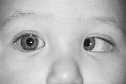
Fig. 3.1
A 5-month old with congenital esotropia
5-month old, constant large esotropia since first few weeks of life.
Strong fixation preference OD
Ductions—trace limitation to abduction, versions—no oblique dysfunction
Full abduction and good saccade to vestibular stimulation (doll’s head maneuver).
Cycloplegic refraction:
OD +1.75 sphere
OS +2.00 sphere
Nsc: ET 60 PD by alternate cover test and Krimsky prism test
Dsc: ET 50 PD by estimation
(D—distance; N—near; sc—without correction)
Diagnosis: Congenital esotropia, strabismic amblyopia left eye, slightly limited abduction probably secondary to tight medial rectus muscles, but not a sixth-nerve paresis because there is a good abduction saccade.
Preoperative treatment: Patch right eye 6 h a day and follow every week until equal fixation preference is achieved, indicating that amblyopia has improved. At 5 months of age, only 1 or 2 weeks of patching are usually needed to improve the amblyopia.
Surgery: Bilateral medial rectus muscle recessions, 6.5 mm for target angle ET 60 PD. (See Appendix A on Surgical Numbers.) Do not prescribe spectacles preoperatively, but if there is a small residual esotropia after surgery, try prescribing the full hypermetropic correction.
3.2.4.4 Surgical Goals
The immediate postoperative goal is a small exotropia (5–10 PD) for infants with possible fusion potential. We naturally have strong, innate fusional convergence (>30 PD), so a small exotropia is “good” because it can be fused. A small esotropia, on the other hand, is difficult to fuse, as our divergence amplitudes are weak (approximately 8 PD).
Dr. Marshall Parks, during his studies on monofixation syndrome and peripheral fusion, found that in order to develop binocular fusion, the eyes must be within 8–10 PD of orthotropia. The goal of surgery is to align the eyes during early infancy to within 8–10 PD, to stimulate the development of binocular fusion. The closer the alignment to orthotropia, the better the sensory outcome. An esotropia larger than 8–10 PD will not allow binocular fusion (not even peripheral fusion). A residual esotropia larger than 10 PD should be considered for further treatment. (See next section.) Patients with a poor prognosis for binocular fusion (e.g., dense irreversible amblyopia, or uncorrected congenital esotropia in a patient more than 2 years old) should be considered for surgery based on cosmetic indications.
Stay updated, free articles. Join our Telegram channel

Full access? Get Clinical Tree


