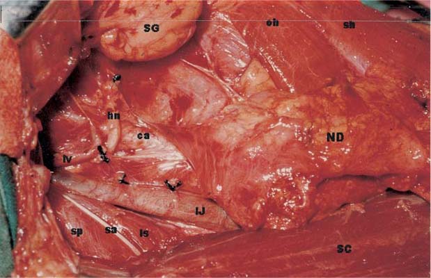
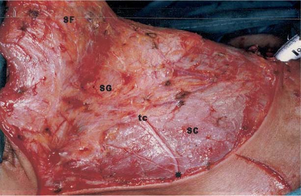
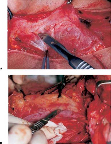
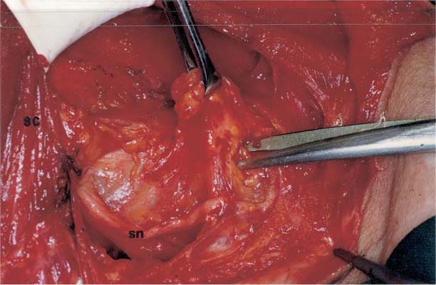
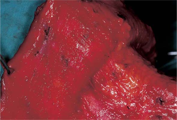
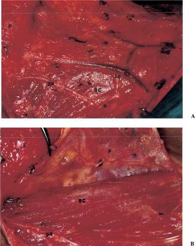
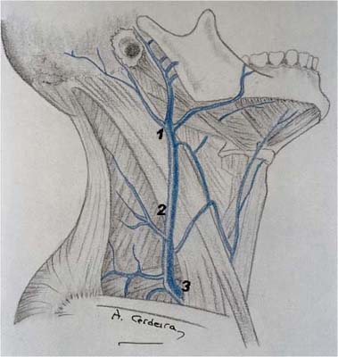
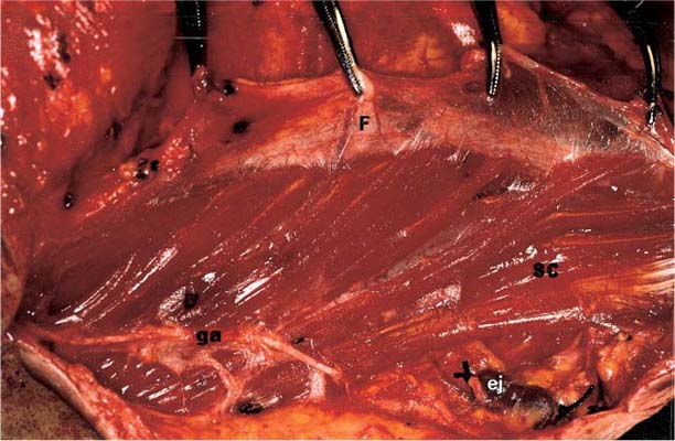
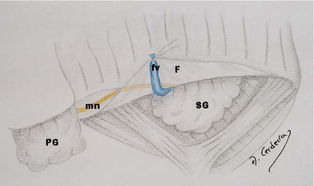
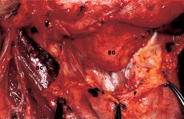
CHAPTER 5 The principle of fascial dissection is more easily achieved when the surgeon uses the knife through fascial planes. For some steps of the operation the scalpel is the best surgical tool whereas for others the scissor is preferred. Elevation of the skin flaps and dissection of the sternocleidomastoid muscle, submandibular fossa, deep cervical muscles, carotid sheath, and strap muscles are best performed using knife dissection. On the other hand, dissection of the area around the spinal accessory nerve, posterior triangle, and paratracheal space is more easily accomplished with the scissor. The main difference between these two groups is the type of tissue that is being dissected. Knife dissection requires firm tissue like muscle or vessels (Fig. 5-1), whereas fibrofatty tissue is more easily dissected with the scissors (Fig. 5-2). Knife dissection requires precise handling of the knife, careful surgical technique, and adequate help from the assistants. The blade of the scalpel must be directed oblique to the tissue that is being dissected and away from the muscle or vessel whose fascia is being removed. This protects the structures, especially the veins, from being injured by the knife blade. To be appropriate, knife dissection must be carried all the way up and down the surgical field, avoiding the creation of holes along the dissected structure. The knife blade is much more efficient when cutting over tense tissue. Thus, the assistants must apply adequate tension to the surgical field to increase the effectiveness of knife dissection. Clear vision of the different structures in the surgical field is of paramount importance. Blood obscures the field and makes identification of structures more difficult. A bloodless field must be maintained throughout the operation. In addition, washing the field regularly with warm saline greatly contributes to cleaning the working area (Fig. 5-3). Figure 5-1 The knife is preferred for the dissection of firm tissue like muscle (A) or vessels (B). Safe knife dissection requires placing the tissue under traction, as seen in the photos. The superficial layer of the cervical fascia must remain intact after the flaps have been raised. This may pose a problem to the novice surgeon, who usually finds it difficult to preserve the integrity of this fascial layer. The best way to achieve this goal is by cutting with the scalpel over the deep face of the platysma muscle. As for any other type of neck dissection, the platysma muscle is included with the skin flaps because it provides additional blood supply that protects the skin and assists in the healing process. The proper sequence for an adequate incision will be to mark the skin incision, incise the skin, cut the platysma muscle, and start raising the flap, keeping the deep face of the platysma under vision. If the muscular fibers of the platysma are seen throughout the elevation of the skin flaps (Fig. 5-4), preservation of the superficial layer of the cervical fascia is assured. Figure 5-2 Fibrofatty tissue is best dissected with the scissors. Here an example of the dissection of the supraclavicular fossa on the right side. SC, sternocleidomastoid muscle retracted medially; sn, supraclavicular nerve. Figure 5-3 Regularly washing the field allows better visualization of the anatomical structures. SG, submandibular gland; IJ, internal jugular vein; ca, carotid artery; lv, lingual veins; sa, spinal accessory nerve; hn, hypoglossal nerve; SC, sternocleidomastoid muscle; oh, omohyoid muscle; sh, sternohyoid muscle; sp, splenius capitis muscle; ls, levator scapulae muscle; ND, neck dissection specimen. Figure 5-4 Fibers of the platysma muscle after elevation of the skin flaps. The great auricular nerve is used to identify the posterior border of the upper part of the surgical field (Fig. 5-5A). This branch of the cervical plexus rounds the posterior border of the sternocleidomastoid muscle from Erb’s point and courses almost directly upward toward the ear lobule, where it supplies almost all the auricle, the skin over the parotid gland, and the skin over the mastoid process. Whenever possible, the great auricular nerve should be preserved to avoid numbness of the ear, which is especially disturbing in female patients (Fig. 5-5B). The external jugular vein begins in the substance of the parotid gland. It is most often formed by the union of the retromandibular (posterior facial) and the posterior auricular veins. It runs vertically downward across the superficial surface of the sternocleidomastoid muscle to pierce the fascia of the posterior triangle of the neck just above the clavicle. The external jugular vein terminates in the subclavian or in the internal jugular vein after receiving several tributaries throughout its cervical course (Fig. 2-12). During functional neck dissection, the external jugular vein is found at different stages of the operation and should be ligated and divided at different levels. In a complete functional neck dissection, there are three places where the external jugular vein must be ligated and divided (Fig. 5-6). From topdown the vein must be transected at the tail of the parotid gland during the dissection of the upper part of the surgical field at the level of the posterior border of the sternocleidomastoid muscle during the dissection of the muscle and within the fibrofatty tissue of the supraclavicular fossa while dissecting the posterior triangle of the neck. Figure 5-5 Identification and preservation of the great auricular nerve on the right side. (A) The great auricular nerve crosses the external face of the sternocleidomastoid muscle from Erb’s point toward the ear lobule. (B) The fascia over the sternocleidomastoid muscle is incised anterior to the great auricular nerve in order to preserve innervation of the ear lobule. ga, great auricular nerve; tc, transverse cervical branch of the cervical plexus; SC, sternocleidomastoid muscle; P, platysma muscle; F, fascia dissected from the sternocleidomastoid muscle; *; Erb’s point. Figure 5-6 The external jugular vein must be ligated and divided at different levels during the operation. 1, tail of the parotid gland; 2, posterior border of the sternocleidomastoid muscle; 3, supraclavicular fossa. During the ligature of the external jugular vein and other large veins in the neck special attention should be directed to distal venous stumps. Open distal venous stumps may be responsible for air embolism. Thus, careful closure of all distal stumps must be ensured before closure of the wound. To facilitate the complete dissection of the fascia surrounding the sternocleidomastoid muscle the initial incision must be made as close to the posterior border of the muscle as possible (Fig. 5-7). The reason for this is that the fascia is more easily dissected off the sternocleidomastoid muscle in a forward direction. Making the incision close to the posterior border of the muscle leaves no remaining fascia to be dissected posteriorly and facilitates the complete isolation of the muscle from its surrounding fascia. It is cosmetically important to preserve the marginal mandibular branch of the facial nerve. The mandibular nerve courses just deep to the superficial layer of the cervical fascia but superficial to both the anterior facial vein and artery. Identification of the nerve is time consuming and may require nerve stimulation for the novice surgeon to confirm the exact location of this thin branch of the facial nerve. Figure 5-7 Lateral view of the dissection of the sternocleidomastoid muscle on the right side. The fascia is incised over the posterior border of the muscle and dissected forward. The great auricular nerve has been preserved. Note the external jugular vein ligated at the posterior border of the sternocleidomastoid muscle and above the clavicle. SC, sternocleidomastoid muscle; F, fascia dissected from the sternocleidomastoid muscle; ga, great auricular nerve; ej, external jugular vein. It is much easier, and equally safe, to identify, ligate, and divide the anterior facial vein at the inferior border of the submandibular gland. The superior ligature, which is left long, is reflected superiorly with a hemostat (Fig. 5-8). Although the mandibular nerve is usually not seen during this maneuver, it will be automatically reflected superiorly with the skin flap, thus preventing its injury. As already mentioned, removal of the submandibular gland is not a routine surgical step of functional neck dissection. The gland must be included in the specimen when the location of the primary tumor dictates its removal or when metastatic disease is suspected in the submandibular triangle. In the remaining situations the submandibular gland may be preserved. This is the case with cancer of the larynx and hypopharynx, where the lymph nodes in the submandibular and submental region (area I) are usually not involved and, additionally, there is no need to approach the primary tumor through the submandibular triangle. When a submandibular gland preserving functional neck dissection is performed, the dissection of the upper border of the surgical field must be modified with respect to the procedure described in the previous chapter. After the flaps have been raised, the submandibular gland can be seen through the superficial layer of the cervical fascia in the upper part of the surgical field (Fig. 5-9). The fascia is then incised from the midline to the tail of the parotid gland, at the level of the lower border of the submandibular gland as for a gland-removing procedure. Then the facial vein is ligated and divided, reflecting upward the superior ligature to preserve the marginal mandibular branch of the facial nerve. The retromandibular vein and the external jugular vein are also ligated and divided. Figure 5-8 Protection to the marginal mandibular branch of the facial nerve is obtained with the maneuver of the facial vein (right side). fv, facial vein; mn, marginal nerve; F, fascia retracted upwards; SG, submandibular gland; PG, parotid gland. Now, instead of including the submandibular gland within the specimen, its fascia is dissected inferiorly while the gland is retracted superiorly (Figs. 5-10 and 5-11). The contents of the submandibular fossa are now exposed. The fibrofatty tissue containing the submandibular nodes is grasped and dissected off the submandibular triangle, preserving the gland. The dissection may be continued medially to include the submental nodes, but this is seldom required in tumors that allow preservation of the submandibular gland. Figure 5-9 Lateral view of the surgical field after elevation of the skin flaps, preserving the deep layer of the cervical fascia (right side). SG, submandibular gland; SC, sternocleidomastoid muscle; tc, transverse cervical nerve; SF, superior skin flap; *; Erb’s point. Figure 5-10 The fascia is incised in the upper boundary of the surgical field and dissected inferiorly over the submandibular gland. SG, submandibular gland; SC, sternocleidomastoid muscle; F, fascia dissected inferiorly. The dissection then continues over the digastric and stylohyoid muscles. The muscles are retracted superiorly (Fig. 5-12
Hints and Pitfalls
KNIFE DISSECTION AND THE FUNCTIONAL APPROACH
WASHING THE FIELD REGULARLY
RAISING THE FLAPS
IDENTIFICATION OF THE GREAT AURICULAR NERVE
MANAGEMENT OF THE EXTERNAL JUGULAR VEIN
INCISION OF THE FASCIA OVER THE STERNOCLEIDOMASTOID MUSCLE
THE MARGINAL MANDIBULAR BRANCH OF THE FACIAL NERVE
PRESERVING THE SUBMANDIBULAR GLAND
![]()
Stay updated, free articles. Join our Telegram channel

Full access? Get Clinical Tree


