Purpose
To evaluate whether the handheld in vivo reflectance confocal microscopy that has been recently developed for the study of skin tumors is suitable for the diagnosis of conjunctival tumors.
Design
Prospective study, observational case series.
Methods
We prospectively evaluated the reflectance confocal microscopy features of 53 conjunctival lesions clinically suspicious for tumors of 46 patients referred to the University Hospital of Saint-Etienne (France) by using the handheld device. Twenty-three lesions were excised (3 nevi, 10 melanomas, 5 squamous cell carcinoma, 2 lymphomas, and 3 pinguecula/pterygium) while the other 30, presenting no reflectance confocal microscopy malignant features, were under follow-up for at least 1 year. Clinical reflectance confocal microscopy and histologic diagnosis were compared.
Results
In vivo reflectance confocal microscopy diagnosis was in agreement with the histologic diagnosis in all cases and none of the lesions that were not excised show any clinical progression under follow-up.
Conclusion
In vivo reflectance confocal microscopy with a handheld dermatology-dedicated microscope can play a role in the noninvasive diagnosis of conjunctival lesions. Further studies should be performed to better define the diagnostic ability of this technique.
The clinical diagnosis of conjunctival tumors is often challenging, and in case of clinical doubt of malignant tumor excision is mandatory. However, a surgical excision in this area may have functional or aesthetic consequences for the patient and may be technically complex for the surgeon. New imaging techniques are of high interest in the diagnosis of conjunctival tumors to reduce the number of unnecessary surgical excisions. In vivo reflectance confocal microscopy is a noninvasive, high-resolution imaging technique that has been demonstrated to be useful for the diagnosis of conjunctival tumors. However, this technique is not extensively used, mainly owing to the difficulty of acquiring the images, especially because reflectance confocal microscopes dedicated to ophthalmology have limited numbers of degrees of freedom and limited possibilities of mechanical displacement. We investigated whether the handheld reflectance confocal microscope that has been developed for the cutaneous tumors, and which has been demonstrated to be applicable to the study of the genital and the oral mucosa with easy and quick examinations, could play a role in the diagnosis of conjunctival tumors. We present a series of conjunctival tumors described from a clinical standpoint and the first that has been investigated by the handheld in vivo reflectance confocal microscope.
Methods
Forty-six patients (24 female, 22 male, average age 53 years, range 13-94 years) presenting with a total of 53 conjunctival lesions suggestive of tumors were recruited at the Dermatology Department of the University Hospital of Saint-Etienne, France, between January 1, 2011 and June 30, 2013. Clinical and in vivo reflectance confocal microscopy diagnosis were prospectively established together by 2 ophthalmologists (D.G., E.M.) and 3 dermatologists (E.C., J.-L.P., B.L.). Tumors confined to the eyelid margin were excluded owing to the particular features of this area of transition between the tarsal conjunctiva and the skin.
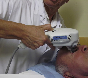
A slit-lamp examination was performed to establish the clinical diagnosis. In vivo reflectance confocal microscopy examination was carried out with the handheld microscope dedicated to skin (VivaScope 3000; Caliber, Rochester, New York, USA, distributed in Europe by MAVIG GmbH, München, Germany) ( Figure 1 ), which uses an 830-nm class 1B (classification CDRH—Center for Devices and Radiological Health) diode laser not harmful to eyes, with a wavelength that does not induce ocular glare. This system has a high optical resolution (horizontal and vertical axis: 1.25 μm and 5 μm, respectively) and allows visualization of tumors up to 250 μm in depth from the epithelial surface of the conjunctiva. Each image corresponds to a horizontal section of 1 mm × 1 mm. Before the ocular examination, topical anesthesia was performed with oxybuprocaine hydrochloride 1.6 mg/0.4 mL and tetracaine hydrochloride 1% applied into the lower conjunctival sac of the eye and a transparent ophthalmic gel of Carbomer 974P (Gel larme; Théa, Clermont-Ferrand, France) was placed on the ocular region to be examined. A disposable sterile transparent film (Visulin; Paul Hartmann AG, Heidenheim, Germany) was applied on the reflectance confocal microscope tip. In some cases an eyelid retractor was used to prevent blinking. Examinations were performed on patients in supine position. Reflectance confocal microscopy examination time per lesion was around 5-10 minutes.
An institutional review board approval was obtained (institutional review board at the University Hospital of Saint-Etienne, France, number IORG0007394; study filed under reference number IRBN332014/CHUSTE). A patient informed consent was always obtained orally during the first consultation and before this examination.
Before imaging the tumors, the normal conjunctiva of the contralateral eye was also observed under in vivo reflectance confocal microscopy and features of normal conjunctiva were assessed. We examined both benign and malignant conjunctival tumors, including nevi, primary acquired melanosis (PAM), melanomas, extrascleral growth of uveal melanomas, squamous cell carcinomas (SCC), and lymphomas. Moreover, some cases of pinguecula and pterygium were also observed when in doubt of SCC. Two cases of nevus of Ota were also examined to exclude the presence of areas of malignant transformation.
The in vivo reflectance confocal microscopy diagnosis of nevus was determined in the presence of junctional and/or stromal hyperreflective homogeneous medium-sized (10-20 μm), roundish cells organized in nests, with the absence of (1) pagetoid cells, (2) atypical cells at the epithelium-stromal junction, and (3) disarrangement of the epithelial layers. The presence of stromal pseudocyst-like structures partly filled with monomorphous material allowed diagnosis of the epithelial-cystic nevus. Lesions with hyperreflective cells confined to the basal layer of the epithelium and/or small (<20 μm) pagetoid dendritic cells at in vivo reflectance confocal microscopy were diagnosed as PAM without atypia, whereas lesions with hyperreflective cells throughout the epithelium and large pagetoid dendritic cells (>20 μm) were diagnosed as PAM with atypia. The in vivo reflectance confocal microscopy diagnosis of conjunctival melanoma and extrascleral growths of uveal melanomas were established in the presence of large (>20 μm) dendritic or roundish hyperreflective cells at the epithelial-stromal junction and/or in the stroma associated with the possible presence of large pagetoid cells.
The in vivo reflectance confocal microscopy diagnosis of SCC was made in the presence of large (>20 μm) epithelial cells and/or a disarranged pattern of the epithelium. The presence of horizontal and dilated blood vessels and the presence of abundant dendritic cells was an additional criterion that confirmed the SCC diagnosis. Pinguecula and pterygium were diagnosed in the absence of epithelial atypia and the subepitehlial presence of degenerated stromal collagen that presented with a coiled shape and a fibrovascular proliferation, respectively. Increased leukocytes (small hyperrefractive roundish homogeneous cells) in the stroma were an additional finding.
Mucosa-associated lymphoid tissue (MALT) lymphoma was diagnosed in the presence of a normal epithelium and abundant small hyperrefractive roundish cells corresponding to lymphocytes in the stroma.
A surgical excision and a histopathologic examination were performed in 20 cases clinically and/or in vivo reflectance confocal microscopy suspicious for malignant tumors (melanoma, SCC, MALT lymphoma). In addition, the first 3 cases of nevi were excised to perform a histopathologic examination considering that the in vivo reflectance confocal microscopy aspect of the conjunctival nevi had never been described using the handheld camera.
The correlation between in vivo reflectance confocal microscopy and the histopathologic examination was evaluated. Lesions that were not biopsied because considered benign at in vivo reflectance confocal microscopy examination were then monitored for at least 12 months.
Results
In vivo reflectance confocal microscopy features of the normal conjunctiva were the presence of (1) 3-6 layers of polygonal medium-size (10 μm) cells with hyperreflective membranes, hyporeflective cytoplasm, and medium reflective round nuclei in the epithelium, (2) a flat epithelial-stromal junction and (3) elongated hyperreflective structures organized in a dense meshwork (corresponding to collagen fibers) and prominent capillaries in the stroma ( Figure 2 ). The movement of blood cell flow could be observed in real time inside blood capillaries.
The clinical features, the in vivo reflectance confocal microscopy, and the histopathologic diagnosis of the lesions are reported in the Table . Considering all the 23 excised lesions ( Figures 3 and 4 ), 12 lesions were clinically challenging, and 3 cases of melanomas were clinically diagnosed as benign lesions, whereas in vivo reflectance confocal microscopy diagnosis was in agreement with the histologic diagnosis in all cases, including 10 melanomas, 5 SCC, and 2 MALT lymphomas.
| Patient Number | Sex | Age | Location | Size (mm) | Clinical Feature | Evolution (Years) | Clinical Diagnosis | Confocal Microscopy Diagnosis | Histologic Diagnosis | Confocal Microscopy/Histology Correlation |
|---|---|---|---|---|---|---|---|---|---|---|
| 1 | M | 78 | Bulbar | 2 × 2 | Dark brown flat papule | 0.5 | Melanoma? | Melanoma | Melanoma | Yes |
| 2 | F | 73 | Bulbar/tarsal | 5 × 3 | Multiple macules, light brown | 10 | Melanoma | Melanoma | Melanoma | Yes |
| 3 | F | 94 | Bulbar | 4 × 3 | Multiple macules, dark brown | 10 | Melanoma | Melanoma | Melanoma | Yes |
| 4 | F | 42 | Bulbar | 3 × 2 | Light brown macule | 2 | Melanoma | Melanoma | Melanoma | Yes |
| 4 | F | 42 | Tarsal | 4 × 2 | Light brown macule | 2 | Melanoma | Melanoma | Melanoma | Yes |
| 5 | M | 64 | Bulbar | 3 × 3 | Red macule | 9 months | Pinguecula? | Melanoma | Melanoma | Yes |
| 5 | M | 64 | Tarsal | 3 × 4 | Brown macule | 9 months | Nevus? | Melanoma | Melanoma | Yes |
| 5 | M | 64 | Bulbar | 2 | Brown macule | 3 months | Melanoma? | Melanoma | Melanoma | Yes |
| 5 | M | 64 | Tarsal | 3 × 6 | Brown macule | 10 months | Melanoma | Melanoma | Melanoma | Yes |
| 6 | F | 66 | Limbal | 6 × 8 | Black flat papule | 7 | Nevus? | Melanoma | Melanoma | Yes |
| 7 | M | 60 | Caruncle | 4 × 3 | Pink papule | NA | Nevus | Compound nevus | Compound nevus | Yes |
| 8 | F | 19 | Limbal | 1 × 1 | Brown macule | 1 | Nevus | Subepithelial nevus | Subepithelial nevus | Yes |
| 9 | M | 59 | Bulbar | 10 × 6 | Brown macule | 10 | Nevus | Epithelial-cystic nevus | Epithelial-cystic nevus | Yes |
| 10 | M | 81 | Limbal | 3 × 2 | Flat rose papule | 0.5 | SCC? | SCC | SCC in situ | Yes |
| 11 | F | 55 | Limbal/bulbar | 9 × 10 | Red macule | 1 | SCC | SCC | SCC in situ | Yes |
| 12 | F | 47 | Limbal | 5 × 5 | Pink macule | NA | SCC | SCC | SCC | Yes |
| 13 | M | 73 | Limbal | 5 × 5 | Reddish papule | 1 month | SCC | SCC | SCC | Yes |
| 14 | M | 77 | Limbal | 5 × 5 | Pink nodule | 0.5 | SCC? | SCC | SCC | Yes |
| 15 | F | 59 | Limbal | 3 × 3 | Whitish nodule | 10 | SCC? | Pinguecula | Pinguecula | Yes |
| 16 | F | 69 | Limbal | 4 × 3 | Whitish nodule | 2 | SCC? | Pterygium | Pinguecula | Yes |
| 17 | M | 71 | Limbal | 3 × 4 | Pink papule | 1 | SCC? | Pinguecula | Pinguecula | Yes |
| 18 | M | 73 | Tarsal | 8 × 5 | Pink patch | 1 month | MALT lymphoma? | MALT lymphoma | MALT lymphoma | Yes |
| 19 | F | 64 | Tarsal | 4 × 3 | Pink papules | 0.5 | MALT lymphoma? | MALT lymphoma | MALT lymphoma | Yes |
| 20 | M | 51 | Bulbar | 3 × 2 | Brown macule | 3 months | Melanoma? | Junctional nevus | NA | NA |
| 21 | M | 56 | Limbal | 1 × 1 | Light brown macule | Congenital | Nevus | Dermal nevus | NA | NA |
| 22 | F | 71 | Caruncle | 2 × 2 | Light brown papule | 5 | Nevus | Subepithelial nevus | NA | NA |
| 23 | M | 17 | Limbal/bulbar | 8 × 5 | Brown-yellow patch | Congenital | Nevus | Compound nevus | NA | NA |
| 24 | F | 23 | Caruncle | 3 × 1 | Brown macule | NA | Nevus | Junctional nevus | NA | NA |
| 25 | M | 58 | Bulbar | 7 × 6 | Flat brown papule | 48 | Nevus | Epithelial-cystic nevus | NA | NA |
| 26 | F | 33 | Bulbar | 4 × 4 | Macule with a light and dark brown pigmentation | Congenital | Nevus | Junctional nevus | NA | NA |
| 27 | F | 28 | Bulbar | 5 × 5 | Brown patch | 10 | Epithelial-cystic nevus | Epithelial-cystic nevus | NA | NA |
| 28 | F | 32 | Bulbar | 1.5 | Brown papule | 2 | Nevus | Epithelial-cystic nevus | NA | NA |
| 29 | F | 84 | Bulbar | 2 × 4 | Light brown macule | 1 | Nevus | Epithelial-cystic nevus | NA | NA |
| 30 | M | 61 | Caruncle | 15 × 7 | Brown macule | 45 | Nevus | Epithelial-cystic nevus | NA | NA |
| 31 | M | 31 | Limbal | Brown macule | 5 | Nevus | Epithelial-cystic nevus | NA | NA | |
| 32 | M | 17 | Limbal | 7 × 5 | Erythematous macule | 10 | Nevus | Subepithelial nevus | NA | NA |
| 33 | M | 68 | Bulbar | 5 × 3 | Flat light brown papule | 10 | Epithelial-cystic nevus | Epithelial-cystic nevus | NA | NA |
| 34 | M | 51 | Bulbar | 3 × 2 | Brown papule | 10 | Epithelial-cystic nevus | Epithelial-cystic nevus | NA | NA |
| 35 | M | 41 | Bulbar | 3 | Light brown macule | 10 | Racial melanosis | Compound nevus | NA | NA |
| 35 | M | 41 | Bulbar | 2 | Light brown macule | 10 | Racial melanosis | Compound nevus | NA | NA |
| 35 | M | 41 | Bulbar | 2 | Light brown macule | 10 | Racial melanosis | Compound nevus | NA | NA |
| 36 | F | 13 | Bulbar | 6 × 4 | Erythematous macule | Congenital, but recently enlarged | Nevus | Subepithelial nevus | NA | NA |
| 37 | M | 27 | Caruncule, conjunctive | 1 | Brown macule | 3 | Nevus | Subepithelial nevus | NA | NA |
| 15 | F | 59 | Bulbar | Brown and gray macules | NA | Nevus of Ota | Nevus of Ota | NA | NA | |
| 38 | M | 48 | Bulbar | 1 × 1 | Brown and gray macules | Congenital | Nevus of Ota | Nevus of Ota | NA | NA |
| 39 | F | 48 | Bulbar | 3 × 4 | Multifocal light brown macule | 10 | PAM | PAM | NA | No |
| 40 | F | 62 | Limbal | 2 × 1 | Light brown macule | 10 | PAM | PAM | NA | NA |
| 41 | F | 66 | Tarsal | 5 | Light brown macule | 0.5 | PAM | PAM | NA | NA |
| 42 | F | 33 | Limbal | 4 | Light brown macule | 10 | PAM | PAM | NA | NA |
| 43 | F | 40 | Bulbar | 10 | Brown macule | 6 | Racial melanosis | PAM | NA | NA |
| 44 | F | 29 | Bulbar | 3 × 4 | Light brown macule | 4 | Racial melanosis | PAM | NA | NA |
| 45 | M | 72 | Limbal | 5 × 2 | Grayish nodule | NA | Pterygium vs SCC? | Pterygium | NA | NA |
| 46 | F | 68 | Limbal | 2 × 2 | Red nodule | 10 | Pterygium vs SCC? | Pterygium | NA | NA |
Stay updated, free articles. Join our Telegram channel

Full access? Get Clinical Tree


