Purpose
To assess the accuracy and reliability of smartphone ophthalmoscopy, we compared the ability of a smartphone ophthalmoscope with that of a slit-lamp biomicroscope to grade diabetic retinopathy (DR) in patients with diabetes mellitus (DM).
Design
Clinical-based, prospective, comparative instrument study.
Methods
This comparative clinical study was performed in 120 outpatients (240 eyes) with type 1 or type 2 DM. After pupil dilation, the patients underwent smartphone ophthalmoscopy with the D-Eye device, followed by dilated retinal slit-lamp examination, to grade DR according to a 5-step scale.
Results
Overall exact agreement between the 2 methods was observed in 204 of 240 eyes (85%) (simple κ = 0.78; CI 0.71-0.84) and agreement within 1 step was observed in 232 eyes (96.7%). Compared to biomicroscopy, the sensitivity and specificity of smartphone ophthalmoscopy for the detection of clinically significant macular edema were 81% and 98%, respectively. Smartphone ophthalmoscopy and biomicroscopy could not be used to examine the fundus and grade DR in 9 eyes (3.75%) and 4 eyes (1.7%), respectively, because of cataract and/or small pupil diameter.
Conclusion
Smartphone ophthalmoscopy showed considerable agreement with dilated retinal biomicroscopy for the grading of DR. The portability, affordability, and connectivity of a smartphone ophthalmoscope make smartphone ophthalmoscopy a promising technique for community screening programs.
In ophthalmology, images are used extensively for disease documentation, treatment monitoring, and educational purposes. Traditionally, fundus images have been obtained with expensive and bulky tabletop units operated by a trained technician in a hospital clinic setting. The pervasive adoption of smartphones by physicians and their ever-improving built-in camera technology has raised much interest in their use for medical and ophthalmic imaging. The portability and immediate connection capabilities of smartphones make them an attractive device for acquiring retinal pictures in remote nonhospital settings. Indeed, telemedicine has the potential to reach patients and communities that currently receive no or suboptimal eye care as a result of geographic and/or sociocultural barriers.
In the past decade, retinal screening programs for common eye diseases, such as diabetic retinopathy (DR), glaucoma, and age-related macular degeneration, have experienced rapid growth. The application of these screening programs in rural, nurse-operated, highly remote primary care facilities highlights the importance of having access to an inexpensive, portable, easy-to-operate, and high-image-quality fundus camera.
Particularly, diabetes mellitus (DM) is a complicated chronic disease that requires continual medical care and patient education to minimize acute and long-term complications. Most clinical practice recommendations suggest that a comprehensive eye examination must be performed at least annually to assess the DR grade in all patients with DM. However, a large percentage of patients with DM (35%-79%) do not receive the recommended level of ophthalmic care.
To capitalize on the potential versatility of smartphones in the screening of DR and other ocular diseases, various prototypes have been created to optically match the smartphone’s camera to a slit-lamp ophthalmoscope, or to use it in conjunction with a condensing lens based on the principle of indirect ophthalmoscopy.
Smartphone ophthalmoscopy can nowadays be performed with the help of the D-Eye system, which is a novel, inexpensive, and very portable optical device designed to be magnetically attached to a smartphone.
The purpose of this study was to validate the efficacy of the D-Eye device to screen for diabetic retinopathy in the community. We compared the ability of smartphone ophthalmoscopy with that of dilated retinal biomicroscopy to grade DR in patients with DM.
Materials and Methods
Study Design
This was a prospective clinic-based comparative study of eyes affected by DR. This study was conducted in the ophthalmic Diabetic Center of “Spedali Civili di Brescia,” according to the ethical principles of the Declaration of Helsinki. The institutional review board of the Eye Clinic (University of Brescia, Italy) approved the study protocol (registered with clinicaltrials.gov , identifier NCT02177747). All study participants provided written informed consent.
Overall, 120 consecutive patients with diabetes, new to the Diabetes Center’s outpatient clinic, underwent dilated retinal digital imaging with a smartphone ophthalmoscope as a part of their routine examination for diabetes. Subsequently, they were referred for a comprehensive dilated retinal biomicroscopy with a slit lamp by a retinal specialist.
Dilating eye drops (0.5% tropicamide and 10% phenylephrine) were administered to outpatients with diabetes who were scheduled for an examination at the Diabetes Center; after 20 minutes; smartphone ophthalmoscopy was performed in these patients by a retinal specialist (L.D.). Subsequently, retinal slit-lamp examination, according to normal clinical practice, was performed by another retinal specialist (A.R.) who was masked to the findings of smartphone ophthalmoscopy. Each ophthalmoscopy procedure was reported using a similar template, in which physicians were asked whether the pupil was dilated and the media clear enough to visualize abnormalities in the fundus. Next, they were asked a series of questions regarding the presence or absence of microaneurysms, dot/blot hemorrhages, hard exudates, soft exudates, intraretinal microvascular abnormalities, venous beading, new vessel formation, fibrous proliferation, vitreous hemorrhage, scars of any previous laser photocoagulation, and clinically significant cystoid macular edema (significant CME, according to the ETDRS criteria ). DR was then graded according to the International Clinical Diabetic Retinopathy Disease Severity scale : no apparent retinopathy, mild nonproliferative DR (NPDR, microaneurysms only), moderate NPDR (more than just microaneurysms but less than severe NPDR), severe NPDR (more than 20 intraretinal hemorrhages in each of the 4 quadrants, definite venous beading in 2 or more quadrants, and prominent intraretinal microvascular abnormalities in 1 or more quadrants), or proliferative DR (neovascularizations and vitreous/preretinal hemorrhage).
Smartphone Ophthalmoscopy
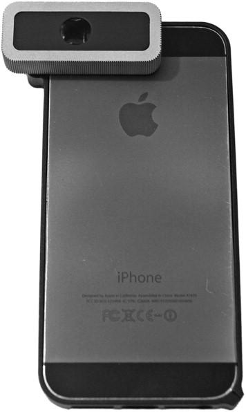
After pharmacologic dilation, a retinal specialist (L.D.) performed a comprehensive dilated fundus examination with a final prototype ( Figure 1 ) of the D-Eye adapter (Si14 S.p.A., Padova, Italy; http://www.d-eyecare.com ) attached to an iPhone 5 (Apple Inc, Cupertino, California, USA). The images were captured on 3264 × 2448 pixels of the camera’s sensor. Thus, direct fundus ophthalmoscopy was performed using live images displayed on the smartphone’s screen (a video showing the acquisition procedure is available as Supplemental Material at AJO.com ).
When the pupil is dilated, the device captures a field of view of approximately 20 degrees in a single fundus image at a distance of 1 cm from the patient’s eye. An acquisition protocol was therefore followed to pan the retina, starting from the posterior pole and then moving to the upper, nasal, inferior, and nasal peripheral retina to the equator. Color digital images and videos of the retina were obtained, encompassing the posterior pole, including the macula, optic disc, and peripheral retina.
Dilated Fundus Biomicroscopy
Twenty minutes after smartphone ophthalmoscopy, a retinal specialist (A.R.), masked to the findings of smartphone ophthalmoscopy, performed a comprehensive dilated fundus examination with a slit-lamp biomicroscope. For this study, dilated fundus biomicroscopy was considered the gold standard for adjudication of differences in DR grading between smartphone ophthalmoscopy and biomicroscopy.
Analysis of Data
Descriptive statistics were used to present the demographic and ocular baseline characteristics. Overall agreement, sensitivity, and specificity of the 2 ophthalmoscopy techniques were calculated and compared. To assess the agreement between smartphone and slit-lamp ophthalmoscopy, the κ statistic was used, which was also adopted by the ETDRS. Statistical analyses were performed using SPSS software version 20 (SPSS Inc, Chicago, Illinois, USA).
Results
Of the 120 patients with DM who underwent smartphone ophthalmoscopy and slit-lamp biomicroscopy, 55 (45.8%) were men and 28 (23.3%) had type I DM. The mean age at examination was 58.8 ± 16.4 years, and mean duration of DM was 11.6 ± 9.7 years.
Grading of Diabetic Retinopathy
The eye fundus was not gradable for DR in 9 eyes (3.75%) by smartphone ophthalmoscopy and in 4 eyes (1.7%) by biomicroscopy because of cataract and/or small pupil diameter.
The clinical level of DR found with both techniques is reported in Table 1 . An exact agreement was found in 204 of 240 eyes (85%) and an agreement within 1 step was observed in 232 eyes (96.7%). Simple κ was 0.78 (95% confidence interval 0.71-0.84; P < .001), showing a substantial agreement for the grading of DR between smartphone ophthalmoscopy and slit-lamp biomicroscopy. In 82% of 1-step disagreements and 93% of disagreements by 2 or more steps, the severity level was higher by biomicroscopy grading.
| Dilated Slit-Lamp Biomicroscopy | |||||||
|---|---|---|---|---|---|---|---|
| Smartphone Ophthalmoscopy | No Apparent DR | Mild NPDR | Moderate NPDR | Severe NPDR | Proliferative DR | Not Gradable | Total |
| No Apparent DR | 110 (45.8%) | 12 (5%) | 0 (0%) | 0 (0%) | 0 (0%) | 0 (0%) | 122 (50.8%) |
| Mild NPDR | 5 (2.1%) | 44 (18.3%) | 5 (2.1%) | 3 (1.2%) | 0 (0%) | 0 (0%) | 57 (23.8%) |
| Moderate NPDR | 0 (0%) | 0 (0%) | 27 (11.2%) | 5 (2.1%) | 0 (0%) | 0 (0%) | 32 (13.3%) |
| Severe NPDR | 0 (0%) | 0 (0%) | 0 (0%) | 11 (4.6%) | 1 (0.4%) | 0 (0%) | 12 (5%) |
| Proliferative DR | 0 (0%) | 0 (0%) | 0 (0%) | 0 (0%) | 8 (3.3%) | 0 (0%) | 8 (3.3%) |
| Not Gradable | 0 (0%) | 3 (1.2%) | 1 (0.4%) | 1 (0.4%) | 0 (0%) | 4 (1.7%) | 9 (3.8%) |
| Total | 115 (47.9%) | 59 (24.6%) | 33 (13.8%) | 20 (8.3%) | 9 (3.8%) | 4 (1.7%) | 240 (100%) |
Stay updated, free articles. Join our Telegram channel

Full access? Get Clinical Tree


