18 Facial Plastic Surgery in Geriatric Patients
 Introduction
Several predictable changes and alterations are typical and normal sequelae of the neck and facial aging process. Unlike other components of the body’s predictable aging changes, aging in the face and neck region carries other significant implications, including those that impact cultural, social, and personal perceptions. Historically there has been an evolution of facial rejuvenation procedures in response to those perceptions. The procedures have gained significant public acceptance and desirability. In the not too distant past facial procedures were often felt to be within the realm of wealthy celebrities or others in the entertainment business. Today these procedures are widely accepted and actively explored by a wide spectrum of social, economic, and ethnic backgrounds. The goal is to achieve an enhanced self-perception of greater fitness and youth coupled with a response to cultural pressures to maintain a youthful appearance related to work, economic, and various social interactions.1,2
Although the geriatric population requires facial plastic surgery for facial trauma and fractures, facial neoplasms, and occasionally persistent congenital defects, the focus of facial plastic surgery in the geriatric population more frequently addresses facial aging processes. An individual’s age is usually judged according to appearance of the skin.3 Although much attention is focused on cutaneous gerontology, the appearance of cutaneous senescence is actually based on a combination of skeletal structure, soft tissue, and skin (Fig. 18.1). The primary changes in a person’s three-dimensional skeletal contour contribute to secondary changes in the overlying soft tissue and skin. It has been said that a youthful face represents that period in time when a particular set of skeletal proportions are ideal for their soft tissue envelope.
What is generally termed facial aging is in reality a combination of the aforementioned factors. Facial wrinkles or rhytids are related to changes in the skin secondary to a variety of factors, including chronological skin aging, ultraviolet (UV) and other environmental factors leading to photoaging, hyperdynamic facial expressions, and skin folding secondary to loss of soft tissue support and skeletal changes. Reversal or techniques to counteract these changes create the foundation for facial plastic surgery in the geriatric patient (Fig. 18.2).
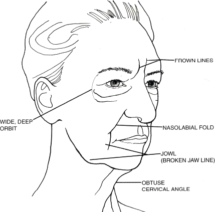
Fig. 18.1 Typical aging changes. (Used with permission from Facial Plast Surg Clin North Am 2001;9(2):179–187.)
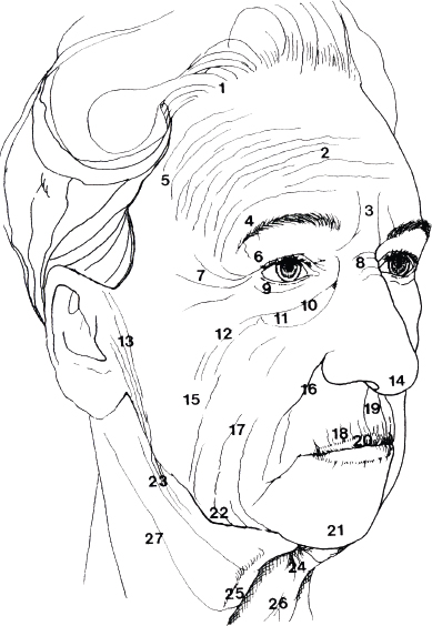
Fig. 18.2 Topographic geriatric facial changes. (Used with permission from Facial Plast Surg Clin North Am 2001;9(2): 179–187.)
 Analysis and Pathophysiology of Facial Aging
Geriatric skin changes are a product of two basic processes. First is chronological aging, sometimes referred to as intrinsic aging, the changes of which are primarily genetic. Second is environmental or extrinsic aging, particularly from such stressors as sun exposure or smoking. Skin changes often seen in the geriatric patient include dyschromia, roughness, and multiple rhytids followed by persistently deeper folds. Structurally this occurs due to dermal atrophy, decreased collagen, and loss of cutaneous fat coupled with loss of inherent elasticity and increased melanogenesis4
The structural changes in soft tissue already noted are variable but can be significant as part of the aging process, two major forces impact these changes. First is chronological aging of the skin related to intrinsic passage of time and second, photoaging related to chronic ultraviolet light exposure. Other components may impact this as well, such as smoking and certain dietetic aspects.5 The skin of geriatric patients shows declining changes with age, which ae further accelerated in UV exposed photoaged skin. Photoaging is responsible for many of the age-associated cosmetic concerns, including dispigmentation, laxity, changes of pores, telangiectasia, a leathery appearance, and wrinkling. Vascular changes increase during this period, creating the tendency for bruising during activities of daily life as well as medical procedures. Loss as well as shifting and redistribution of subcutaneous tissue further contribute to folds and drooping skin. Because of these changes, dealing with soft tissue in the geriatric patient carries additional considerations as compared with more youthful patients:
• Cell replacement
• Immune response
• Injury response
• Barrier function
• Sensory perception
• Sweat production
• Sebum production
• Vitamin D production
The facial changes typically associated with the geriatric patient are a result of a combination of forces related to facial aging including gravity, soft tissue maturation, skeletal remodeling, facial muscular changes, and solar changes to the skin. The aging process comprises a complex and diverse set of elements, each of which leaves a specific and distinctive result on the aging face. The anatomical and tissue changes already noted are intimately interconnected, each having an impact on the other components. Skin, soft tissue, muscle, and skeleton, although affected individually by the aging process, present themselves as an interactive aging process acting in concert to result in the facial appearance typically seen in the geriatric patient.
 Geriatric Skin Changes
The geriatric patient displays several predictable changes related to ongoing aging affect. These changes include secondary results of gravity acting on skin that is becoming progressively thinner, dryer, and less elastic. There are genetic factors also influencing the location of facial wrinkles, and these progress as a result of cutaneous senescence combined with cumulative environmental insults.
Chronic solar exposure and UV interaction constitute the major environmental component contributing to the clinical changes that are typical of aging skin. This is accurately termed photoaging and is distinct from intrinsic or chronological aging.
The relationship of inherent aging changes in the geriatric patient’s skin can be contrasted with those changes that are compounded by photoaging and chronic solar exposure. More photologic changes and sun-protected skin consist primarily of fine wrinkling and laxity with occasional benign neoplasm development. Those who leave their skin exposed to chronic solar damage demonstrate changes of texture, color, and functional capacity. Color changes include dyschromia, often a yellow pallor, lentigines, and the gradual appearance of telangiectasia. Textural changes likewise accompany these variations and include a loss of palpable smoothness, keratosis, and increasing rhytids ending in persistent deep folds and creases. Histological changes associated with aging skin begin with a thinning of the viable epidermis and flattening of the dermal–epidermal junction. This is in contrast to photoaged skin in which the epidermis becomes increasingly thicker with a flattening of the dermal–epidermal junction.
Of the dermal structural elements, elastic fibers are prominently affected by the sequelae of both chronologically damaged and photodamaged skin. Chronically aged skin reveals elastic fibers that are slightly increased in thickness. Photodamaged skin shows striking histological changes with the presence of massive quantities of thickened bundles of degraded elastic fibers or dermal elastosis. The result of these various histological reactions and changes in aging skin is that the skin itself is less stretchable and less resilient. The loss of inheritant elasticity results in skin that is more lax with dependent draping that is prone to wrinkling from gravitational effects. There is also an increase in mechanical fragility and a susceptibility to lateral sheer stress combined with an increase in dryness and skin irritability.6
 Geriatric Facial Musculature
Contractions and sphincteric actions of the facial musculature are responsible for facial expressions exhibiting emotion as well as functional actions including eye closing, lip movement, and numerous others. However, over time and combined with aging skin changes facial rhytids and folds are accentuated by this normal muscular action and begin to contribute to changes in facial appearance associated with aging in the geriatric patient. For example, continued contraction of the orbicularis component of the orbicularis oculi creates concentric folds emanating from the lateral canthus resulting in lines referred to as crow’s feet. The corrugator supercilii lies deep to the frontalis muscle and has it origin on the frontal bone medial to the eyebrows near the superior medial orbital rim. The corrugator acts as a brow abductor drawing the brows inferiorly and medially and thus ultimately producing vertical frown lines. In the same anatomical region, the procerus muscle originates on the lower part of the nasal bone and inserts in the skin overlying the nasion. This likewise pulls the medial brow inferiorly, creating transverse wrinkles over the nasal bridge. The antagonist of the brow depressors is the frontalis muscle, which is ultimately responsible for horizontal forehead creases. All of these actions, which are normal facial movements, ultimately contribute to an aging facial appearance and are key areas addressed, particularly by Botox (Allergan, Irvine, CA), as a treatment modality. Similarly, the normal muscle actions of the lower facial mimetic muscles and the perioral muscles all contribute to rhytids and skin folds related to aging changes in the lower face. Although it is a normal process for these hyperdynamic facial lines, crow’s feet, and glabellar furrows to reflect underlined muscular anatomy, the ultimate result is reflective of what are typically thought of as unaesthetic changes in the face related to aging.
 The Aging Nose
Often overlooked in terms of facial anatomical changes that are recognized as aging in the face are the progressive changes to nasal appearance and its impact on facial aesthetics. The nose at a central location is a key component of facial aesthetics and facial relationships, and the aging changes that affect the nose affect the rest of the facial appearance. Aging alters the nasal skeleton, cartilaginous framework, and soft tissue covering.7 Progressive descent of the nasal tip is often associated with loss of support from changes and attachment of the upper and lower level cartilages, which then lengthen and weaken the nasal tip support, thus lengthening and enlarging the nose. The nasal labia angle becomes increasingly acute in relationship to this loss of tip structure. The relative appearance of a nasal dorsal hump is exaggerated as the tip decreases in its projection. Pyriform remodeling affects the alar base, and, in combination with upper maxillary resorption, it results in superior repositioning and thus further narrowing of the nasal labial angle and accentuation of the tip ptosis. Chin pad ptosis secondary to bony resorption further contributes to the illusion of increased nasal length (Fig. 18.3).
Rhinoplasty of the geriatric patient often not only contributes to a more youthful appearance based on repositioning of the nasal anatomy but also frequently contributes to airway improvement. Aging changes to the nose that impact nasal function include loss of the internal nasal valve support, external valve collapse, and changes of narrow airway openings related to loss of tip support. Rhinoplasty is a frequent requirement in the aging patient because of these functional and airway difficulties in addition to aesthetic goals of attaining greater youthfulness and enhanced aesthetic facial appearance in the geriatric patient.
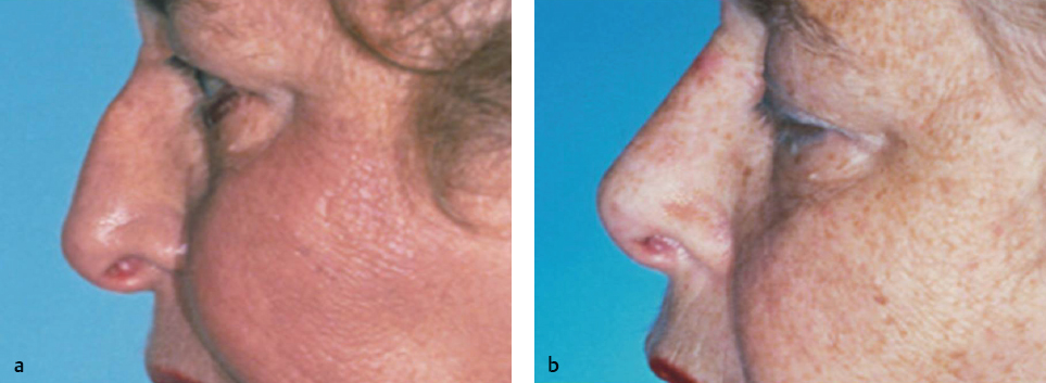
Fig. 18.3 (a) Typical findings in the geriatric nose include soft tissue thickening, loss of tip support, and narrowing of the nasolabial angle. (b) Postoperative improvements following rhinoplasty in the geriatric patient.
 Eyebrows and Eyelids in the Geriatric Patient
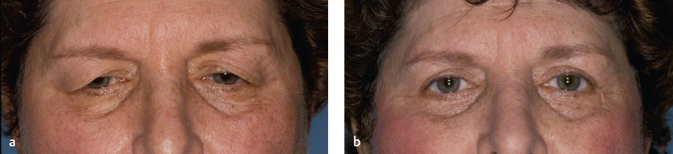
The combination of gravity, loss of tissue elasticity, a loss of subcutaneous tissue, and at times bony resorption lead to inferior displacement of the brow. With aging, typically the eyebrow position descends from above the supraorbital rim to point of some portion below it. This contributes to excess accumulation of upper lid skin that accentuates ongoing skin changes of dermatochalasis with loss of eyelid creases and additional folds of skin over the upper lid. Excess sliding of skin and a weakened orbital septum allow interorbital fat to herniate, creating eyelid bags and protruding adipose tissue. The periorbital soft tissue in the more youthful patient is often shallow and described as an unbroken convex line from the lower eyelid to the cheek. With progressive aging those relationships change the dimensions and become wider and deeper as time progresses. Ptotic cheek fat may descend inferiorly, contributing to a melolabial fold leaving a cheek depression that can be accentuated by buccal fat attenuation. The orbicularis muscle becomes increasingly ptotic; with aging its inferior border becomes increasingly apparent. This creates the malar decent of the zygomatic eminence laterally resulting in deepening of the nasojugal fold, which continues to deepen with aging. Multiple surgical procedures in the aging patient are designed to correct and reverse these predictable changes. Procedures used individually or in combination include upper and lower blepharoplasty, brow lift, and forehead lift. These surgical procedures may be used independently or in combination with other facial aesthetic procedures depending on the severity of the aging changes in the geriatric patient as well as the patient’s aesthetic goals.
 Jowl and Neck in the Aging Patient
The typical changes of the aging neck arise from a combination of changes, including changes in the skin, fat distribution, the platysmal muscle, and the underlying bony cartilaginous framework. The continual downward pull of the platysma creates jowls with the loss of definition of chin and jowl line. The jowl region along the margin of the mandible as created by ptosis of the facial portion of the platysmal muscle skin accordingly can also become lax over the platysma, developing horizontal rhytids. Typically in the aging patient anterior edges of the platysma separate and lose tone, creating the interior neck banding and, particularly in the male patient, the so-called turkey neck deformity. The common entity is development of a large submental fat pad deep to the platysmal bands as well as a similar fat pad in the subcutaneous area above the platysma. These fat accumulations contribute to the double chin appearance in those patients in addition to the sagging skin and loss of muscular support. In many patients the hyoid bone and larynx will gradually descend with age, making the larynx appear more prominent and blunting the cervicomental angle.
A variety of techniques to correct these predictable changes of aging have been described and are frequently used. These include an array of essentially facelift procedures that entail tightening of the skin and subcutaneous tissue, correction of the loosening and displaced musculature support, and elevation of the connective tissue of the face with redraping of redundant skin. These are often combined with soft tissue and facial skin surface modalities of treatment to further smooth the skin surface, which improve the appearance of cutaneous skin rhytides and act as adjuncts to surgical correction of these redundant sagging tissues of the geriatric patient facial anatomy.
 Nonsurgical Treatment in the Geriatric Face
Botulinum Toxin
Injectable botulinum toxin can be of help in correcting certain nonaesthetic anatomical changes in the aging face. Botulinum toxin is available in various types; however, type A (Botox) has become the most frequently used agent. Botox does not offer a permanent solution; however, the relative ease of treatment on an outpatient basis has added to its popularity. Although it may be more useful in the younger patient without additional soft tissue aging changes there are certainly applications for the patient in the geriatric age group, and it is frequently used in this fashion. The toxin disrupts the nerve cells’ ability to release acetylcholine. Facial rhytides or wrinkles that occur naturally over time secondary to repetitive contraction of underlying facial musculature are often felt to be nonaesthetic and a sign of aging. Temporarily weakening or paralyzing the musculature of the face in the area of rhytid formation allows the skin to settle and conform to the face without the dominate influence of muscle contraction. In most cases this will alleviate the appearance of rhytides and thus enhance facial appearance.8
Although there are a variety of types of botulinum toxin available, botulinum toxin type A is the most common neurotoxin used clinically for facial aesthetic treatment. The most common treatment indications are in the area of the upper third of the face and include forehead rhytides, thytids, and brow elevation. The periocular region, particularly in the lateral crow’s feet areas, can also be safely helped in our frequent area of indication. Areas of required facial animation in the lower face, such as the lips, must be approached with great care. Facial animation is vital to expressing human emotions, and lack of activity or emotion in those areas can have a negative effect in terms of interaction and aesthetic result.
There are certain contraindications to the use of botulinum toxins. The materials are certainly contraindicated in those with a known allergic response to any component of the product or with a general history of anaphylaxis. Additionally patients with neuromuscular conditions or peripheral motor neuropathies such as myasthenia gravis, amyotrophic lateral sclerosis, or similar syndromes are not perfect candidates. In the geriatric patient group many of the individuals are on a variety medications for treatments, and this should be explored in depth. The use of botulinum toxin should be avoided in patients using medications that may create difficulties. These would include depolarizing blockers, anticholinesterases and lincosamides, and aminoglycosides. The use of botulinum toxin is also contraindicated in those individuals with autoimmune, dermatologic, or other systemic disorders that predispose to poor wound healing or exuberant inflammatory response. Blood thinners can be considered a relative contraindication due to ecchymosis or potential hematoma formation.
The older geriatric patient should be approached in a very conservative fashion. The treating physician should remember that although the goal with botulinum toxin is to smooth some facial rhytides related to muscle contraction, this may be counterproductive in the geriatric patient. Those patients with significantly loose soft tissues and lax skin may find that, for example, smoothing the forehead rhytides creates a lowering of the upper brow to an unacceptable degree. Injection should generally be avoided within 1 cm of the margin of the ridge of the supraorbital rim and most importantly at the midpupillary line where diffusion can affect the upper eyelid levator. Treatment should be adjusted and individualized for the patient and customized based on the soft tissue anatomy and patient needs. Treatment in the lower half of the face, particularly in the perioral region, although described, should be approached very conservatively. Often at best it can offer only modest relief in specific areas and can lead to both functional and aesthetic negative appearance. Areas of inappropriate muscle weakness can potentially be a result of the injections.
Treatment in the neck area and of the platysmal muscle should likewise be approached very conservatively in the geriatric patient. Although this is described in the literature and used effectively in certain patient categories, senior or geriatric patients may have increased laxity of soft tissue of the neck that is unaesthetic and counteracts any potential improvement in the platysmal bands themselves.
 Facial Peels in the Geriatric Patient
Laser resurfacing has become increasingly popular as technical advances of new modalities have been developed. Chemical peels continue to be a potential adjunct for many patients and are actively used by some treating physicians in the aging patient with specific aging skin issues. These problems for the patient include photoaging, rhytides, keratosis, pigmentary dyschromias, and some level of superficial scarring such as from acne. There may be some improved elastic and tensile strength to the skin with some tightening of the skin. Because of this peels may be beneficial to some geriatric patients but as with all the treatments it should be individualized for the patient. There is a wide array of chemical peel modalities available, and these should be individualized according to the patients’ needs.
Skin texture and skin type can be assessed using the Glogau classification for photoaging as well as the Fitzpatrick classification of sun reactivity type. These are useful when determining which treatment agent to use (Table 18.1).
Most individuals classify chemical peels based on histological depths of reaction and treatment. The knowledge of this reaction of the peeling agent is useful in determining the appropriate selection of the correct peeling method. Superficial light peels exfoliate with fresh skin at the level of the epidermis, whereas medium-depth and deep peels can work in the papillary and the reticular dermis, respectively.
Superficial peels would include the α-hydroxyl acids such as glycolic acid 30 to 70% and Jessner solution or trichloroacetic acid (TCA) peels 10 to 20%. Medium peels, which are used for the papillary upper reticular dermis, will include Jessner solution with TCA, and deep peels would typically use a version of the Baker–Gordon phenol peel. The key ingredients are 88% phenol and croton oil.
It should be recognized that there are indeed complications with the peels, particularly in the medium and deeper depth peel solutions. The postoperative complications can include prolonged erythema, acne and milia, infection, skin dyschromia, and scarring.
As noted earlier, in the geriatric patient, it is important to determine preoperatively any medical conditions or possible interactions with other medications. Likewise, although the peels may be helpful in terms of surface improvement they will not correct significantly redundant flaccid skin, which is often associated with the older patient age group.
 Laser Skin Resurfacing in the Geriatric Patient
The goal of laser resurfacing is to give aging skin a more youthful appearance by improving dispigmentation, wrinkles, and potentially other lesions. Two different types of laser resurfacing have been developed: nonablative and ablative. Generally nonablative resurfacing is used where the surface texture has minimal wrinkling and surface change, and the treatment is usually able to spare the epidermis through adjunctive surface cooling. Ablative laser resurfacing is the more aggressive treatment for skin tightening and textural enhancement, because it removes the entire epidermis and parts of the dermis in an attempt to regenerate smoother-appearing skin in place of photodamaged skin. The treatment effect is much like the ultimate effect from dermabrasion or chemical peels. In general nonablative techniques are used in younger patients with mild and fine rhytides and perhaps early melasma. Ablative laser resurfacing is often more useful in the older patient age group where moderate to severe facial rhytides and wrinkling are present on the facial skin surface.9
Nonablative laser therapy uses a wide array of alternatives for this treatment modality, including light-emitting diode (LED), potassium-titanyl-phosphate (KTP), pulsed dye, neodymium:yttrium-aluminum-garnet (Nd:YAG), and intense pulsed light (IPL), among others. Each wavelength choice tends to offer certain advantages at the expense of some disadvantages, and the physician must balance those aspects when selecting the treatment modality. In general the nonablative skin rejuvenation treatments treat earlier photodamage and wrinkling and offer the advantage in appropriate patients of reducing patient downtime and recovery.
Table 18.1 Glogau classification of aging skin
Severity | Typical age | Features |
Mild |
| Smooth skin, minimal keratosis |
Moderate | 35–50 | Early rhytides, wrinkling, early actinic keratosis |
Advanced | 50–65 | Persistent rhytids and wrinkles, discoloration with telangiectasia and actinic keratosis |
Severe | 60–75 | Marked wrinkling and photoaging with dynamic rhytides, active actinic keratosis difficult to cover with makeup |
Complications related to laser resurfacing include prolonged erythema that may persist up to several months or longer. Infection should be rare but can happen and can be of bacterial, viral, or even fungal etiology. Acne and milia are relatively common early on but more minor for most patients in terms of their issues. Hyperpigmentation can occur 3 to 4 weeks after resurfacing and may persist for several months if treatment is not begun. Often exposure to sunlight during the healing period can stimulate melanocyte activity. Sun avoidance and sun screens are key in the postresurfacing period. More severe complications from laser treatment include hypertrophic scarring and unintended soft tissue changes such as lower lid ectropion.
 Blepharoplasty in the Geriatric Patient
The eyes play a key role in expression as well as facial identity and contribute significantly to the overall appearance of the face. The eyes are one of the first areas of the face to demonstrate aging changes and thus are a frequent component of patients’ self-evaluation and request for treatment. Actual changes are first manifest as smile lines or crow’s feet in the lateral canthus region and progress to excess skin (dermatochalasis) and fat pseudoherniation. Both the upper and lower lids typically present with changes effected by these aging sequelae. These changes combined with brow ptosis can contribute to a “tired” as well an aging look.
Upper Eyelid Blepharoplasty
Upper blepharoplasty is performed to address aging changes of the upper eyelid. The steps typically include excision of excess skin from the upper lid and removal of pseudoherniating fat as needed; most often this in the medial fat compartment. The procedure may be done in conjunction with lower lid blepharoplasty and other facial aesthetic procedures.10
The preoperative eyelid evaluation consists of examining for effective skin and fat pseudoherniation of the medial and central compartments. The eyelid crease should be approximately 9 to 10 mm above the lash line for the skin in the female. This crease in the male can be slightly shallower, in the 8 to 10 mm region. The technique begins with preoperative marking, typically done in the preoperative area with the patient in the upright animated position. The inferior level of the incision falls along the tarsal crease and typically extends superiorly as one approaches the orbital rim. Determination of the appropriate amount of redundant skin is then marked on the patient. It is helpful to use forceps to gently pinch the excess skin to determine the appropriate amount.
Once marking has been completed the skin is infiltrated with 1% lidocaine to 1:100,000 epinephrine, typically using 1 to 2 mL per side. The face is prepped and the skin incision performed. A helpful technique is to combine a small amount of hyaluronidase with the injection to allow the skin to be pinched according to the preoperative planning lines that will remain in that pinched state and facilitate excision. In some patients where greater definition is required for the upper lid, a small strip of orbicularis oculi muscle can be resected just above the sulcus. Ophthalmic cautery or bipolar cautery is used throughout to maintain hemostasis. The fat compartment can be opened and gently teased out with cotton-tipped applicator dissection, the base cauterized and excised. The wound is then closed with placement of key 6–0 polypropylene sutures followed by a running 6–0 polypropylene suture (Fig. 18.4).
Lower Lid Blepharoplasty
Lower lid blepharoplasty begins with appropriate marking with the patient in the preoperative holding area in the upright animated position, noting areas with redundant skin as well as possible pseudoherniation of fat pads. A variety of procedures are used depending on the patient’s preoperative status. Those patients with appropriate skin and muscle tightness in the lower lid but with fat protrusion can be approached through a transconjunctival approach. For the patients where it is appropriate to tighten the dermatochalasis of the lower lid, a subciliary incision can be used. Various fat preservation techniques have been described from fat repositioning to camouflaging of the orbital rim and filling the so-called tear trough or nasojugal depression. A variety of fat injections are also appropriate at times.
Typically for lower lid procedures general anesthesia or sedation may be used. For the transconjunctival approach with fat excisions local anesthesia is infiltrated in the conjunctival side of the lid with the lid retracted. An incision is made typically just below the inferior tarsal plate as the lid is retracted appropriately inferiorly. A conjunctival flap is then developed and retracted superiorly with a 5–0 silk suture exposing the lower lid fat compartments. The orbital septum is opened to each fat compartment and the appropriate amount of fat is dissected away with a cotton-tipped applicator, the base cauterized or excised. At this point the flap is released and typically no closure is required. Ophthalmic antibiotic ointment is applied within the sulcus. An iced sterile oval eye pad is then placed as the opposite side is done, and a light cold dressing is maintained postoperatively. In certain patients a transconjunctival approach can be used with fat preservation. In this situation the medial and at times middle fat compartment can be maneuvered into a subperiosteal pocket created at the orbital rim. This fat can be further held in place with a transcutaneous absorbable suture that is left in place for several days postoperatively.
The transcutaneous approach to blepharoplasty follows local injection of the lower lid, and a subciliary incision is made easily leaving several millimeters below the lash line. A submuscular flap is developed and a “skin-muscle” flap is developed and typically extended down to the orbital rim. The medial and lateral fat pads can be identified, the periosteum opened, and fat removed as needed. If orbital fat is preserved the arcus marginalis is identified and the periosteum elevated and the subperiosteal pocket is created as is described in the transconjunctival flap. The excess skin-muscle flap is then redraped and the excess skin excised. It is important to leave edge to edge apposition of the flap so that the excision can be closed without tension so as to prevent ectropion. A lateral muscle suspensory suture is often helpful suturing the orbicularis muscle component of the flap to the lateral orbital rim periosteal tissue near the lateral tarsus. Typically a polydioxanone suture is used for this. The skin is closed with interrupted or running subciliary absorbable sutures using 6–0 fastabsorbing gut, and the lateral component is typically reinforced with several 6–0 polypropylene sutures, which are removed 1 week afterward.
 Facelift in the Geriatric Patient
As in most aesthetic facial procedures, the patient in whom the aging process has been less severe tends to present the best postoperative appearance and most successful facelift procedure. Those patients who are minimally obese and have what is most often described as good bone structure tend to make the best candidates. As in all aesthetic procedures, the patient must have realistic expectations regarding surgery. Psychological evaluation of the patient along with the physical facial findings is an important part of the patient evaluation. The geriatric patient must be counseled that severely aged skin with loss of elasticity and changes in collagen as well as elastic fibers will minimize the amount of correction and longevity of the procedure. The more obese a patient is, the less likely the patient is a favorable candidate. Patients with minimal adipose content and a high cervical angle with high positioning of the hyoid and thyroid complex allows for superior results in the lower face and neck region.