Table 8.1
Stages of Eyelid Development
Cryptophthalmos
Epidemiology
Cryptophthalmos, or “hidden eye,” was first reported in 1872 by Zehender and Manz. It is a rare disorder with varying clinical presentations. Complete cryptophthalmos is most common. Isolated cryptophthalmos can either occur sporadically or in an autosomal recessive pattern. Cryptophthalmos can also be associated with two related syndromes, both of which display autosomal recessive inheritance patterns. The first is Fraser syndrome, caused by mutations in the FRAS1 and FREM2 genes. It is characterized by cryptophthalmos, syndactyly, genitourinary tract malformations, craniofacial dysmorphism, orofacial clefting, mental retardation, and musculoskeletal anomalies.2 The second is Manitoba-oculotrichoanal syndrome, which is caused by a FREM1 gene mutation and characterized by cryptophthalmos, triangular growths of hair on the face, a bifid or broad nasal tip, and gastrointestinal anomalies.3
Pathogenesis
Cryptophthalmos represents failure of appropriate formation and migration of the eyelid folds. The features of each type of cryptophthalmos can be explained from an embryologic standpoint. The lens develops from invaginating surface ectoderm, a process that occurs before the eyelid fold forms. Therefore, the cryptophthalmic eye may or may not have a lens, depending on whether the developmental error occurred before or after the invagination of ectoderm. The superior cornea may be clear at birth and keratinize postnatally if not adequately lubricated. Alternatively, the superior cornea may be keratinized at birth because of poor eyelid coverage in utero, resulting in incomplete development of the corneal layers (which occurs at week 8 to month 5 gestation), and exposure of the superior cornea to renal excretory contents within the amniotic fluid.4
Classification
In 1969, Francoise proposed a scale, still used today, that divided cryptophthalmos into three general types: complete (total), incomplete (partial), or abortive/congenital symblepharon variant.5 Subramanian et al. recently provided an expanded classification of the congenital symblepharon variant into mild, moderate, and severe based on the degree of eyelid, conjunctival fornix, and corneal integrity.6
Differential Diagnosis
Although there are underlying pathogenic features common to cryptophthalmos, microphthalmos, and clinical anophthalmos, these entities can usually be distinguished clinically.
Clinical Features
Complete cryptophthalmos is characterized by forehead skin that extends over the globe and onto the cheek without differentiation into distinct upper and lower eyelids.5 The intraocular structures are grossly disorganized. A skinfold that covers the medial aspect of the interpalpebral fissure characterizes incomplete cryptophthalmos, which has distinct lateral eyelid tissues. There may be a coloboma in the upper eyelid, intraocular contents are usually disorganized and the bony orbit may be contracted. In both complete and incomplete forms, an ocular cyst may be present. In the congenital symblepharon variant, there are usually formed eyelids, but skin from the upper eyelid is fused with the superior aspect of the cornea or globe (Fig. 8.2). There may be corneal keratinization in the area of the superior symblepharon, but the inferior cornea is usually clear.

According to Subramanian’s series of 25 patients with cryptophthalmos over the past 22 years, all patients with congenital symblepharon variant had associated nasal defects (including bifid and bulbous nose tip and elevation of the nasal ala), defects of the eyebrow (varying from medial to lateral), and a strip of hair extending downwards from the scalp.6 Normal adnexal components of the eyelids (i.e., tarsus and Meibomian glands) may be absent or rudimentary. The affected orbital cavity is often hypoplastic in forms of complete and incomplete cryptophthalmia.6
Investigations
The underlying ocular abnormalities are not always apparent through the overlying epithelialized surface. Electrophysiologic evaluation with electroretinography (ERG) and visual evoked potential (VEP) can be helpful in evaluating the visual potential of an affected eye. Ultrasonography can be useful in assessing the organization of intraocular contents. Computed tomography (CT) evaluates the orbital structure but should not be used unnecessarily in children, given the risk of radiation exposure. A history of consanguineous parents can be ascertained and, in children with other anomalous features (i.e., syndactyly, etc.), evaluation by a medical geneticist is warranted for screening of associated systemic conditions such as Fraser syndrome or Manitoba-oculotrichoanal syndrome.
Management
Reconstruction must include consideration of the four major structural and functional components of the eyelids: mucous membrane for lubrication and smooth posterior surface; tarsus for eyelid framework; eyelid retractor and protractor muscles for blink function; and skin to cover the external surface. Many approaches for reconstruction have been described. In their review of 25 patients with crypthophthamos, Subramanian et al. described good results with the Mustarde lid switch flap in cases of congenital symblepharon variant.6 Other local skin flaps or grafts may be used to reconstruct the anterior lamella. In children, eyelid-sharing procedures are generally avoided if the eye has useful vision. Amniotic membrane grafts can be used to cover the exposed cornea and conjunctival surfaces.7 Conjunctival, mucous membrane grafts from the mouth or nose, scleral grafts, or cadaveric dermis can also be used to reconstruct the posterior lamellar surface.6,8,9
Course, Complications and Prognosis
Attempts at visual rehabilitation can be made in all forms of cryptophthalmos, although they are generally more successful in the incomplete and congenital symblepharon variants. The results of surgical reconstruction for complete cryptophthalmos have not been encouraging.10 Overall, visual potential is poor, and the rationale for surgical correction is usually good cosmesis with good lid support for possible future placement of an ocular prosthesis. None of the tissue substitutes for the posterior lamella contains the elements of normal tear secretion, and it is a constant challenge to maintain a clear corneal surface after successful eyelid reconstruction. As in the case of other forms of ocular dysgenesis with no visual function (i.e., microphthalmia), a buccal mucous membrane graft can be used to cover the eye, allowing for a prosthesis to be worn comfortably and provide better overall cosmesis.11
Eyelid Coloboma
Epidemiology
Coloboma has an overall prevalence of 0.7 per 10,000 births.12 It may occur sporadically, be associated with corneopalpebral adhesions or with syndromes, including Fraser syndrome, Goldenhar syndrome, Treacher Collins syndrome (TCS), CHARGE (Coloboma, Heart defect, Atresia choanae, Retarded growth and development, Genital abnormality, and Ear abnormality) syndrome, Manitoba-oculotrichoanal syndrome, and other rare variants. Isolated coloboma is sporadic in occurrence, and the exact prevalence is unknown.13
Pathogenesis
The term eyelid coloboma describes an anatomic disruption or void along the eyelid margin. There are several possible causes of eyelid coloboma. Embryologically, it may result from an error in ectodermal or mesodermal migration. Mechanical forces such as amniotic membrane bands may cause compressive tissue destruction of the developing eyelid. In the case of Goldenhar syndrome, the presence of a limbal dermoid may exert pressure on the eyelid, resulting in a focal failure of eyelid development. Movement of the fetal growth plates during embryogenesis can explain variation in coloboma location. In order of descending frequency, colobomas occur at the junction of the middle and inner one-third of the upper eyelid, the central upper eyelid, the junction of the lateral and middle one-third of the lower eyelid, and at the junction of the middle and inner one-third of the lower eyelid14 (Table 8.2).
Classification
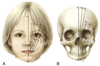
Eyelid colobomas fall under the schema of craniofacial clefting, which was first described by Tessier in 197613 (Fig. 8.3). This is a purely anatomic method of classification. Tawfic et al. recently grouped eyelid colobomas into two distinct categories: (1) isolated colobomas, including those associated with corneal palpebral adhesions and those occurring in isolation with no associated adhesions; and (2) colobomas that are part of a systemic syndrome, including Fraser syndrome, Goldenhar syndrome, and other rare syndromic variants.13
Clinical Features
While colobomas associated with Goldenhar syndrome, Fraser syndrome, or TCS are often bilateral, isolated simple colobomas are strictly unilateral, have no associated systemic signs, and do not usually present with significant corneal pathology. The size of the colobomatous lid defect may be overestimated in the clinical setting because of tissue elasticity and muscle tone that pulls the coloboma edges to either side (Fig. 8.4). Associated ocular findings can include limbal dermoid in the case of Goldenhar syndrome, microphthalmos, and iris coloboma. Facial abnormalities can include loss of ipsilateral eyebrow hair, a poorly formed supraorbital rim, cleft and lip palate, and a bifid nose. TCS may be associated with either true colobomas (those that fit the description above) or “pseudo-colobomas,” where there is continuity of the anterior lamella but discontinuity of the posterior lamella.10 Additional clinical features of Goldenhar syndrome, Fraser syndrome, and TCS are described elsewhere in Chapter 8.13

Investigations
The pediatrician or a neonatal team should be involved in the initial evaluation of a child with an eyelid coloboma to screen for syndromes. When applicable, referral to a geneticist for screening and counseling is warranted. A careful ophthalmic examination should be performed, with special attention given to the condition of the cornea.
Management
Early surgical intervention is often guided by the degree of corneal exposure. Regardless of the size of the coloboma, the medical team should institute an aggressive lubricating regimen at the time of initial presentation. Occlusive dressings with plastic wrap over the exposed cornea can also be employed. However, an approach that involves either an occlusive dressing or frequent lubricating ointment can be amblyogenic. In the case of a small colobomatous defect (<25%) without evidence of keratopathy and a clear indication of Bell’s phenomenon, watchful observation is favored until 2 to 4 years of age when more tissue is available for reconstruction and the risk of amblyopia has abated to some degree.15 Defects that involve 25% to 50% of the eyelid can be closed primarily or be coupled with release of the lateral canthus. Even if minimal tension at the closure site is observed at the time of repair, postoperative ptosis can occur (in the case of upper eyelid colobomas) with primary closures.
For eyelid defects involving more than 50% of the eyelid margin, several approaches are available: (1) lid-sharing technique, (2) rotational flap, (3) tarsomarginal graft, or (4) preoperative skin expansion. Eyelid sharing techniques can be used in older children but are amblyogenic in young children. When faced with uncontrollable corneal breakdown and the threat of corneal ulceration, a lid-sharing procedure can be done in a younger child, along with aggressive postoperative amblyopia treatment. In these cases, division of the transposed eyelid may be performed as early as 2 weeks postoperatively.10 In addition to amblyopia, another disincentive to the lid-sharing approach is the lack of eyelashes in the reconstructed area. A rotational, or switch, flap may provide a better cosmetic outcome, which can also be separated in 2 to 3 weeks. With a tarsomarginal graft, tarsoconjunctiva can be harvested from the unaffected upper eyelid or hard palate to reconstruct the posterior lamella, and skin–muscle advancement can be used to reconstruct the anterior lamella.
Course, Complications and Prognosis
Unlike in cases of cryptophthalmos, clinical anophthalmos, and microphthalmos, the eye is usually normal in isolated presentations of eyelid coloboma. As such, large eyelid colobomas may represent one of the few oculoplastic emergencies, since the cornea in a normally sighted eye is at risk for permanent damage related to exposure and infection. As long as the cornea is protected, long-term prognosis is quite good in patients with colobomas. However, it is important to counsel families that more than one operation may be required to achieve optimal results.
Ankyloblepharon
Epidemiology
Isolated ankyloblepharon and ankyloblepharon filiforme adnatum (AFA) are of unknown etiology and occur sporadically. AFA has an incidence of 4.4 per 100,000 births.16 Cleft palate and/or lip may be an associated clinical feature. Several syndromes have been associated with ankyloblepharon, including ankyloblepharon–ectodermal defects–cleft lip/palate syndrome, Edward syndrome (trisomy 18), and CHANDS (curly hair, ankyloblepharon, and nail dysplasia syndrome).17
Pathogenesis
Ankyloblepharon occurs when the fused eyelid folds fail to separate in utero. The child is born with completely or partially conjoined eyelids18,19 (Fig. 8.5). AFA occurs when there are single or multiple strands of fibrous tissue connecting the upper and lower eyelids. Because embryologic development up to the usual point of eyelid separation has occurred normally, the eyelid and eye structures are normal. Secondary forms of ankyloblepharon can occur as a result of infection, ocular trauma, Stevens-Johnson syndrome (SJS), or toxic epidermal necrolysis and silicone tube intubation of the nasolacrimal system.20

Clinical Features
Iridogoniodysgenesis and the development of juvenile glaucoma can be associated with AFA. Systemically, AFA can be associated with cleft lip, cleft palate, hydrocephalous, meningomyelocele, imperforate anus, cardiac defects, and syndactyly.21
Investigations
A detailed ophthalmic examination should be performed, checking closely for elevations in intraocular pressure and ocular stigmata of hydrocephalus. Genetic screening may be warranted.
Management
Given the risk of amblyopia in both forms of congenital ankyloblepharon, the eyelids should be surgically separated along the normal division, which is often easy to appreciate, given normal eyelid structures.
Course, Complications and Prognosis
If separation of the eyelids is achieved in a timely manner, visual prognosis is excellent. Cosmetic appearance of the eyelids after separation is also usually very good.
Distichiasis
Epidemiology
Distichiasis may be either congenital or acquired. The congenital form is almost always associated with lymphedema distichiasis syndrome (LDS), an autosomal dominant condition (FOXC2 gene at 16q24.3) that is characterized by two parallel rows of cilia at the eyelid margin and lymphedema of the extremities.22 LDS exhibits incomplete penetrance, with marked variability of expression, although distichiasis is almost always present. Lymphedema has a later onset (after age 10 years) and is generally not noticeable at the time of presentation (Fig. 8.6). Isolated congenital distichiasis is extremely rare. The acquired form is more common than its congenital counterpart. It is seen most often in the setting of chronic inflammatory conditions such as blepharitis, ocular cicatricial pemphigoid and SJS, and following chemical injury.

Pathophysiology
The eyelashes are a dermal appendage and differentiate in association with the glands of Zeis. These pilosebaceous units, as well as the Meibomian glands, form by invagination of surface ectoderm along the eyelid margin.10 Distichiasis results from a developmental error in which cilia form within or very close to the Meibomian gland orifices. The presence of cilia in this aberrant location may be asymptomatic or may cause irritation of the ocular surface from chronic lash touch. In the case of acquired distichiasis chronic inflammation leads to metaplasia, which results in lash growth from the Meibomian gland orifices.
Differential Diagnosis
Trichiasis is also an acquired lash abnormality in which the normal pilosebaceous units anterior to the Meibomian glands become malpositioned. This can occur secondary to chronic inflammation and cicatrization. Eyelashes rooted in the normal position grow toward the ocular surface and result in similar corneal and conjunctival irritation. Careful examination at the slit lamp allows for differentiation between trichiasis and distichiasis.
Clinical Features
Clinically, distichiatic eyelashes can be seen emanating from the Meibomian gland orifices and usually traverse the entire length of the affected eyelid (Fig. 8.7). Acquired distichiasis is more likely to be symptomatic than congenital LDS may be associated with other congenital abnormalities, including ptosis, congenital heart defects, extradural spinal cysts, and cleft palate.22

Investigations
Given the association between congenital distichiasis and adolescent onset lymphedema, the pediatrician should be made aware of the clinical finding of distichiasis to coordinate routine follow-up through adolescence. Genetic testing may be helpful.
Management
Regardless of etiology, treatment is pursued based on the extent that the eyelashes come into contact with the ocular surface. If the lash does not touch the cornea or conjunctiva, no treatment is necessary. When the lashes touch only the conjunctiva, intervention may alleviate discharge or discomfort. Conservative treatment with ocular lubricants can be initiated, but if symptoms persist, definitive surgical intervention should be pursued. Corneal lash touch creates a higher risk of vision loss from corneal irregularity, ulceration and scarring, and surgical treatment should be planned.
There are several surgical options for correction of distichiasis. More conservative approaches include electroepilation or cryotherapy. However, these treatments can be associated with depigmentation and structural distortion at the lid margin (especially with cryotherapy) and have a considerable rate of recurrence. Split-thickness eyelid resection with removal of lash follicles offers a more permanent solution but can be associated with trichiasis and entropion. Cryotherapy, using a double freeze–thaw technique to the posterior lamella, coupled with split-thickness eyelid resection and advancement of the posterior lamella, tends to have a high success rate with few complications related to eyelid malposition or skin pigmentation.23
Course, Complications and Prognosis
If treatment is initiated promptly, before permanent damage to the cornea occurs, prognosis is quite good. Families should be made aware that there can be recurrence of distichiatic lashes after surgical intervention and that repeat surgery may be required.
Epiblepharon
Epidemiology
Epiblepharon is a relatively common eyelid malposition, estimated to occur in up to 12.6% of East Asian children (Chinese, Korean and Japanese) aged 7 to 14 years.24 It is rare in non-East Asian children. It usually affects the lower eyelid, although it can be seen in the upper eyelid as well.24,25 Although epiblepharon is mostly known as a pediatric condition, it is becoming more frequently reported in adults as well.25 Acquired epiblepharon has been reported in thyroid eye disease and other conditions of proptosis.26
Pathogenesis
Epiblepharon is characterized by a horizontal skinfold extending along the eyelid margin and may be associated with several causes, including poor development of the eyelid retractors, failure of the retractors to gain access to skin, hypertrophy of the orbicularis muscle, and pretarsal orbicularis muscle insertion close to the eyelid margin with weak attachment of orbicularis and skin to underlying tarsus.25,27,28 These underlying processes can result in pretarsal orbicularis muscle and eyelid skin that overrides the eyelid margin, causing inward rotation of the cilia and potential ocular surface irritation (Fig. 8.8). Gentle pressure on the eyelid fold usually momentarily re-establishes the normal position of the eyelid cilia. In the case of thyroid eye disease–related proptosis causing acquired epiblepharon, the etiology is felt to be increased intraorbital pressure leading to an overriding anterior lamella.25

Differential Diagnosis
Epiblepharon of the lower lid can have a clinical appearance similar to entropion. However, in entropion, the eyelid margin itself turns inward (see below). Epicanthus can result in a similar-appearing skinfold close to the eyelid margin. If a semicircular epicanthal fold is also present, it can contribute to additional medial canthal tension and worsen epiblepharon.27 If epiphora persists after treatment of the epiblepharon, congenital nasolacrimal duct obstruction should be considered. Allergic conjunctivitis should also be considered in cases of chronic bilateral eye irritation, redness, and tearing.
Clinical Features
Epiblepharon is typically bilateral and generally affects the medial lower eyelids. Similar to other eyelid malpositions in which there is contact of lashes with the ocular surface, symptoms can range from none to significant, with foreign body sensation, discharge, photophobia, eye rubbing, epiphora, itching, frequent blinking, and eye redness. The severity of epiblepharon symptoms can be associated with the type and number of eyelashes.25 Fine eyelashes, as well as scant lashes, are often less symptomatic than thick and plentiful lashes. This variability may account for some later presentations of symptomatic epiblepharon as eyelashes change with aging. Decreased visual acuity from induced astigmatism can occur.28,30
Stay updated, free articles. Join our Telegram channel

Full access? Get Clinical Tree


