 Eyelid Cystic Lesions Simulating Neoplasms
Eyelid Eccrine Hidrocystoma
General Considerations
There are a number of cutaneous cystic lesions that can simulate neoplasms. In this section we discuss some of the more important ones that can affect the eyelids, beginning with eccrine hidrocystoma (1,2,3,4,5,6,7,8).
Eccrine hidrocystoma is a ductal retention cyst of an eccrine sweat gland (1). It appears to be more common in the eyelid region than elsewhere in the body (2). It usually occurs on the cheeks and eyelids of adult women and may be similar clinically to other cutaneous cysts. In contrast to apocrine hidrocystoma, which is almost always solitary, eccrine hidrocystoma is more likely to be multiple. Eccrine hidrocystomas develop from retention of sweat. Heat, humidity, and perspiration can cause them to become larger, more numerous, and more symptomatic (3). Hence, they tend to increase in number and size in summer and decrease in winter.
Clinical Features
Clinically, eccrine hidrocystoma characteristically appears as a clear cystic translucent lesion, usually near the eyelid margin. The overlying skin is usually smooth and shiny (1,2,3,4,5). It may be indistinguishable clinically from apocrine hydrocystoma. It can occasionally have a bluish color, a finding that is more common with apocrine hidrocystoma (to be discussed). Ultrasound biomicroscopy has been used to detect the cystic nature of the lesion and to differentiate it from melanoma and other solid tumors (8).
Pathology
Management
Management includes observation or local excision. The cystic structure collapses if traumatized or surgically incised.
Selected References
1. Font RL. Eyelids and lacrimal drainage system. In: Spencer WH, ed. Ophthalmic Pathology. An Atlas and Textbook. 4th ed. Philadelphia: WB Saunders; 1996:2343.
2. Geist CE. Benign epithelial tumors. In: Albert DM, Jakobiec FA, eds. Principles and Practice of Ophthalmology. 2nd ed. Philadelphia: WB Saunders; 2000:3351-3352.
3. Griffith DG, Salasche SJ, Clemons DE, eds. Cutaneous Abnormalities of the Eyelid and Face. New York: McGraw-Hill; 1987:236.
4. Smith JD, Chernosky ME. Hidrocystomas. Arch Dermatol 1973;108:676-679.
5. Cordero AA, Montes LF. Eccrine hidrocystoma. J Cutan Pathol 1976;3: 292-293.
6. Elder D, Elenitsas R, Ragsdale BD. Tumors of the epidermal appendages. In: Elder D, Elenitsas R, Jaworsky C, et al., eds. Lever’s Histopathology of the Skin. Philadelphia: Lippincott-Raven; 1997:777-778.
7. Yasaka N, Iozumi K, Nashiro K, et al. Bilateral periorbital eccrine hydrocystoma. J Dermatol 1994;21:490-493.
8. Furuta M, Shields CL, Danzig CJ, et al. Ultrasound biomicroscopy of eyelid eccrine hydrocystoma. Can J Ophthalmol, in press 2007.
Eyelid Eccrine Hidrocystoma
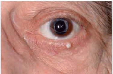
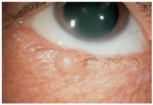 Figure 11.1. Solitary eccrine hidrocystoma of lower eyelid in a 62-year-old man. |
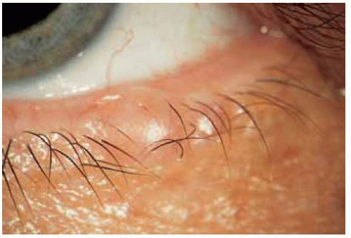 Figure 11.2. Solitary eccrine hidrocystoma of lower eyelid in a 67-year-old woman. |
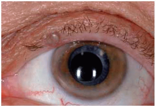 Figure 11.3. Slightly blue eccrine hidrocystoma of the left upper eyelid. |
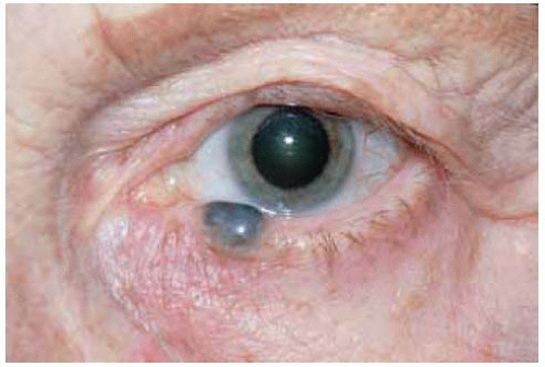 Figure 11.4. Blue-black eccrine hidrocystoma in a 75-year-old woman. This lesion appears pigmented, simulating a melanoma, a finding also common in apocrine hidrocystoma. Histopathologic studies confirmed eccrine hidrocystoma. |
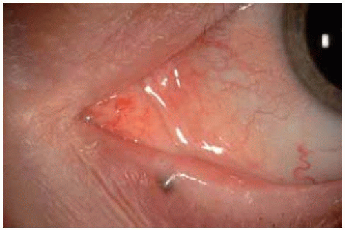 Figure 11.5. Blue-black cystic lesion on lower eyelid of a middle-aged woman. The lesion was excised. |
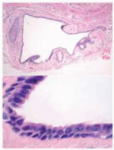 Figure 11.6. Photomicrograph of lesion shown in Figure 11.5, showing features of eccrine hidrocystoma. Upper picture shows entire cyst removed intact. (Hematoxylin-eosin 10.) Lower picture shows double layer of epithelium lining the cyst. (Hematoxylin-eosin 75.) |
Eyelid Apocrine Hidrocystoma
General Considerations
Apocrine hidrocystoma (apocrine cystadenoma; sudoriferous cyst) is a retention cyst of an apocrine gland (1,2,3,4,5,6,7,8,9,10,11,12,13,14,15,16). It can develop from the apocrine glands of the eyelids (glands of Moll), in which case it usually occurs near the eyelid margin, corresponding to the orifices of Moll’s glands. It is more common near the medial canthus. There is no gender predisposition. It generally occurs in adults, with a mean age of 55 years, but is occasionally seen in children (2,5,6).
Clinical Features
Apocrine hidrocystoma is a smooth or multiloculated cystic lesion that can develop on the eyelid, eyebrow, or near the medial or lateral canthus (1,2,3,4,5,6,7). It frequently has a bluish color and may resemble a blue nevus or melanoma. It can range from 1 mm to more than 1 cm in diameter.
Schopf-Schulz-Passarge Syndrome
There is a variant of hereditary ectodermal dysplasia in which some cases have multiple apocrine hidrocystomas affecting both the upper and lower eyelids bilaterally. These patients can also demonstrate hypodontia, palmar-plantar hyperkeratosis, and onychodystrophy (8,9,10,11,12,16). This constellation of findings is called the Schopf-Schulz-Passarge syndrome (8,9,10,11,12,16). Some cases appear isolated; others have an autosomal-dominant or -recessive pattern (12). This entity is described in more detail by Font et al (1,9).
Pathology
Microscopically, apocrine hidrocystoma is a cystic lesion with a clear lumen lined by a double layer of cells (1,2,3,4,5,6,7). The innermost layer has apical snouts that project into the lumen, a characteristic feature of apocrine cells. The outermost layer consists of flattened myoepithelial cells. The cells also contain PAS-positive, diastase-resistant granules on their apical surfaces. Some authorities regard apocrine hidrocystoma as a form of papillary cystadenoma rather than a retention cyst, because the secretory cells do not appear flattened, as seen with a true retention cyst (1,4).
Stay updated, free articles. Join our Telegram channel

Full access? Get Clinical Tree


