7
Eyelid Anomalies
Kammi B. Gunton 
ANKYLOBLEPHARON
Etiology
 Ectodermal dysplasia, otherwise unknown
 In ankyloblepharon–ectodermal dysplasia– clefting (AEC) patients, a defect in the TP63 gene, an epidermis intercellular junction regulator protein, is deficient. AEC is inherited in autosomal dominant fashion.
Symptoms
 Poor eyelid opening
Signs
 Partial or complete fusion of eyelid margins along portion of their length by webs of skin (Fig. 7-1)
 Shortened palpebral fissure
 Internal ankyloblepharon has fusion along inner canthus
 External ankyloblepharon has fusion of margin along outer canthus and is more common
 In ankyloblepharon filiforme adnatum variant, fine strands of skin connect margins.
 Associated with cleft lip, cleft palate, and ectodermal defects called AEC. Patients have ankyloblepharon filiforme adnatum; ectodermal defects such as sparse wiry hair, skin erosions, pigmentary changes, nail dystrophy, or dental abnormalities; and cleft lip or palate.
Differential Diagnosis
 Cryptophthalmos, failure of differentiation of eyelid structures; the cornea is attached to eyelid skin
 Congenital coloboma, defect within the eyelid, small notch or entire eyelid absent
 Epiblepharon, with extra fold of orbicularis, in the lower eyelid, turning eyelashes inward; no attachment between eyelids
Diagnostic Evaluation
 External examination of the eyelids reveals strands or a web of skin attaching the eyelid margin.
 Underlying cornea and eye structures are intact and unaffected.
 In AEC, molecular genetic testing for TP63 mutations
Treatment
 Spontaneous resolution with ankyloblepharon filiforme adnatum is possible.
 Otherwise, hemostat to connective tissue followed by excision or skin strands or webs. Edges of conjunctiva and eyelid margin are apposed with sutures to prevent readhesion of skin.
Prognosis
 Excellent prognosis with treatment
 Prevent readhesion of skin
REFERENCES
Lopardo T, Loiacono N, Marinari B, et al. Claudin-1 is a p63 target gene with a crucial role in epithelial development. PLoS One. 2008;3(7):e2715.
Sumita S, Mridula M, Rainath, et al. What’s your diagnosis? Diagnosis of ankyloblepharon filiforme adnatum. J Pediatr Ophthalmol Strabismus. 2010;47:139, 177.
Sutton VR, Bree AF, van Bokhoven H. Ankyloblepharon-ectodermal defects-cleft lip/palate syndrome. In Pagon RA, Bird TC, Dolan CR, et al, eds. GeneReviews. Seattle: University of Washington; 2008.
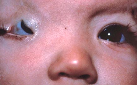
Figure 7-1. Ankyloblepharon with coloboma, right eyelid. (Courtesy of Robert Penne, MD, Department of Oculoplastics, Wills Eye Hospital, Philadelphia.)
BLEPHAROPHIMOSIS SYNDROME
Etiology
 Autosomal dominant inheritance
 Associated with FOXL2 mutations
Symptoms
 Present congenitally
 Severe ptosis may cause ametropic amblyopia with blurred vision.
Signs
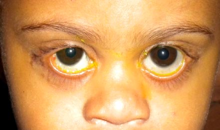
 Shortened palpebral length with three associated major signs: telecanthus (widened intercanthal distance), epicanthus inversus, and severe ptosis (Fig. 7-2)
 Additional signs include lower eyelid entropion, a poorly developed nasal bridge, hypoplasia of superior orbital rim, low-set ears, a short philtrum, lacrimal duct anomalies, refractive errors, and hypertelorism. Type I blepharophimosis syndrome (BPES) is associated with ovarian dysfunction, leading to premature ovarian failure.
 Type II BPES without ovarian dysfunction and premature ovarian failure
Differential Diagnosis
 Congenital ptosis; would occur with absence of other features
 Epicanthus, isolated finding
Diagnostic Evaluation
 Based on the presence of four classic signs: blepharophimosis, ptosis, epicanthus inversus, and telecanthus
 Testing for FOXL2 may help in the diagnosis.
 Evaluation for ovarian dysfunction is needed.
Treatment
 Staged surgical treatment of signs, including multiple Z-, Y-, or V-plasties and intranasal wiring of medial canthus tendons to correct telecanthus at age 3 to 5 years followed 1 year later by bilateral frontal sling or levator resection as indicated for ptosis. With bilateral ametropic amblyopia, ptosis repair may need to be expedited.
 Simultaneous medial canthoplasty and blepharoptosis correction in select patients
Prognosis
 With surgical correction, improved cosmesis
 Visual prognosis is guarded because of amblyopia development. The timing of surgical correction is critical to prevent amblyopia.
 Ovarian dysfunction is treated with hormone replacement therapy, and reproductive issues may be addressed with reproduce technologies, including embryo or egg donation.
REFERENCES
Beysen D, DePaepe A, DeBaere E. FOXL2 mutations and genomic rearrangements in BPES. Hum Mutat. 2009;30:158–169.
DeBaere E. Blepharophimosis, ptosis, and epicanthus inversus. In: Pagon RA, Bird TC, Dolan CR, Stephens K, eds. GeneReviews. Seattle: University of Washington; 2009.
Huang WQ, Qiao Q, Zhao R, et al. Surgical strategy for congenital blepharophimosis syndrome. Chin Med J. 2007;120:1413–1415.
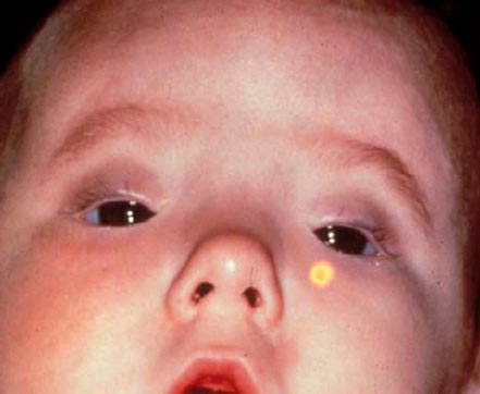
Figure 7-2. Child with blepharophimosis syndrome. Note blepharophimosis with telecanthus (widened intercanthal distance) epicanthus inversus, and severe ptosis. (Courtesy of Robert Penne, MD, Department of Oculoplastics, Wills Eye Hospital, Philadelphia.)
CONGENITAL ECTROPION
Etiology
 Congenital; classified as primary or secondary
 Primary resulting from absence or atrophy of tarsal plate
 Secondary resulting from paralytic, cicatricial, or mechanical causes in childhood with vertical shortage of anterior lamella such as congenital malformations with skin retraction, trauma, burns, ichthyosis (cicatricial), inflammatory conditions from medications, birth trauma, allergies with orbicularis spasm, or tumors (mechanical)
 May occur in association with BPES, euryblepharon, microphthalmos, orbital cysts, and Down’s syndrome
Symptoms
 Chronic epiphora, conjunctival injection, foreign body sensation, photophobia, reduced vision
Signs
 Eversion of eyelid margin; the lower eyelid is more commonly involved because of a vertical deficiency of skin (Fig. 7-3)
 Exposure keratitis and conjunctivitis
Differential Diagnosis
 Congenital tarsal kink: upper eyelid bent back with 180-degree fold of the upper tarsal plate
 Congenital entropion: distal portion of lower tarsal plate bent inward
 Euryblepharon: downward displacement of temporal portion of lower eyelid caused by enlargement of the lateral aperture
Diagnostic Evaluation
 Based on external examination with eversion of the eyelid
Treatment
 Mild cases require lubrication with artificial tears or ointments.
 With corneal exposure, surgical repair is required with lateral tarsorrhaphy or lateral canthoplasty to eliminate horizontal eyelid laxity to reposition the eyelid to the globe.
 In severe cases, a full-thickness skin graft is required.
 In tarsus agenesis, auricular cartilage may be used in the graft.
Prognosis
 With surgical correction, and skin graft in cases of vertical deficiency of skin, good prognosis
 Must prevent permanent corneal scarring from exposure keratitis, with resulting amblyopia
REFERENCES
Bedran EG, Pereira MV, Bernandes TF. Ectropion. Semin Ophthalmol. 2010;25:59–65.
Hintschich C. Correction of entropion and ectropion. Dev Ophthalmol. 2008;41:85–102.
Piskiniene R. Eyelid malposition: lower lid entropion and ectropion. Medicina (Kaunas). 2006;42:881–884.
Figure 7-3. Ectropion. Note eversion of lower eyelids. (Courtesy of Jacqueline Carrasco, MD, Department of Oculoplastics, Wills Eye Hospital, Philadelphia.)
Stay updated, free articles. Join our Telegram channel

Full access? Get Clinical Tree


