1
Abnormalities Affecting
the Eye as a Whole
Judith B. Lavrich 
ANOPHTHALMIA
Anophthalmia, also known as anophthalmos, is a congenital anomaly that is characterized by the complete absence of ocular tissue within the orbit. Primary or true anophthalmia is a very rare condition and can involve one or both eyes. Extreme microphthalmos is far more common and can be mistaken for this condition. Anophthalmia has a prevalence of 0.18 per 10,000 births and has no racial or sexual predilection.
Etiology
During embryogenesis, there is an arrest in the development of the neuroectoderm of the primary optic vesicle, which stems from the anterior neural plate of the neural tube.
Anophthalmia is most frequently idiopathic and sporadic but can be inherited as a dominant, recessive, or sex-linked trait. It is associated with maternal infections during pregnancy (e.g., toxoplasmosis, rubella) as well as syndromes with craniofacial malformations (e.g., Goldenhar, Hallerman-Streiff, Waardenburg syndromes). It is linked with genetic defects, including trisomies 13 to 15; chromosomal deletion in band 14q22-23 with associated polydactyly; and deletions involving SOX2, SIX6, and STRA6.
Signs
 The eye is the stimulus for proper growth of the orbital region; therefore, an infant born with anophthalmia has the following:
 Orbital findings
 Small orbital rim and entrance
 Reduced size of bony orbital cavity
 Globe is completely absent
 Extraocular muscles are usually absent
 Lacrimal gland and ducts may be absent
 Small or maldeveloped optic foramen
 Eyelid findings
 Narrow palpebral fissures
 Foreshortening of the eyelids
 Shrunken conjunctival fornices
 Levator function is decreased or absent with poor eyelid folds
 Contracture of the orbicularis oculi muscle
Symptoms
 Unilateral or bilateral blindness because of absence of the globe(s)
Differential Diagnosis
 Microphthalmos, which includes:
 Secondary anophthalmos: the development of the eye begins but gets arrested, resulting in only residual eye tissue or extreme microphthalmos.
 Degenerative anophthalmos: there is formation of the optic vesicle, but subsequent degeneration occurs from lack of blood supply or other causes.
 Cryptophthalmos: abnormal fusion of the entire eyelid margin with absence of the eyelashes
 Cystic eye: a cyst of neuroglial tissue lacking normal ocular structures
Diagnostic Evaluation
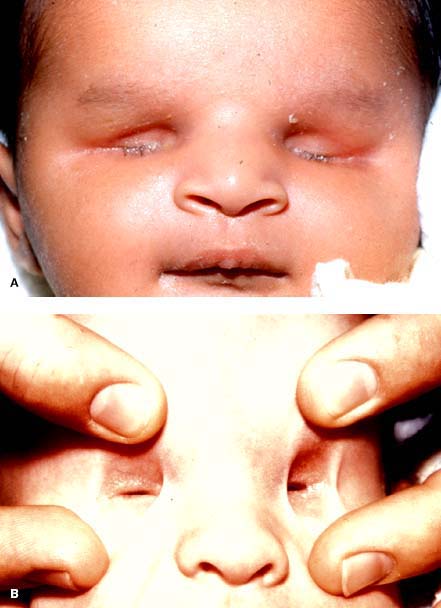
 Anomalous eyelid and orbital features (Fig. 1-1)
 Ultrasound imaging. B-scan ultrasonography of the orbit will show a complete absence of the globe. After 22 weeks’ gestation, transvaginal ultrasonography can detect eye malformations but its sensitivity in the detection of anophthalmia is not known.
 Magnetic resonance imaging (MRI) scan of the head and orbits. MRI will show the soft tissue within the orbital cavity (Fig. 1-2). Associated intracranial abnormalities can also be evaluated. Individuals with bilateral anophthalmos may have a related hypoplastic or absent optic chiasm as well as agenesis or dysgenesis of the corpus callosum.
 Computed tomography (CT) scan of the head and orbits. CT scan will image the bony changes and intracranial and craniofacial abnormalities seen with anophthalmia.
Treatment
 Medical care
 Orbital conformers can be placed in the orbital cavity to stimulate growth of the bony orbit (Fig. 1-3). As the orbit grows, the conformers are changed and progressively increased in size to further expand the orbital cavity. This serial augmentation takes time and cooperation from both the patient and parents.
 Contraction and reversal of the benefit often occur if the conformer is left out of the orbit for a significant amount of time. With unilateral anophthalmos, the family should be aware that, most likely, the final result will not mirror the normal healthy orbit.
 An ocular prosthesis can be fitted over the conformer to simulate the eye and improve appearance.
 Surgical care
 The small bony cavity is both a cosmetic deformity and may not allow proper fitting of a prosthesis. Therefore, surgery may be indicated for either of these problems.
 Inflatable tissue expanders are used if conformers are not well tolerated or cannot be fit. The inflatable silicone expander is surgically positioned deep in the orbit and is accessed through a tube placed at the lateral orbital rim. The expander is filled with saline and gradually reinflated on a weekly or biweekly schedule. Compared with solid conformers, inflatable expanders may allow more rapid and extensive expansion of the bony orbit. When the desired volume is achieved, the port and bladder need to be removed and replaced with a permanent implant.
 Hydrogel (methylmethacrylate and N-vinylpyrrolidone) expanders are self-expanding hydrophilic expanders that are implanted in the orbital tissue in their dry, contracted state through a small incision. The implant gradually expands in size by osmotic absorption of surrounding tissue fluid. The benefit of this method is the controlled self-expansion, reducing the risk of tissue atrophy, and without the need for repeat fittings or surgery.
 Dermis fat grafting involves biocompatible grafts that grow slowly over time can be a good option to restore volume to the hypoplastic orbit. The graft is harvested from a second surgical site, typically the buttocks. However, the graft compatibility and growth can be variable. In some cases, the fat can atrophy. Rarely, the fat can hypertrophy, necessitating debulking.
 Injectable calcium hydroxylapatite (Radiesse) is a semipermanent dermal filler that has been reported as a new, simple, cost-effective technique to treat volume deficiency in the anophthalmic orbit in adults. Augmentation is accomplished with serial injections of the filler until adequate volumization is achieved. The results have demonstrated a lasting effect in the orbit of 1 year or more.
 Orbito-cranial advancement surgery is used for orbital expansion if conformers and expanders are unsuccessful. This method involves multiple osteotomies to divide the periocular bones and advancing them forward and outward with bone grafts and plates.
 Because the foreshortening of the eyelids may limit the passage of a large conformer, a lateral canthotomy or cantholysis may be needed to increase the horizontal length of the palpebral fissure. Other methods to lengthen the eyelids may include skin, mucosal, or cartilage grafts.
Prognosis
 Severe cosmetic deformities can result from anophthalmia, especially if not treated early. Even with proper treatment, the results are often cosmetically suboptimal with incomplete expansion of the orbit, malformations and immobility of the eyelids, and complete immobility of the ocular prosthesis.
 Psychosocial issues caused by absence of an eye and facial disfigurement can result. Referral for psychological counseling may be indicated for these children.
REFERENCES
Bardakiian T, Weiss A, Schneider AS. Anophthalmia/microphthalmia overview. In Pagon RA, Bird TC, Dolan CR, Stephens K, eds. GeneReviews. Seattle: University of Washington; 2007:1993–2004.
Bernardino R. Congenital anophthalmia: A review of dealing with volume. Middle East Afr J Ophthalmol. 2010;17:156–160.
http://www.geneclinics.org/profiles/anophthalmia-ov/index.html.
Verma AS, Fitzpatrick DR. Anophthalmia and microphthalmia. Orphanet J Rare Dis. 2007;2:47.
FIGURE 1-1. A. External examination of bilateral anophthalmia. B. Clinical examination of bilateral anophthalmia showing empty orbits. (Courtesy of Leonard B. Nelson, MD.)
Stay updated, free articles. Join our Telegram channel

Full access? Get Clinical Tree


