Eyelid
6.1 Ptosis
Symptoms
Drooping upper eyelid, visual loss often worse with reading and at night. Concerning associated symptoms include diplopia, headache, and neck pain.
Signs
(See Figure 6.1.1.)
Critical. Drooping upper eyelid.
Other. Concerning associated signs include anisocoria, proptosis, and ocular motility deficits. See individual entities.
Etiology
Horner syndrome.
Third cranial nerve (CN) palsy (complete, partial, or aberrant CN III regeneration).
Myasthenia gravis.
Superior eyelid or orbital malignancy.
Chronic progressive external ophthalmoplegia (CPEO) (particularly Kearns–Sayre syndrome, SEE 10.12, CHRONIC PROGRESSIVE EXTERNAL OPHTHALMOPLEGIA).
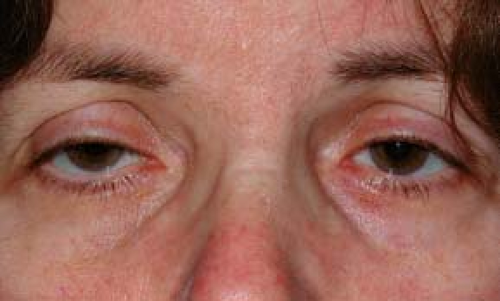 Figure 6.1.1 Ptosis. |
Categories:
Myogenic: Congenital myogenic ptosis is present at birth with poor levator function and a poor or absent lid crease due to dysgenesis of the levator muscle, fibrosis, and its replacement with adipose tissue. A poor Bell phenomenon, lagophthalmos in downgaze, and upgaze limitation may indicate a double elevator palsy. Acquired myogenic ptosis is uncommon and may be seen with muscular dystrophy and CPEO.
Aponeurotic: This is characterized by a high or absent eyelid crease, moderate degree of ptosis, good levator function (10 to 15 mm). May worsen in downgaze. Levator stretching or dehiscence can be due to normal aging, repetitive eye rubbing, use of rigid contact lenses, or previous intraocular surgery.
Neurogenic: Third CN palsy (often complete ptosis, never an isolated abnormality; congenital, compressive, vasculopathic, SEE 10.5, ISOLATED THIRD NERVE PALSY); Horner syndrome (subtle upper and lower eyelid ptosis, SEE 10.2, HORNER SYNDROME); myasthenia gravis (variable ptosis, worsens with fatigue, SEE 10.11, MYASTHENIA GRAVIS); Marcus Gunn jaw-winking syndrome (ptotic eyelid elevates with jaw movement); ophthalmoplegic migraine; multiple sclerosis.
Mechanical: Retained contact lens in upper fornix; upper eyelid or forniceal inflammation (chalazion, giant papillary conjunctivitis, posttraumatic or postsurgical edema) or neoplasm.
Traumatic: History of eyelid laceration with levator transection, contusion injury to the levator, tethering or ischemia within an orbital roof fracture, late dehiscence or cicatricial changes.
Pseudoptosis: Contralateral eyelid retraction or proptosis, ipsilateral enophthalmos or hypertropia, microphthalmia, phthisis bulbi, dermatochalasis, brow ptosis, chalazion or other eyelid tumor, eyelid edema, blepharospasm, Duane syndrome.
Work-Up
History: Determine the onset and duration of ptosis. Present since birth? Acute onset? Old photographs (e.g., driver’s license) and family members’ opinions are useful adjuncts to the history. History of surgery in either eye? Orbital or eyelid trauma? Variability with fatigue? Associated with diplopia, headache, or neck pain? Any history of autoimmune disease (lupus, Sjögren syndrome) or corneal abnormalities (may predispose the patient to postoperative exposure keratopathy)?
Mandatory documentation: Must carefully check and document pupillary size and extraocular motility, even if normal. If anisocoria is present, measurements should be documented under light and dark conditions. Additional pharmacologic testing may be indicated (SEE 10.1, ANISOCORIA). If extraocular muscle dysfunction is noted, additional testing with prism bars may be indicated.
Complete orbital examination of both eyes: Measure and compare globe position, margin reflex distance, levator function (full upper eyelid excursion while preventing frontalis muscle assistance), and upper eyelid crease position of both eyes. Is there lagophthalmos? Associated lower eyelid “ptosis” (elevation of ipsilateral lower eyelid) is often seen in Horner syndrome. Proptosis or eyelid lag may masquerade as contralateral ptosis. Exophthalmometry measurements are useful. Any sign of aberrant eyelid movements like jaw winking, variability and/or fatigue, orbicularis weakness, eyelid retraction with adduction and/or infraduction? Palpate the superior orbit to rule out a mass or superior orbital rim deformity.
Complete ocular examination: Flip upper eyelid to examine conjunctival surface and superior fornix. Dilated fundus examination to look for pigmentary changes in adolescents and young adults who present with ptosis, poor levator function, and external ophthalmoplegia (possible CPEO).
Corneal protective mechanisms. Document the presence or absence of preoperative lagophthalmos, orbicularis function, Bell phenomenon, and tear production. Check the cornea carefully for any abnormalities or dystrophies, which may predispose the patient to postoperative keratopathy.
Other tests
Ice test: Apply ice pack to ptotic eye(s) for 2 minutes and reassess the degree of ptosis. Improvement with ice is highly suggestive of myasthenia gravis.
Neosynephrine test: Instill one drop of 2.5% phenylephrine in the ptotic eye(s) and reassess the degree of ptosis. Patients with improvement of ptosis after 5 to 7 minutes may be good candidates for ptosis correction by an internal approach.
Apraclonidine, cocaine, and hydroxyamphetamine tests. SEE 10.2, HORNER SYNDROME.
 NOTE: In recent years, limited access to ophthalmic cocaine and hydroxyamphetamine has reduced their utility in the clinical setting.
Imaging studies: In very select cases where a systemic or neurologic cause is suspected:
Computed tomography (CT) or magnetic resonance imaging (MRI) of orbit if a superior orbital mass is suspected.
Emergent CT/computed tomography angiogram (CTA) or MRI/magnetic resonance angiogram (MRA) of the head and neck if carotid artery dissection is suspected in a patient with a painful Horner syndrome. Imaging of the head alone is inadequate. SEE 10.2, HORNER SYNDROME.
Emergent CT/CTA, MRI/MRA, or conventional angiography if intracranial aneurysm causing a third CN palsy with pupillary involvement or a partial third CN palsy is suspected. SEE 10.5, ISOLATED THIRD NERVE PALSY.
Ancillary studies:
If myasthenia gravis is suspected, acetylcholine receptor antibody (binding, blocking, and modulating) testing, single-fiber
electromyography (including the orbicularis muscle), and/or edrophonium chloride testing under monitored conditions may be indicated. SEE 10.11, MYASTHENIA GRAVIS.
Urgent ECG and cardiology consult if Kearns–Sayre syndrome is suspected. These patients can have heart block, resulting in sudden death.
If severe dry eye is found, consider an autoimmune work-up to rule out lupus and Sjögren syndrome.
Treatment
Depends on the underlying etiology (SEE 10.2, HORNER SYNDROME; 10.5, ISOLATED THIRD NERVE PALSY; 10.11, MYASTHENIA GRAVIS).
Nonsurgical options: Observation. Taping upper lids open and eyelid crutches attached to glasses in neurogenic and myogenic ptosis. Management of chalazion with warm compresses and/or topical or intralesional steroid/antibiotic.
Surgical options: Excision of eyelid and/or orbital lesions (e.g., chalazion, neoplasm), transcutaneous levator advancement, transconjunctival levator advancement, frontalis muscle suspension, Fasanella–Servat procedure, Müller muscle resection. Surgical approach depends on preoperative evaluation and the underlying etiology of ptosis.
Follow-Up
Congenital: Close follow-up is required to monitor for the presence of amblyopia, caused either by occlusion or by refractive error secondary to induced corneal astigmatism, abnormal head positioning, and exposure keratopathy.
Traumatic: Observation for 6 months before considering surgical intervention. Many spontaneously improve or completely resolve.
Neurologic: Reevaluate based on particular entity.
Postoperative (after ptosis repair).
Acute: Monitor for infection and hemorrhage.
Subacute: Monitor for exposure keratopathy and for asymmetry that may require postoperative readjustment. Mild lagophthalmos is common for 2 to 3 weeks after surgical repair and usually resolves.
Chronic: Monitor for ptosis recurrence and exposure keratopathy.
6.2 Chalazion/Hordeolum
Symptoms
Acute or chronic eyelid lump, eyelid swelling, tenderness.
Signs
(See Figure 6.2.1.)
Critical. Visible, or palpable, well-defined subcutaneous nodule in the eyelid. In some cases, a nodule cannot be identified.
Other. Blocked meibomian orifice, eyelid swelling and erythema, localized eyelid tenderness, associated blepharitis or acne rosacea. May also note “pointing” of mucopurulent material.
Definitions
Chalazion: Area of focal inflammation within the eyelid secondary to the obstruction of a meibomian gland or gland of Zeis.
Hordeolum: Acute infection; can be external (abscess of gland of Zeis on lid margin, classic “stye”) or internal (abscess of meibomian gland). Usually involves Staphylococcus species and occasionally evolves into a preseptal cellulitis.
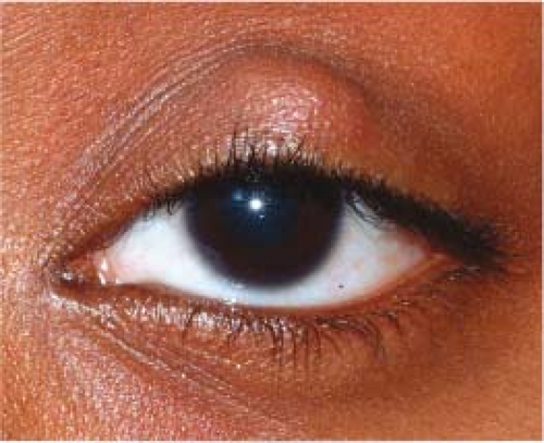 Figure 6.2.1 Chalazion. |
Differential Diagnosis
Preseptal cellulitis: Eyelid and periorbital erythema, edema, and warmth. SEE 6.10, PRESEPTAL CELLULITIS.
Sebaceous carcinoma: Suspect in older patients with recurrent chalazia, eyelid thickening, madarosis, or chronic unilateral blepharitis. SEE 6.11, MALIGNANT TUMORS OF THE EYELID.
Pyogenic granuloma: Benign, deep-red, pedunculated lesion often associated with chalazia, hordeola, trauma, or surgery. May be excised or treated with a topical antibiotic–steroid combination such as neomycin/polymyxin B/dexamethasone q.i.d. for 1 to 2 weeks. Intraocular pressure must be monitored if topical steroids are used.
Work-Up
History: Previous ocular surgery or trauma? Previous chalazia or eyelid lesions?
External examination: Palpate the involved eyelid for a nodule.
Slit lamp examination: Evaluate the meibomian glands for inspissation and evert the eyelid. Assess for madarosis (lash loss), poliosis, and ulceration to rule out other etiologies.
Treatment
Warm compresses for 10 minutes q.i.d. with light massage over the lesion.
Consider a short course of topical antibiotic if lesion is draining or for associated blepharitis (e.g., bacitracin or erythromycin ointment b.i.d. for 1 to 2 weeks). Consider systemic therapy with doxycycline 100 mg p.o. daily to b.i.d. for its antibacterial and anti-inflammatory properties (e.g., multiple hordeola, ocular rosacea).
If a hordeolum worsens, consider incision and drainage and management as per preseptal cellulitis (SEE 6.10, PRESEPTAL CELLULITIS).
If the chalazion fails to resolve after 3 to 4 weeks of appropriate medical therapy and the patient wishes to have it removed, incision and curettage are performed. Occasionally, an injection of steroid (e.g., 0.2 to 1.0 mL of triamcinolone 40 mg/mL usually mixed 1:1 with 2% lidocaine with epinephrine) into the lesion is performed instead of minor surgery, especially if the chalazion is near the lacrimal apparatus. The total dosage depends on the size of the lesion. All recurrent or atypical chalazia must be sent for pathology.
Follow-Up
Patients are not routinely seen after instituting medical therapy unless the lesion persists beyond 3 to 4 weeks. Patients who have a procedure such as incision and curettage are usually reexamined in 1 week or as needed.
6.3 Ectropion
Symptoms
Tearing, eye or eyelid irritation. May be asymptomatic.
Signs
(See Figure 6.3.1.)
Critical. Outward turning of the eyelid margin.
Other. Superficial punctate keratopathy (SPK) from corneal exposure; conjunctival injection, thickening, and eventual keratinization from chronic conjunctival dryness. Scarring of skin may be seen in cicatricial cases. Facial hemiparesis and lagophthalmos may be seen in paralytic cases.
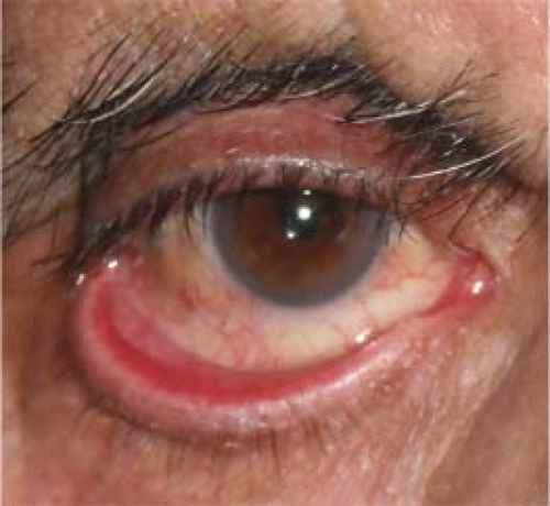 Figure 6.3.1 Ectropion. |
Etiology
Involutional: Horizontal eyelid laxity related to aging. Most common.
Paralytic: Seventh CN palsy.
Cicatricial: Shortening of the anterior lamella from burn injury, prior surgery/trauma, eyelid laceration scar, actinic damage, chronic inflammation, skin diseases (e.g., eczema, ichthyosis), and others.
Mechanical: Due to herniated orbital fat, eyelid tumor, and others.
Allergic: Contact dermatitis.
Congenital: Facial dysmorphic syndromes (e.g., Treacher Collins syndrome) or isolated abnormality.
Work-Up
History: Previous surgery, trauma, chemical burn, or CN VII palsy?
External examination: Check orbicularis oculi function and assess horizontal lid laxity and punctal location. Look for an eyelid tumor, eyelid scarring, herniated orbital fat, etc.
Slit lamp examination: Check for SPK due to exposure and evaluate conjunctival integrity.
Treatment
Treat exposure keratopathy with lubricating agents. SEE 4.5, EXPOSURE KERATOPATHY.
Treat an inflamed, exposed eyelid margin with warm compresses and antibiotic ointment (e.g., bacitracin or erythromycin t.i.d.). A short course of combination antibiotic–steroid ointment (e.g., neomycin/polymyxin B/dexamethasone) may be helpful if close follow-up is ensured.
Taping the eyelids into position may be a temporizing measure.
Definitive treatment usually requires surgery. Surgery is delayed for 3 to 6 months in patients with a seventh nerve palsy because the ectropion may resolve spontaneously (SEE 10.9, ISOLATED SEVENTH NERVE PALSY). Corneal exposure may make repair more urgent.
Follow-Up
Patients with signs of corneal or conjunctival exposure are examined as needed based on the symptoms and integrity of the cornea and conjunctiva. If the tissues are relatively healthy, follow-up is not urgent.
6.4 Entropion
Symptoms
Ocular irritation, foreign-body sensation, tearing, redness.
Signs
(See Figure 6.4.1.)
Critical. Inward turning of the eyelid margin that pushes otherwise normal lashes onto the globe.
Other. SPK from eyelashes contacting the cornea, conjunctival injection. In severe cases, corneal thinning and ulceration are possible.
Etiology
Involutional: Age-induced horizontal lid laxity, retractor disinsertion, and orbicularis override.
Cicatricial: Due to conjunctival scarring in ocular cicatricial pemphigoid, Stevens–Johnson syndrome, chemical burns, trauma, trachoma, and others.
Spastic: Sustained orbicularis contraction due to surgical trauma, ocular irritation, or blepharospasm.
Congenital.
 Figure 6.4.1 Entropion. |
Work-Up
History: Previous surgery, trauma, chemical burn, or infection (trachoma, herpes simplex, and varicella zoster)?
Slit lamp examination: Check for corneal involvement as well as conjunctival and eyelid scarring.
Treatment
If blepharospasm is present, SEE 6.7, BLEPHAROSPASM.
Aggressive lubrication and antibiotic ointment (e.g., erythromycin or bacitracin ophthalmic t.i.d.) for SPK.
Everting the eyelid margin away from the globe and taping it in place with adhesive tape may be a temporizing measure.
Stay updated, free articles. Join our Telegram channel

Full access? Get Clinical Tree


