Purpose
To evaluate in vitro the amoebicidal effects of riboflavin and ultraviolet A (UVA) collagen cross-linking.
Design
Experimental study, laboratory investigation.
Methods
Two different strains of Acanthamoeba species were tested identically. Four treatment groups were considered: group 1 consisted of 0.1% riboflavin and 30-minute UVA irradiation; group 2 consisted of 0.1% riboflavin and 60-minute UVA irradiation; group 3 consisted of no riboflavin and no UVA exposure; group 4 consisted of 0.1% riboflavin and no UVA exposure. The application of UVA was performed under the parameters used for in vivo corneal collagen cross-linking.
Results
In all cases, cysts and trophozoites were detected 24 hours after treatment at a radial distance from the center of the seeding point more than 5 mm, indicating that the amoebae were viable. All treated and untreated groups of amoebae from the 2 strains exhibited growth (radii of 14 to 15 mm in groups 1, 3, and 4; radius of 12 mm in group 2). The final morphologic features of the 2 strains of trophozoites that received treatment were similar to those of the initial seeding group and the untreated control group.
Conclusions
The results obtained in our study show that a single dose (30 or 60 minutes) of cross-linking cannot achieve eradication in the 2 different Acanthamoeba strains examined. However, in vitro results do not always indicate in vivo efficacy, so future studies should test the validity of this treatment for Acanthamoeba keratitis.
Acanthamoeba is one of the most commonly isolated amoebae in environmental samples. It has a cosmopolitan distribution and can act as both an opportunistic and nonopportunistic pathogen. Acanthamoeba is a recognized cause of serious corneal infection, especially in contact lens wearers. Contact lenses have been noted as the most important risk factor in all recent case series. The Acanthamoeba life cycle includes 2 morphologic forms: an amoeba or trophozoite stage, in which the amoeba is active and divides, and a resistant cyst stage that protects the amoeba from adverse environmental conditions.
Acanthamoeba keratitis (AK) has presented a medical challenge for most ophthalmologists because it is a frustrating and often devastating infection. Diagnostic dilemmas usually delay appropriate treatment, which is one of the most important factors affecting visual prognosis. Persistent infection is related to the presence of cysts, and only agents that take action against the latter can be expected to be an effective therapy.
There are no standardized algorithms for AK; treatment practices involve topical treatment with monotherapy or multiple therapeutic agents (including oral antifungals) and aggressive surgical intervention with penetrating keratoplasty for severe active AK infection. Diamidines and biguanides are the most effective cysticidal antiamebics in vitro, and their use is supported by substantial case series. Nonetheless, toxicity complications have been attributed to the topical use of these drugs. Therapeutic keratoplasty usually is used in unresponsive active cases when drug therapy fails.
During the last decade, there has been a significant increase in the incidence of AK, and new therapies are being considered that may help to overcome the infection when organisms are present that are resistant to standard treatments. Recent cases have documented clinical improvement using ultraviolet A (UVA) as an adjunct to other antiamebic treatments in cases of large ulcers associated with edema and corneal infiltration, but cases of amebic infection after treatment with cross-linking also have been reported.
Riboflavin UVA cross-linking increases the biomechanical rigidity of the human cornea with secondary keratocyte apoptosis. Based on the cytotoxic effect of UVA cross-linking on corneal cells, we may suppose that the treatment has a cytotoxic effect on other pathogenic microorganisms as well. Recent in vitro studies have shown an antimicrobial effect of riboflavin and UVA cross-linking on a number of bacterial and fungal pathogens, but its effectiveness in vitro against protozoa has not been proven in published studies.
The aim of this study was to evaluate the in vitro viability of 2 different strains of Acanthamoeba after UVA cross-linking application. To this end, we designed a method of assessing the growth of the amoebae in a solid medium, which allowed us to reproduce the conditions of in vivo treatment.
Methods
Amoebic Isolates
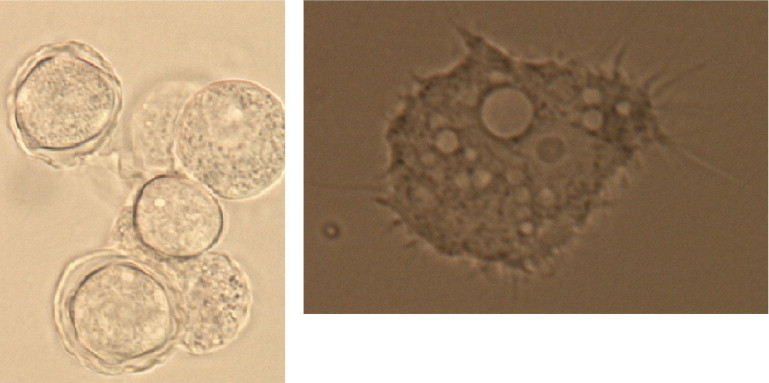
The following strains of Acanthamoeba species were used as amebic isolates: Acanthamoeba species 65 was used as an environmental amoeba isolated from superficial water, and Acanthamoeba species 7376 was used as a clinical strain isolated via corneal scraping from a patient with keratitis. The 2 isolates were identified through the microscopic examination of their trophozoites and cysts ( Figure 1 ) . The keys reported by Poussard and Pons and by Page were used. To confirm the identification of the amoebae, an Acanthamoeba species-specific polymerase chain reaction analysis was performed, including primer pair JDP1/JDP2 targeted toward 18S ribosomal DNA stretch ASA.1, DNA was extracted from cultured organisms in protease peptone 2.0 g, yeast extract 0.5 g, glucose 0.5g/100 mL distillate water following the method described by Ausubel. The polymerase chain reaction analysis confirmed that both Acanthamoeba strains belong to genotype T4, which is the strain most commonly found in the environment and also is a cause of AK.
Preparation of Acanthamoeba Culture
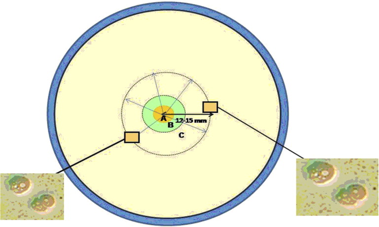
One milliliter of a 7-day axenic culture in protease peptone 2.0 g, yeast extract 0.5 g, glucose 0.5g/100 mL distillate water was centrifuged at 200 g for 6 minutes. The pellet was resuspended in 50 μL Page amoeba saline solution, and the number of trophozoites and cysts was determined using a Neubauer chamber. Three microliters of suspension containing 1000 amoebae (both trophozoites and cysts) were deposited on the center of nonnutrient agar plates that previously had been streaked with Escherichia coli as a food source for the amoeba. The drop of suspension did not exceed 5 mm in diameter, because this ensured a drop smaller than the treatment diameter and that all amebic cells received UVA radiation.
Experimental Design
The overall procedure was performed under aseptic conditions that prevented the culture plates from being exposed to air and light (with less than 3 hours until the experiment). The assay was conducted in triplicate. Plates seeded with Acanthamoeba remained hermetically sealed until seconds before the test. The 2 different strains of Acanthamoeba were tested in an identical manner. Four groups were created as follows. In group 1, 0.1% riboflavine (Ricrolin; Sooft Italia, Montegiorgio (AP), Italy) was dropped directly onto the strains, and it was allowed to soak for 10 minutes in the agar media before UVA irradiation. When the presence of the yellow tinction was observed in the agar media , the culture plates were irradiated with UVA (3 mW/cm 2 , 370 nm) for 30 minutes. During exposure to UVA, 1 drop of riboflavine was applied onto the agar media every 5 minutes. In group 2, the treatment was similar to that of group 1, but the period of exposure to UVA was 30 minutes longer (for a total of 60 minutes). The control group (group 3) received no treatment. Group 4 received only 0.1% riboflavine (Ricrolin; Sooft Italia) using the same method used for group 1.
The application of UVA was performed using the parameters used for in vivo corneal collagen cross-linking, and the diameter of the radiation was 9 mm in all cases, thus exceeding the limits of seeded protozoa (less than 5 mm in diameter). The ultraviolet source was CBM-X-LINKER (CSO, Scandicci Firenze, Italy). The diode lamp was calibrated before each experiment to ensure that the value was between 2.7 and 3.3 mW/cm 2 . Subsequently, the agar plates were sealed and incubated at 30 C. The migration distance of surviving and growing amoeba was measured after 24 hours of incubation using a Nikon AZ-100 microscope (×5 objective, ×100 magnification; Nikon Corp, Kanagawa, Japan) and was photographed using a Jenoptik camera (Jenoptik AG, Jena, Germany).
Assessment of Amoeba Viability
The evaluation of amoeba viability in a solid medium usually is based on the observation of trophozoites and cysts in the agar plate. The presence of trophozoites beyond the limits of the seeding area indicates growth. If there is no growth within 7 days, the piece of agar containing the drop with the amoeba usually is cut and placed in a new plate. Again, this plate is incubated, and it is expected that trophozoites will be observed within a maximum of 15 days, indicating that the amoeba remains viable.
In our experiment, the seeding plates for both species of amoebae for each group (including the control group) were evaluated after 24, 48, and 72 hours of incubation at 30 C. If no growth was detected, the reculture method explained above was applied. The migration radius (from the center of the point of application of the amoeba to the point where trophozoites had migrated) was measured to quantify the results for the different groups. We assumed that trophozoites, cysts, or both survived the treatment if the distance was more than 5 mm.
Results
In all groups and for both isolates studied (environmental and pathogenic strains), cysts and trophozoites were detected at distances from the center of the point of application of more than 5 mm after incubation at 30 C in the first evaluation (24 hours), so no re-evaluation or reculture was necessary because of the early growth. This indicated the viability of the amoebae tested. Trophozoites were found at a similar distance from the center of the point of application for all treatment groups and for the control group for both strains of amoebae (14 to 15 mm); the only exception was the group exposed to cross-linking and riboflavin for 60 minutes (group 2), for which the distance traveled was slightly lower (12 mm; Figure 2 ) . After 72 hours, the trophozoites had spread across the agar in all groups.
Stay updated, free articles. Join our Telegram channel

Full access? Get Clinical Tree


