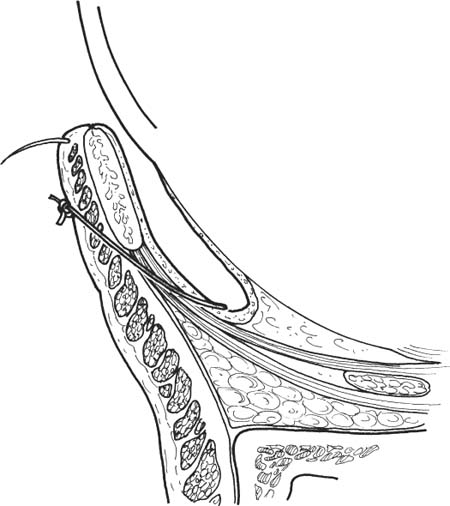
51 Involutional entropion results from aging changes in the lid. Such changes include: (1) horizontal lid laxity, (2) lower lid retractor disinsertion and laxity, and (3) overriding of the pretarsal by preseptal orbicularis. Numerous procedures and variations have been designed to correct entropion by ameliorating one or more of these pathological conditions. The specific operation undertaken will depend upon the individual case and the personal preference of the surgeon. The less commonly encountered entropion due to cicatricial conjunctival disease (e.g., cicatricial ocular pemphigoid) will require a lid splitting procedure or mucous membrane graft. Note: These chapters will deal only with the surgical correction of involutional entropion. The management of cicatricial entro-pion will not be discussed. The following describes the most commonly used procedures for the correction of routine cases of involutional entropion. Entropion sutures prevent overriding of the orbicularis while reinforcing the lower lid retractors. This technique is useful in cases where permanent repair may not be necessary (e.g., spastic entropion) or where a simple procedure is desired (e.g., debilitated patients). Entropion sutures may also be used as an adjunct to other lid procedures. Combined horizontal lid splitting and lid shortening is useful for entropion with horizontal lid laxity. This technique tightens the lid while reinforcing the lower lid retractors and preventing orbicularis override. Most involutional entropion results from horizontal eyelid laxity and generalized laxity or dehiscence of the lower eyelid retractors from the normal tarsal attachments. Tightening of the lower eyelid retractors may be accompanied by horizontal shortening of the eyelid by tarsal strip procedure or by wedge resection. See Introduction in this chapter. See Chapter 3. 1. Discontinue aspirin and nonsteroidal anti-inflammatory agents for 10 days before surgery. Discontinue warfarin 2–3 days preoperatively, if medically possible. 2. Query patient about bleeding tendencies. A useful screening question is asking if the patient had unusual bleeding after dental extraction. Obtain hematological evaluation if bleeding tendency is suspected. Figure 51.1 1. Locate suture placement (Fig. 51.1): a. Place medialmost mattress suture at junction of medial third and lateral two thirds of lower lid (avoid eversion of punctum). b. Proceed laterally, separating each mattress suture by ~5 mm, for a total of three sutures. c. The lateralmost suture should end ~5 mm from the lateral canthus. Figure 51.2 2. Technique of suture placement (Fig. 51.2): a. Grasp lid with forceps and pass each end of double-armed 4–0 chromic gut suture i. through conjunctiva deep in inferior fornix ii. upward toward inferior edge of tarsus, imbricating lower lid retractors and passing just below inferior edge of tarsus, and iii. out through skin ~2 mm below lash line (pull skin inferiorly when passing needle through it). b. Separate the two arms of the suture by 3 mm. c. Titrate tightness of suture to provide for slight over-correction (slight ectropion). 3. Mechanism of suture action. a. Formation of adherence of pretarsal orbicularis to lower eyelid retractors preventing the preseptal muscle from overriding the pretarsal orbicularis. b. Tightening of lower lid retractors and transfer of their pull to the anterosuperior tarsus, causing tarsal eversion. 4. Apply antibiotic ointment into lower conjunctival fornix and onto external sutures. 1. Apply ice packs to decrease swelling. 2. Keep head of bed elevated 30 degrees to decrease swelling. 3. Apply antibiotic ointment twice daily to suture line. 4. Remove sutures in 2 to 3 weeks. 1. Undercorrection 2. Loss of correction with time Note: Entropion sutures may be repeated as necessary. See Introduction section at the beginning of this chapter. 1. Entropion with horizontal lid laxity.
Entropion Repair
 Introduction
Indications
Horizontal Lid Splitting and Lid Shortening
Reinsertion of Lower Eyelid Retractors (with full-thickness horizontal tightening)
 Entropion Sutures
Indications
 Temporary correction of mild or intermittent ectropion (e.g., spastic ectropion)
 Correction of ectropion in debilitated patients who are unable to undergo a more complex and time-consuming procedure
 May be used as an adjunct to other entropion procedures.
Preoperative Procedure
Instrumentation
 Suture (double-armed 4–0 chromic gut with long needles)
 Needle holder
 Toothed forceps
Operative Procedure
Postoperative Procedure
Complications
 Horizontal Lid Splitting/Lid Shortening
Indications
Stay updated, free articles. Join our Telegram channel

Full access? Get Clinical Tree


