Fig. 31.1
Diagram of standard transmission electron microscopy (TEM) and scanning electron microscopy (SEM), showing major components: 1 electron optical column, 2 electron gun, 3 magnetic lenses, 4 magnetic coil, 5 apertures, 6 detector, and 7 digital imaging systems
1.
Electron optical column
2.
Electron gun that consists of an electron source to produce electrons, such as a tungsten filament
3.
Magnetic lenses to demagnify the beam
4.
Magnetic coils to control and modify the beam
5.
Apertures to define the beam and prevent electron spray
6.
Detectors to collect, detect, and display the signal
7.
Digital imaging systems that produce an image from the signal
8.
Vacuum systems consisting of vacuum pumps and a vacuum chamber
There are two basic types of electron microscopy, transmission electron microscopy (TEM) and scanning electron microscopy (SEM).
TEM uses an electron beam that transmits through ultrathin (60–90 nm) sections that are glutaraldehyde-fixed and usually double-stained. TEM produces two-dimensional images on a fluorescent screen, photographic film, or CCD (charge-coupled device) camera. TEM can detect structures by the transmission of the electron beam and discriminate details of 0.2 nm. However, the quality of the obtained image mainly depends on the preparation of the biological sample (Ross and Pawlina 2011).
SEM obtains topographic, three-dimensional images with a resolution of about 2 nm. The lens system of SEM produces a small focused spot of electrons that are then scanned over the specimen surface by deflection coil. SEM is able to produce an image by detecting secondary electrons and backscattered electrons generated from the specimen. A secondary electron detector in the SEM builds the image by mapping the signals of a finely focused electron beam that is scanned on the sample surface (Bozzola and Russell 1999; Potter and Love 1999; Ross and Pawlina 2011).
Both TEM and SEM can provide only black-and-white images. Although the original image is monochrome, micrographs can be colored digitally to emphasize details. In recent years, digital imaging systems provide incredible opportunities to obtain high-quality electron micrographs.
Biological materials usually require processing before being viewed by electron microscopy (Bozzola and Russell 1999; Ross and Pawlina 2011). The tissue preparation technique varies depending on the type of microscope and the type of specimen. Low-vacuum SEMs and the environmental scanning electron microscope (ESEM) overcome both these limitations (Slowko 2001).
The stages for tissue preparation for TEM are as follows:
(a)
Fixation: can be achieved by perfusion and microinjection or immersion using various fixatives including aldehydes.
(b)
Post fixation: performed in OsO4.
(c)
Dehydration: done with a graded series of alcohol.
(d)
Epoxy resin block preparation: treat with propylene and embed in epoxy resin.
(e)
Semi-thin sectioning: One- to two-micrometer-thick semi-thin sections obtained from the epoxy resin blocks should be stained with methylene blue or azure for light microscopy.
(f)
Ultrathin sectioning: Ultramicrotome, an instrument for cutting extremely thin sections, is used for ultrathin sectioning of tissue. Ultrathin sections are obtained from selected areas and then double-stained with uranyl acetate/lead citrate.
During SEM sample preparation, after the fixation step, the specimens must be dried. Electron microscopists prefer to use Critical Point Drying. By removing carbon dioxide after the transition from the liquid to the gas phase at the critical point, the specimen can be dried without structural damage. Specimens must be mounted onto a holder that can be inserted into the scanning electron microscope. The last step prior to sample imaging is coating the samples. The objective of this coating is to increase its conductivity in the scanning electron microscope and to prevent the buildup of high-voltage charges on the specimen. Typically, specimens are coated with a thin layer of gold, gold palladium, or platinum (Bozzola and Russell 1999).
In addition to visual inspection, a grading system can be used to quantitatively evaluate the samples to compare different experimental conditions. The data can then be analyzed statistically to make further evaluations. This grading system was established based on similar principles of methods used for evaluating different tissue samples (Taşdemir et al. 1993; Kırkalı et al. 1995; Kaptanoğlu et al. 2000; Görgülü et al. 2001; Hazer et al. 2010).
It is important to pay close attention during the sample preparation to get small-sized (less than 2 mm) biopsy materials and to fix them in seconds to prevent autolysis and obtain optimum diffusion of the fixatives (Bozzola and Russell 1999).
31.1.2 Microscopic Anatomy of the Nose
The nose humidifies, filters, and warms the air we breathe as well as providing the sense of olfaction. The nose is considered to have two parts: the external nose and the nasal cavity.
The external nose consists of skin and a framework of compact bone and hyaline cartilage that forms a projection covered by skin. Electron microscopic observation of this part does not show any regional specifications. The skin consists of two main layers. The outer layer is the epidermis, which is composed of a keratinized and stratified squamous epithelium (Figs. 31.2 and 31.3) (see Sect. 31.1.2.1). The inner layer is the dermis, which is dense connective tissue including epithelial derivates of the skin such as hair follicles and sweat and sebaceous glands (Jones 2001). The supporting framework is composed of nasal bones, frontal process of the maxillae, and the nasal part of the frontal bone and septum, as well as major and minor alar cartilages. Bone is also a connective tissue characterized by a mineralized extracellular matrix containing mainly type I collagen along with other non-collagenous matrix proteins (Ross and Pawlina 2011). Bones of the external nose consist of layers of relatively thick compact bone with a layer of spongy bone covered by periosteum, which is a sheath of dense fibrous connective tissue containing osteoprogenitor cells.
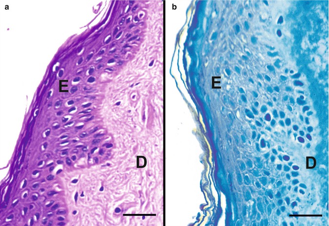
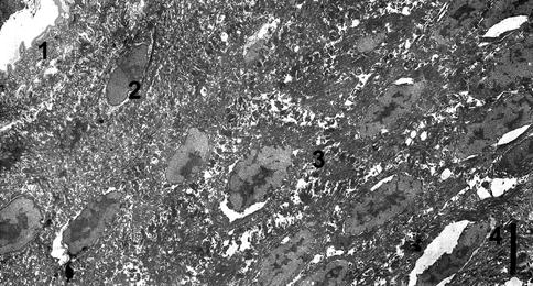
Fig. 31.2
The epidermis, which is composed of a keratinized stratified squamous epithelium (E), and the dermis (D) (scale bar: 25 μm). (a) Light micrograph of the epidermis of the external nose (paraffin block, stain: H&E). (b) Light micrograph of the epidermis of the external nose (Araldite block, stain: methylene blue). The specimens were obtained from a fresh frozen cadaver from a microscopic anatomy lab at Hacettepe University, Faculty of Medicine, Department of Anatomy. H&E hematoxylin and eosin
Fig. 31.3
Electron micrograph (TEM) keratinized stratified squamous epithelium of the epidermis of the external nose (scale bar: 5 μm) (Araldite block, stain: uranyl acetate/lead citrate). The specimen was obtained from a fresh frozen cadaver from a microscopic anatomy lab at Hacettepe University, Faculty of Medicine, Department of Anatomy. TEM transmission electron microscopy, 1 Stratum corneum, 2 stratum spinosum, 3 stratum granulosum and 4 stratum basale
The type of the cartilage that contributes to the framework of the nose is hyaline cartilage (Fig. 31.4). The matrix of the hyaline cartilage consists of collagen, predominantly type II fibrils and other cartilage-specific collagen molecules (Ross and Pawlina 2011). The chondrocytes are either rounded or ellipsoidal (Figs. 31.4 and 31.5). The plasma membrane is folded into a moderate number of microvilli. Numerous cytoplasmic filaments and coarse granules of glycogen are prominently present in the cytoplasm. The Golgi complex is also prominent and frequently contains dilated vesicles enclosing small dense particles. A small amount of rough endoplasmic reticulum is present, while unattached ribosomes are not numerous. There are a few lipid droplets in the cytoplasm. The nuclei are ovoid and usually contain a single large nucleolus (Fig. 31.5). Mitochondria are small and not very numerous. The matrix is composed mostly of collagen fibrils and matrix granules. Frequently, granules appear to be linked together by extremely fine intergranular fibrils, usually less than 50 Å thick, which connect the projections of adjacent granules. Infrequently, clusters of membrane-bounded matrix vesicles are observed between collagen fibrils of the matrix (Anderson and Sajdera 1971)
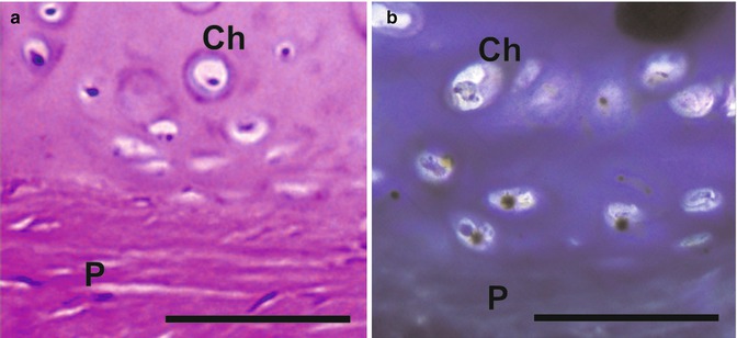
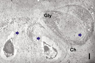
Fig. 31.4
The hyaline cartilage, rounded or ellipsoidal chondrocytes (Ch) and fibroblast-like cells of the perichondrium (P). (a) Light micrograph (paraffin block, stain: H&E) (scale bar: 100 μm). (b) Light micrograph (Araldite block, stain: methylene blue) (scale bar: 100 μm). H&E hematoxylin and eosin
Fig. 31.5
Electron micrograph (TEM) of the hyaline cartilage, rounded or ellipsoidal chondrocytes (Ch) (scale bar: 2 μm) (Araldite block, stain: uranyl acetate/lead citrate). The specimen was obtained from a fresh frozen cadaver at a microscopic anatomy lab at Hacettepe University, Faculty of Medicine, Department of Anatomy. TEM transmission electron microscopy, Arrows cytoplasmic filaments, Gly coarse granules of glycogen
The perichondrium, a firmly attached dense connective tissue composed of fibroblast-like cells, surrounds the hyaline cartilage (Fig. 31.4).
The nasal cavity is divided into paired chambers separated by a bony and cartilaginous septum. Each chamber is divided into three regions:
(a)
Vestibule of the nasal cavity
(b)
Respiratory region
(c)
Olfactory region
31.1.2.1 Vestibule of the Nasal Cavity
The nasal cavity extends from the nares anteriorly to the choanae posteriorly. Just behind the nares, the nasal cavity widens and forms the vestibule (Stenberg 1997). It is lined with keratinized stratified squamous epithelium and the dermis (Fig. 31.6) that contains connective tissue elements, many hair follicles (hairs in this region are called vibrissae) (Figs. 31.7 and 31.8), and sebaceous glands and sweat glands (Figs. 31.9 and 31.10) (see Sect. 31.1.2).
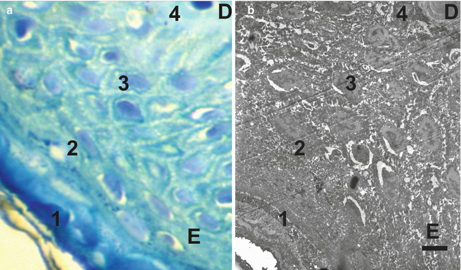
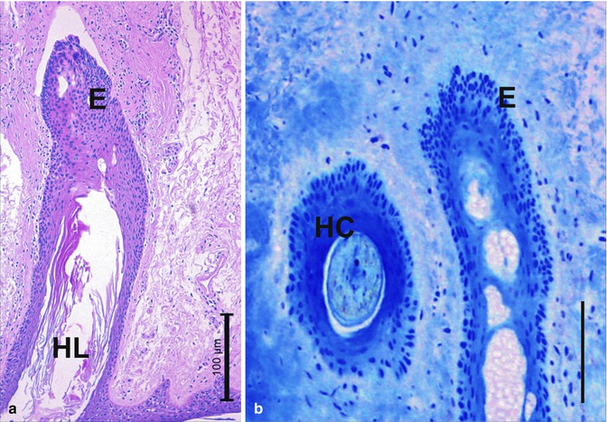
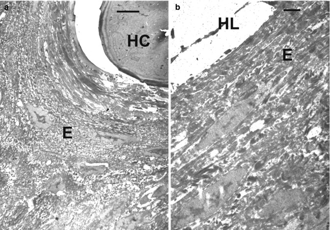
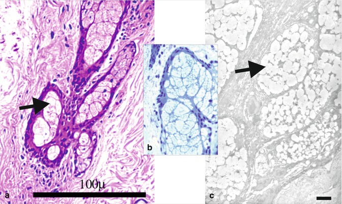
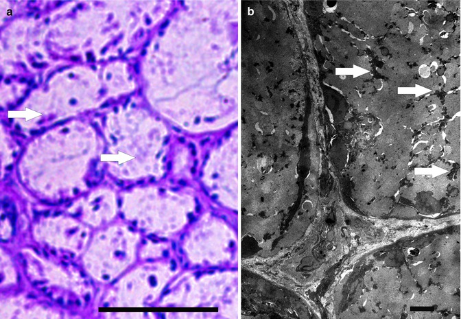
Fig. 31.6
(a) Light micrograph of the epidermis of the vestibule (scale bar: 5 μm) (Araldite block, stain: methylene blue). (b) Electron micrograph (TEM) of the epidermis of the vestibule (scale bar: 5 μm) (Araldite block, stain: uranyl acetate/lead citrate). Both specimens were obtained from fresh frozen cadavers at a microscopic anatomy lab at Hacettepe University, Faculty of Medicine, Department of Anatomy. TEM: transmission electron microscopy. E epidermis, D dermis, 1 stratum corneum, 2 stratum spinosum, 3 stratum granulosum and 4 stratum basale
Fig. 31.7
Light micrograph from the dermis of the vestibule (scale bar: 100 μm). (a) Hair follicle and vibrissae, longitudinal section (paraffin block, stain: H&E). (b) Hair follicle and vibrissae, cross (HC) and longitudinal section (HL) (Araldite block, stain: methylene blue) (scale bar: 100 μm). The specimens were obtained from fresh frozen cadavers at a microscopic anatomy lab at Hacettepe University, Faculty of Medicine, Department of Anatomy. H&E hematoxylin and eosin, E epithelial cells
Fig. 31.8
Electron micrographs (TEM) from the dermis of the vestibule. (a) Cross and (b) longitudinal sections of the vibrissae (scale bar: 5 μm) (Araldite block, stain: uranyl acetate/lead citrate). The specimen was obtained from a fresh frozen cadaver at a microscopic anatomy lab at Hacettepe University, Faculty of Medicine, Department of Anatomy. TEM transmission electron microscopy E epithelial cells
Fig. 31.9
The sebaceous glands (arrows) from the dermis of the vestibule (scale bar: 5 μm). (a) Light micrograph (paraffin block, stain: H&E). (b) Light micrograph (Araldite block, stain: methylene blue). (c) Electron micrographs (TEM) (Araldite block, stain: uranyl acetate/lead citrate). H&E hematoxylin and eosin, TEM transmission electron microscopy
Fig. 31.10
The sweat glands and their myoepithelial cells (arrows) from the dermis of the vestibule. (a) Light micrograph (paraffin block, stain: H&E) (scale bar: 5 μm). (b) Electron micrographs (TEM) (Araldite block, stain: uranyl acetate/lead citrate) (scale bar: 100 μm). H&E hematoxylin and eosin, TEM transmission electron microscopy
Stratified squamous epithelium consists of several layers of cells. The flattened cells form its outer layer and the deepest cells are columnar. The epidermis is composed of four distinct layers. These are the stratum corneum, stratum spinosum, stratum granulosum, and stratum basale (stratum germinativum) (Fig. 31.6). The cells of the stratum corneum are anucleate corneal cells (squamous), called corneocytes or cornified cells. The corneocytes are flattened cells that lack nuclei and cytoplasmic organelles. The cells contain aggregated keratin filaments. The upper spinous layer and granular cell layer also contain smaller lamellate granules called lamellar, membrane-coating granules (MCGs or Odland bodies). These are numerous within the upper spinous layer. They play an important role in providing the barrier and intercellular cohesion functions of the stratum corneum. They release their lipid components into the intercellular space. The basal and spinous cells together are called the Malpighian layer, which includes cells such as melanocytes, Langerhans cells and Merkel cells. When outer cells become damaged, cell division occurs within the basal layer. The cells move outwards to the stratum corneum, passing through the stratum spinosum. The characteristics of these cells then transdifferentiate to become the cells of the stratum corneum. There are biochemical and signaling interactions between the epithelial cells, including desmosomes, adherens junctions, gap junctions, and tight junctions (McGrath et al. 2010).
Stay updated, free articles. Join our Telegram channel

Full access? Get Clinical Tree


