Purpose
To investigate the in vitro effect of pH, osmolarity, solvent, and light interaction on currently used and novel dyes to minimize dye-related retinal toxicity.
Design
Laboratory investigation.
Methods
Retinal pigment epithelium (RPE) human cells (ARPE-19) were exposed for 10 minutes to different pH solutions (4, 5, 6, 7, 7.5, 8, and 9) and glucose solutions (2.5%, 5.0%, 10%, 20%, 40%, and 50%) with osmolarity from 142 to 2530 mOsm, with and without 0.5 mg/mL trypan blue. R28 cells were also incubated with glucose (150, 310, and 1000 mOsm) and mannitol used as an osmotic control agent in both experiments. Dye-light interaction was assessed by incubating ARPE-19 for 10 minutes with trypan blue, brilliant blue, bromophenol blue, fast green, light green, or indigo carmine (0.05 mg/mL diluted in balanced saline solution) in the presence of high-brightness xenon and mercury vapor light sources.
Results
Solutions with nonphysiologic pH, below 7 and above 7.5, proved to be remarkably toxic to RPE cells with or without trypan blue. Also, all glucose solutions were deleterious to RPE ( P < .001) even in iso-osmolar range. No harmful effect was found with mannitol solutions. Among the dyes tested, only light green and fast green were toxic to ARPE-19 ( P < .001). Light exposure did not increase RPE toxicity either with xenon light or mercury vapor lamp.
Conclusions
Solutions containing glucose as a dye solvent or nonphysiologic pH should be used with care in surgical situations where the RPE is exposed. Light exposure under present assay conditions did not increase the RPE toxicity.
The use of vital dyes during vitreoretinal surgery, “chromovitrectomy,” allows a better visualization of thin, transparent preretinal tissues in the vitreoretinal interface. Currently, the available vital dyes for chromovitrectomy include indocyanine green, trypan blue, brilliant blue, and bromophenol blue. However, conflicting results regarding retinal toxicity associated with the application of these dyes are still observed in the literature. Engelbrecht and associates reported retinal pigment epithelium (RPE) changes and visual field defects even with a low-concentrated indocyanine green (0.125%)–assisted internal limiting membrane (ILM) peel in patients with macular hole. Indeed, postoperative atrophic RPE changes and the poorer visual outcome after macular hole or rhegmatogenous retinal detachment surgery may be related to the direct exposure of the dye solution to the RPE or dye migration into the subretinal space.
Although dye concentration has a recognizable effect on cellular viability, this result has drawn attention to other elements that could be related to RPE toxicity, such as pH and osmolarity. Sippy and associates reported that hypo-osmotic indocyanine green solution plus light promotes a marked reduction in enzymatic activity in RPE cells. Subsequently, Stalmans and associates found a correlation between severity of damage to RPE cells and duration of indocyanine green incubation, light exposure time, and osmolarity of the solvent.
Dye solubilization is another important factor in toxicity. Balanced saline solution (BSS) and distilled water are mainly used as solvents in chromovitrectomy. However, glucose solution has also been considered an alternative solvent. Lesnik Oberstein and associates achieved a proper epiretinal membrane (ERM) staining using trypan blue and 5% glucose with no toxic effects. The same technique was reported for ILM staining in patients with full-thickness macular hole with no clinical sign of retinal toxicity. The combination of glucose and dyes, a low-cost and well-known technique, results in a dense solution, which helps to concentrate the dye in the posterior pole, avoiding contact with the lens posterior capsule. However, it has been mentioned that high glucose concentrations (40%) may cause severe damage to retinal function.
The dye-stained retina may also affect light absorption during vitrectomy. Previous in vitro studies by our group showed a remarkable spectral overlap with different vitrectomy light sources and vital dyes. Among the tested fiber optics, xenon dual-output illumination with an integrated laser pathway showed the greatest, and mercury vapor lamp the least, wavelength overlap. Furthermore, Haritoglou and associates demonstrated histologic and functional damage to the retina in the presence of indocyanine green and halogen and xenon light sources.
The aim of this study was to provide a detailed investigation of factors that may contribute to dye toxicity and highlight the importance of dye solution preparation to the surgical practice. We performed in vitro experiments using human retinal cultured cells (ARPE-19), examining a wide range of pH, osmolarity, glucose, and mannitol concentrations and an evaluation of glucose effect on rat retinal neural cell line (R28) viability. Experiments with xenon and mercury light, in the presence or absence of dyes (trypan blue, fast green, light green, indigo carmine, brilliant blue, and bromophenol blue), were also executed. All dyes and solutions were submitted to a meticulous preparation in terms of pH and osmolarity to minimize deleterious conditions related to unexpected variations, which could influence toxicity found in the experiments.
Methods
An experimental study prospectively approved by the Institutional Review Board and Ethics Committee of the Federal University of Sao Paulo, the project evaluated osmolarity and pH proprieties of vital dyes for vitreoretinal surgery and its effect on human retinal cultured cells (ARPE-19) and rat retinal neural cell line (R28). The dyes fast green, light green, indigo carmine, and brilliant blue were purchased from Merck (Darmstadt, Germany), and bromophenol blue, 3-4,5 dimethylthiazol-2,5 diphenyl tetrazolium bromide (MTT), and other cell culture reagents were purchased from Sigma-Aldrich (Munich, Germany). Trypan blue, glucose, and mannitol were obtained from Ophthalmos (São Paulo, Brazil) and BSS from Alcon Laboratories (Fort Worth, Texas, USA).
Dye Solubilization
Initially, 5 mg of each dye was measured using an analytical balance (Mettler-Toledo Inc, Columbus, Ohio, USA) and dissolved in sterile BSS to obtain stock solutions. Trypan blue was solubilized at 0.5 mg/mL or 0.05 mg/mL and the solutions sonicated for 15 minutes, using an Ultrasound Ultracleaner (Unique Ind, Idaiatuba, Brazil). Brilliant blue, bromophenol blue, indigo carmine, fast green, and light green were solubilized at the concentration of 0.5 mg/mL. Afterwards, the pH and osmolarity of each dye solution were determined using a previously calibrated pH meter (Quimis, Diadema, São Paulo) and osmometer (Advanced Instruments Inc, Norwood, Massachusetts, USA).
Cell Culture
The cytotoxic effects of the solutions were determined using the human RPE cell line, ARPE-19, and R28 was used to complement the osmolarity experiments. The ARPE-19 cell line was cultured in a mixture of DMEM/F-12 (Gibco, Carlsbad, California, USA) containing 10 U/mL penicillin and 10 μg/mL streptomycin, supplemented with 10% fetal bovine serum. R28 cells were derived from postnatal rat retina and cultured in DMEM high glucose (Gibco) with 10% fetal bovine serum, as described by Seigel. This cell line expresses genes characteristic of neurons, as well as functional neuronal properties.
The cells were seeded at a concentration of 5 × 10 3 cells/mL in 96-well, flat-bottom tissue culture plates in 200 μL culture medium and grown for 24 hours before the experiments in a humidified incubator in 5% CO 2 at 37 C. Subsequently, the cell culture medium was removed and the cells washed once with BSS. Next, cells were incubated for 10 minutes in the presence of BSS or different dyes solutions (100 μL/mL). The 10-minute incubation was chosen to simulate the period of macular surgery from dye injection into the vitreous cavity to its complete removal after ILM peel and the air-fluid exchange. After incubation, cells were washed 3 times with 200 μL BSS and incubated for 4 hours in the presence of MTT reagent at a final concentration of 0.5 mg/mL. The supernatant was discarded and 200 μL/mL of dimethyl sulfoxide was added to dissolve the MTT formazan product. The results were obtained by measuring absorbance at 570 nm. All the experiments were performed in triplicate.
pH and Osmolarity Influences on Cell Viability in the Absence or Presence of Trypan Blue
pH analysis was performed using ARPE-19 cells and BSS at pH 4, 5, 6, 7, 7.5, 8, and 9, in the absence or presence of trypan blue (0.5 mg/mL), and osmolarity between 317 and 339 mOsm. To investigate the influence of osmolarity, with or without trypan blue (0.5 mg/mL), ARPE-19 cells were treated with 100 μL of different glucose stock concentrations: 2.5% (142 mOsm), 5% (270 mOsm), 10% (577 mOsm), 20% (1165 mOsm), 40% (2470 mOsm), and 50% (2530 mOsm), prepared in BSS and the pH corrected to 7. In addition to the glucose analysis, mannitol was selected as another solution for osmolarity investigations. Thus, 100 μL of mannitol at different stock concentrations, 1% (74 mOsm), 2.5% (150 mOsm), 5% (309 mOsm), 10% (464 mOsm), 15% (567 mOsm), and 20% (658 mOsm), was also tested on ARPE-19 cells. Additionally, for the osmolarity assay, R28 cells were exposed to 100 μL of the following solutions: glucose 2.7% (150 mOsm), 5.5% (310 mOsm), and 18% (1000 mOsm) with the respective mannitol solutions of 2.5% (150 mOsm), 5.6% (310 mOsm), and 18.2% (1000 mOsm). The cell culture images were obtained using Axio Observer Z1 (Carl Zeiss, Göttingen, Germany).
Light Toxicity Investigations in the Presence of Different Dyes
ARPE-19 cell viability was also studied using the 6 dyes (trypan blue, brilliant blue, bromophenol blue, indigo carmine, light green, and fast green) in the absence or presence of high-brightness xenon and mercury vapor lamp for 10 minutes in a dark room. Thus, cells were submitted to 100 μL of the dye stock solutions at 0.05 mg/mL (final concentration of 0.005 mg/mL), pH 7, and osmolarity of 290-313 mOsm. Both 20-gauge fiber optics, with 2760 lumens measured by a lux meter, were positioned at 20 mm from the cell culture. According to the inverse square law, the power of the light will be inversely proportional to the square of the distance. Thus, just one-quarter of the original power, 690 lumens, was delivered to the cell culture. The luminous flux at this distance on a previously determined surface was approximately 5.5 × 10 5 lux/mL. After illumination, the cells were washed 3 times with BSS and incubated with MTT (0.5 mg/mL). BSS was used as experimental control.
Statistical Analyses
Statistical analyses were performed using Graphpad Prism 5 software (Graphpad, La Jolla, California, USA). The ANOVA test was used for statistical analysis of cell viability results and a P value of less than .05 was considered statistically significant.
Results
Influence of pH on ARPE-19 Viability
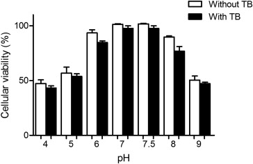
The viability assay using ARPE-19 and 10-minute incubations in the presence of different pH solutions demonstrated that values from 7 to 7.5, with and without trypan blue ( P < .001), showed optimal conditions for retinal cell viability. The solutions were increasingly more toxic from pH 6 to 4 ( P < .001) and from pH 8 to 9 ( P < .001). No significant differences were seen between BSS and BSS with trypan blue at the same pH, and therefore, the dye caused no additional toxicity ( Figure 1 ).
Osmolarity Toxicity Analysis
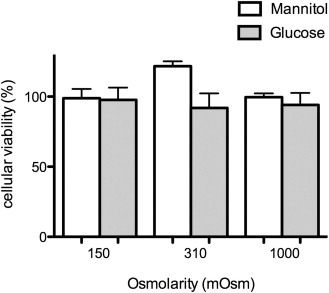
ARPE-19 cells incubated in the presence of glucose solutions with and without trypan blue displayed a marked decrease in cell viability when compared with the BSS control ( P < .001) ( Figure 2 , Top). Even the iso-osmotic solution of 5% glucose (270-277 mOsm), independent of trypan blue, was significantly more toxic than the control ( P < .001). Moreover, in the absence of trypan blue, no differences were observed between 2.5% and 5.0% glucose and also 40% and 50% glucose ( P > .05). Similar findings were observed with the dye solution, where no statistical difference was detected between 2.5% and 5.0%, 5.0% and 10%, and 40% and 50%. High glucose concentrations (20%, 40%, and 50%) were significantly more deleterious than low-concentration glucose (2.5%) ( P < .001). The presence of the dye did not lead to any additional decrease in cell viability at any concentration ( P > .05) ( Figure 2 ). However, after 10 minutes of incubation, no glucose toxic effect was observed in R28 as well as for mannitol solution in both cell lines, when compared with the BSS control ( P > .05) ( Figures 2 and 3 ). With mannitol as the osmolarity control, the assay revealed that glucose itself had a role in the RPE toxicity.
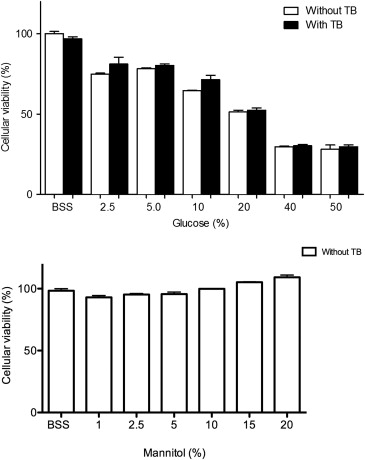
ARPE-19 morphologies were also analyzed using the microscope Axio Observer Z1 (Carl Zeiss) after 10 minutes of incubation with BSS, glucose (310 mOsm), and mannitol (310 mOsm). Glucose solutions caused modifications in cell structure, showing toxicity effect and morphologic characteristics similar to apoptosis on RPE cells. BSS and mannitol did not show significant cellular structural changes in both cell lines ( Figure 4 ).
Toxic Effect of Dyes, Light Exposure, and Dye-Light Interactions
To evaluate cell viability in the presence of different dyes (trypan blue, brilliant blue, bromophenol blue, indigo carmine, fast green, and light green) and light sources (high-brightness xenon and mercury vapor lamp), we first tested 2 different concentrations (0.05 mg/mL and 0.5 mg/mL) of the dyes on ARPE-19 cells. Considering that no toxicity was observed at the highest value, except for fast green and light green, which showed about 20% decrease in cell viability (data not shown), we chose 0.05 mg/mL for the mercury and xenon light analysis. Regarding the effect of the light sources on cell viability without dye incubation, no decrease was observed compared with BSS ( P > .05) and no difference between the lights was seen ( P > .05). Indeed, the cultured RPE cells exposed to the 6 dyes in the presence of the light sources revealed that in the absence or presence of xenon or mercury light, only fast green and light green showed a similar ( P > .05) and significant deleterious effect ( P < .001) ( Figure 5 ). Comparing xenon and mercury light exposure, no statistical difference was found between the lights with all dyes tested ( P > .05). These results indicated that both lights, when the light source is held 20 mm from tissue, and the dyes brilliant blue, bromophenol blue, indigo carmine, and trypan blue at pH 7 and osmolarity of 290-313 mOsm were safe to the RPE cells.


