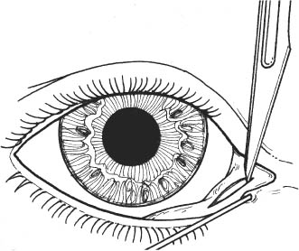
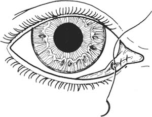
50 Involutional ectropion results, in general, from aging changes of the lid and, in particular, from generalized laxity and descent of the eyelid structures and midface. Seventh nerve palsy will manifest a similar but more pronounced appearance. Numerous procedures and variations have been designed to correct ectropion. The specific operation undertaken depends on the anatomic etiology of a particular case and the personal preference of the surgeon. Following are some of the commonly used procedures for correcting routine cases of involutional, cicatricial, and paralytic ectropion. This technique may be used alone or in combination with another ectropion procedure to reposition a punctum that is everted from the globe secondary to medial ectropion. Horizontal lid shortening (described in Chapter 46, “ Repair of Full Thickness Lid Margin Lacerations and Defects”) is easy to carry out and is occasionally useful for central lower lid ectropion. It may be combined with a blepharoplasty-type skin excision in cases where, in addition to horizontal lid laxity, excess skin is present (see Chapter 52). The lateral tarsal strip technique is indicated for generalized ectropion and horizontal lid laxity, lateral ectropion, and ectropion with lateral canthal tendon laxity. It may be used in cases of seventh nerve palsy—although the long-term result varies from patient to patient. Patients with ectropion and vertical deficiency of skin may benefit from full-thickness eyelid shortening with midface elevation with or full-thickness eyelid shortening with skin graft skin. These patients may have a general history of actinic damage and skin cancer. Some patients have a specific history of removal of lower eyelid or cheek skin cancers, prior lower eyelid transcutaneous blepharoplasty, or facial trauma. In most cases, examination shows smooth, relatively unwrinkled skin. Pull the ectropic lateral eyelid up to a normal position at the lateral canthal angle. If there is significant pull at the angle of the mouth or below, there may be descent of the midfacial structures or deficiency of skin. Cicatricial ectropion is worse in upgaze, as the skin cannot allow superior movement of the eyelids. Select patients with ectropion may benefit from elevations of the midfacial structures together with full-thickness eyelid tightening. Patients with ectropion and vertical deficiency of skin may benefit from full-thickness eyelid shortening with skin graft skin. This treatment is usually curative, but the mismatch of skin between the donor and recipient beds may be objectionable to some patients. For all procedures, discontinue aspirin and nonsteroidal anti-inflammatory agents for 10 days before surgery. Discontinue warfarin 2–3 days preoperatively, if medically possible. See Introduction section at the beginning of this chapter. 1. Treatment of epiphora secondary to everted inferior punctum. 2. May be combined with other ectropion procedures in cases with prominent punctal eversion. See Chapter 3. 1. Document medical indications for surgery. 2. For oculoplastic procedures, discontinue aspirin and nonsteroidal anti-inflammatory agents for 10 days before surgery. Discontinue warfarin 2–3 days preoperatively, if medically possible. 1. Apply topical anesthetic. 2. Subconjunctival infiltration in the medial lower eyelid beneath the canaliculus: a. 50:50 mixture of lidocaine 2% plus 1:100,000 epinephrine and 0.75% bupivacaine. 3. Prep and drape in the usual sterile manner. 4. Place scleral shield. 5. Place “0” lacrimal probe in inferior canaliculus to identify canaliculus during procedure. 6. Evert lid with chalazion clamp or forceps. (Plate of chalazion clamp may facilitate initial incisions.) Figure 50.1 7. Excise a horizontal ellipse of tissue including conjunctiva and a portion of the underlying lid retractor complex using scalpel or scissors (Fig. 50.1). a. Center excision below punctum. b. Apex of superior incision should be 2 mm below inferior canaliculus. c. Excision should be ~5 mm long and 3 mm wide. 8. Remove lacrimal probe. Figure 50.2 9. Close defect with interrupted 7–0 Vicryl sutures (Fig. 50.2). a. Pass suture deep enough to imbricate the edges of the tarsus and lower lid retractors. b. Bury knots in wound. 10. Remove scleral shield. 11. Apply antibiotic ointment. 12. Apply chilled compress. 1. Apply ice packs to decrease swelling. 2. Elevate head of bed 30 degrees to decrease swelling. 3. Apply topical antibiotic ointment twice daily for 3 days or until patient has no conjunctival discomfort or irritation. See Introduction section at the beginning of this chapter. See Chapter 3. Document medical indications for surgery.
Ectropion Repair
 Introduction
Indications
Repair of Punctal Ectropion
Full-Thickness Eyelid Shortening by Wedge Resection
Full-Thickness Eyelid Shortening by Lateral Tarsal Strip
Treatments of Cicatricial Ectropion
Midface Elevation with Tarsal Strip to Treat Involutional and Cicatricial Ectropion
Skin Graft with Tarsal Strip to Treat Cicatricial Ectropion
 Repair of Punctal Ectropion
Indications
Preoperative Procedure
Instrumentation
 Lacrimal probe
 Scleral shield
 Toothed forceps
 Chalazion clamp
 Needle holder
 Sutures (7–0 Vicryl)
 Scalpel (e.g., # 15 Bard-Parker blade)
 Scissors (e.g., Westcott).
Operative Procedure
Postoperative Procedure
Complications
 Overcorrection
 Undercorrection
 Ectropion Repair: Lateral Tarsal Strip
Indications
 Generalized ectropion with horizontal lid laxity.
 Lateral ectropion.
 Ectropion secondary to seventh nerve palsy.
Preoperative Procedure
Instrumentation
 Scleral shield
 Needle holder
 Sutures (5–0 Dexon on half-circle (SS-2) needle, 6–0 Vicryl, 6–0 Prolene)
 Scissors (e.g., Stevens)
Stay updated, free articles. Join our Telegram channel

Full access? Get Clinical Tree


