13 Diseases of the External Ear
The external ear is affected by diseases related to its components: skin, adnexa, cartilage, and bone. It is also a host to conditions related to its unique architecture. It is both an external structure and an internal structure; unique in that a skin-bearing unit penetrates deeply inside the skull. The external ear is among the most exposed parts of the human body making it subject to conditions of exposure such as actinic damage, cold, and injury.
Conditions of the Skin
Cutaneous Lesions of the Auricle
The auricle is among the most exposed regions of the integument system. As such, it is subject to trauma and environmental exposures to sun and cold. It is also in full view conferring high cosmetic importance. Treatment will mirror that of other cutaneous regions except where the unique anatomic features of the pinna dictate. The epidermis on the anterior side of the ear is firmly attached to the convoluted elastic cartilage of the auricle. The posterior surface has a thicker subcutaneous layer that is less firmly attached.
Herpes Simplex and Varicella Zoster Virus (Herpes Zoster Oticus and Ramsey Hunt Syndrome)
After initial infection, herpes viruses remain dormant in nerve ganglia and can activate later in life during periods of stress or immunocompromise. Otitis externa presenting with the characteristic dermatomal distribution of herpetic vesicles on the auricle or in the external meatus (occasionally even on the tympanic membrane) can either be due to the varicella zoster virus (VZV) or herpes simplex virus, though ear involvement is more often due to VZV. The virus is in the geniculate and/or trigeminal ganglia.
Reactivation of VZV may cause a condition called herpes zoster oticus. Clinically, patients present initially with otalgia and then vesicles arise and crust over in the external auditory canal (EAC). Treatment requires a topical antibiotic/steroid and oral viral suppressants (famciclovir, valacyclovir) and ear care. The viral suppressants are important in preventing postherpetic neuralgia often seen in herpes zoster infections. When the spread of herpetic lesions is associated with a facial nerve paralysis, it is called Ramsay Hunt syndrome. When the eighth cranial nerve is involved, some patients will present with sensorineural hearing loss and possibly vertigo in addition to the facial paralysis and severe pain. In cases where facial nerve paralysis occurs in the absence of the skin rash, this is named zoster sine herpete. High-dose steroids are given in addition to the topical and antiviral regimen when there is cranial nerve involvement (seventh or eighth).
Keloid
Keloids are a form of excessive scarring that extend beyond the borders of an incision or injury (Fig. 13.1). They are differentiated from hypertrophic scars, which are excessive scars that do not extend. They are fibrotic dermal lesions that likely stem from aberrant wound healing. There appears to be a genetic predisposition and they are more common in dark-skinned races. Several treatments have been described for effective management of keloid scars. They include mechanical compression, radiation, cryosurgery, topical immunomodulator application, intralesional injections of steroids, and 5-fluorouracil. Excisional surgery is still the traditional treatment and is often combined with intralesional steroids or other adjuvant therapy to reduce a significant recurrence rate. The surgeon is often challenged to preserve the general architecture of the pinna.
Gout
Tophaceous gout is the end result of chronic hyperuricemia. The elderly, women, and those with chronic renal damage are most prone present with tophi as the initial presentation of gout. The ear is one of the most common sites for tophus formation. Tophi appear as pale, nontender nodules. Auricular tophi are usually asymptomatic but can become inflamed and ulcerated through the thin skin that overlies them. A chalky, yellow-white material can be seen extruding from an ulcerated tophus. Gout is definitively diagnosed by histologically showing negatively birefringent needle-like crystals. Symptomatic or deforming tophi can be excised.
Trauma (Hematoma, Lacerations and Abrasions, Frostbite, Burns)
Auricular Hematoma
Hematomas generally form between perichondrium and cartilage after blunt trauma to the ear (Fig. 13.2). After prompt hematoma evacuation, the perichondrium must be reapposed to the cartilage. Early initiation of treatment is important to avoid neocartilage formation that may lead to “cauliflower ear” appearance. Depending on the size of the hematoma, aspiration or drainage can be done, followed by application of an antibiotic-coated, contoured dressing of dental rolls, or other form-fitting pressure dressing. The dressing is left for 1 to 2 weeks, usually accompanied by systemic antibiotics active against Staphylococci.
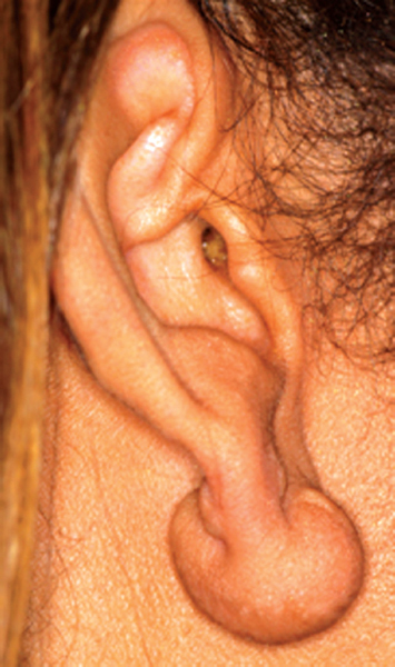
Figure 13.1 Keloid scar of lobule.
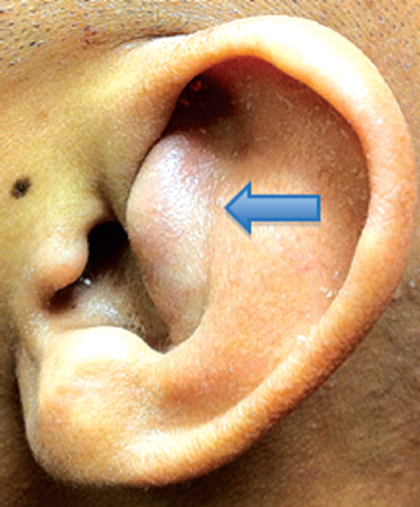
Figure 13.2 Auricular hematoma of the cymba concha (arrow).
Abrasions and Lacerations
Abrasions and lacerations should be treated such that all parts of the auricle are realigned, and infection, notching, tattooing, and other such complications are prevented. First, the wound must be anesthetized and thoroughly cleaned by saline irrigation. Gentle scrubbing will remove small particles.
Any bites, human or animal, that have been present for more than 5 hours must immediately be cleaned, dressed, and antibiotics given for several days before any closure of the wound. Recommended antibiotics are intravenous ampicillin/sulbactam or piperacillin/sulbactam, followed by oral amoxicillin/clavulanate). In addition, rabies vaccination and immunoglobulins and tetanus prophylaxis should be considered. Any highly contaminated or contused ears must also be considered for delayed closure; this helps avoid a suppurative chondritis.
Abrasions
Superficial abrasions should be debrided, with all damaged tissue carefully removed and the ear treated with topical antibiotics then lightly dressed for 2 to 3 weeks, until repithelialization is complete. Deep abrasions may require split thickness skin grafting.
Lacerations
Skin lacerations of the pinna require careful realignment. Skin to skin suture repair achieves adequate results if all skin edges are aligned. Cartilage to cartilage sutures may be required for deeper damage. Exposed cartilage must be covered by vascularized tissue.
Frostbite
A frostbitten auricle should be rapidly rewarmed and protected from further trauma. Thawing of the ear is accomplished by applying sterile pledgets soaked in 38 to 42°C water. Dry heat is not recommended because it is difficult to regulate. The rewarming process is painful and analgesics should be used. Swelling and blisters are likely to form and pressure will rupture the bullae, causing further damage. A topical antimicrobial agent should be applied and tetanus prophylaxis administered. For deep tissue infections, antibiotics are needed. Gangrene may develop in which case debridement should only be done after demarcation.
Burns
Superficial or deep burns can cause deformity or loss of the ear. Burns of the ear must be debrided and covered with healthy tissue as early as possible, before exposure of cartilage. Exposed cartilage, especially in the helix and antihelix, can cause chondritis and irreversible deformity. Early treatment greatly minimizes risk of infection. In patients with extensive body burns, treatment of burned ears is often delayed due to other priorities. But, early surgical intervention can stop progression of damage and save the shape of the extensively burned ear.
Otitis Externa
Bacterial
Acute otitis externa (AOE) is an infection of the EAC primarily caused by bacteria. The EAC is normally protected by continuous outward sloughing of the outer keratin layer. Pooling of moisture, sometimes from cerumen accumulation, serves as a culture medium for infection. Other risk factors such as water sports increase moisture and can cause swelling and damage to the epithelial cells. This can lead to invasion by bacteria such as Pseudomonas aeruginosa a pathogen associated with water, hence the term “swimmer’s ear” associated with otitis externa. Similarly, a foreign object or cotton swab in the ear canal may introduce pathogens, by removing the hydrophobic layer and traumatizing the epithelium. Environmental factors such as hot, humid conditions, obstruction of ear canal, such as by a hearing aid or ear plug also increases susceptibility to AOE.
The most common pathogen in otitis externa is Pseudomonas followed by Staphylococcus and Streptococcus species; diphtheroids, gram-negative rods, and fungal species can also cause infection.
Clinical Manifestation
Stay updated, free articles. Join our Telegram channel

Full access? Get Clinical Tree


